The main constitutive macromolecules of cardio-vascular tissues, essential to the cohesion and resiliency, are also active components which evolve with the physiology and pathology. Their functionality is connected to their internal dynamics over various scales of time and length, in close correlation with water. Vibrational, thermal and dielectric techniques, which have showed their ability to characterize synthetic polymers, deserve to be adapted to the study of proteins and biological tissues. Through some examples, we will show how these techniques are concretely used to answer cardio-vascular problematics.
Fourier Transform Infrared (FTIR) spectroscopy is a powerful technique that has provided accurate results with a high reproducibility in many areas including cardiovascular research [1-3]. Fast analyses can be performed directly on tissues, without the need of staining. Another suitable technique to characterize biological tissues at the submicronic scale is Differential Scanning calorimetry (DSC). It allows to evaluate the hydric organization of tissues [4], as well as proteins thermal stability [3,5,6] directly in aortic tissues [7] or biomaterials [8,9]. It has been also successfully applied to characterize protein components of muscle tissues, such as myosin, actin and sarcoplasmic proteins [10-12] .
Due to the polar character of biological tissues, dielectric techniques are well suited to the analysis of their various levels of organization in interaction with water or in the freeze-dried stat tissues [13]. The molecular mobility of proteins in tissues can be scanned from the nanometer range to some tens of nanometers. In the very low frequency region, Thermo Stimulated Current (TSC) gives information on the molecular origin of the relaxation modes, while Dynamic Dielectric Spectroscopy (DDS) follows protein/water dynamics over a very broad frequency range.
The combination of FTIR, DSC, TSC and DDS on the pure components of cardio-vascular tissues has allowed us to establish a set of structural, dynamic and chemical indicators on the main macromolecules constitutive of these tissues [14,15] . These vibrational, thermal and dielectric markers were then successfully used to determine the evolution of these main proteins produced by lipid- loading smooth vascular cells [16] and cardiomyocytes in culture [3].
In the next section, we will focus on the application of these biophysical techniques to characterize cardio-vascular tissues themselves.
Left ventricle remodeling post-myocardial infarction
Adverse cardiac remodeling after Myocardial Infarction (MI) causes impaired ventricular function and heart failure. If histopathology is commonly used to detect the location, size and shape of MI sites, information about chemical composition and physical structure of infarct zones post-MI is limited. FTIR/ATR can evidence alteration of key cardiac components post-MI in a mice model [17] . As shown in figure 1, the FTIR spectra of freeze-dried mice left ventricles shows amide I and II as major absorptions bands. Moreover, collagen possesses a specific band at 1338 cm-1 [18] that can be used to compile a collagen/proteins indicator. Finally, the sur-resolution of FTIR spectra by the Fourier Self Deconvolution (FSD) method in the amide I/II zone is useful to determine the secondary structure of proteins [19]. The increase of collagen indicator associated with the predominance of triple helical conformation of proteins in infarcted myocardium evidences the deep remodeling of post-MI tissues (collagen deposition in the scar maturation of infarcted zones).
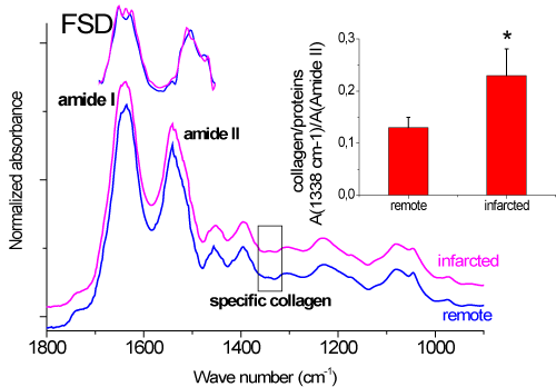
Figure 1. Vibrational characterization (FTIR/ATR) of remote and infarcted zones in mice ventricles.
Ventricular remodeling in tachycardia-induced dilated cardiomyopathy
In non-ischemic Dilated Cardiomyopathy (DCM), the main clinical manifestations are progressive heart failure, ventricular and supraventricular arrhythmias, thromboembolisms and sudden death. It also constitutes the most common cause of heart failure reported for cardiac transplantation. Biophysical markers can be used to identify molecular and conformational alterations of cardiac remodeling in a pig model of DCM [20]. Using FTIR, it is shown that the myofiber/collagen ratio is reduced in ventricles from dilated hearts, while the carbohydrate/lipid ratio is upregulated in left and right ventricles (in a greater extent in the right ventricle). Additional information is provided from calorimetric analysis; in peculiar the depression of the onset melting temperature of freezable water, is indicative of an alteration in the tissue architecture of dilated ventricles, while the shift of the protein denaturation temperature evidences newly synthesized collagen and protein of lower stability. The combination of these data supports that both accumulation of collagen and thermal instability of myofibers and ECM proteins contribute to the imbalance in the myofiber/collagen ratio and to the accumulation of unorganized and agglomerated collagen in dilated ventricles.
Abdominal aortic aneurysms (AAA)
The pathogenesis of AAA, responsible for 2% of human deaths in industrialized nations, remains uncertain, and the lack of definitive insight suggests a complex, multifactorial process. For this thermal/dielectric study [21] , specimens were collected from the anterior aspect of the infrarenal abdominal aorta during operation for non-specific Abdominal Aortic Aneurysms (AAA) in 4 patients. DSC thermograms of freeze-dried atheroma plaques are plotted in figure 3A. It is noteworthy that in contrast with safe aortic wall which is characterized by both elastin and collagen thermal answers (elastin glass transition at 205°C and collagen denaturation at 230°C), in atheroma plaques the collagen signal is predominant. Moreover, the collagen denaturation signal is multiple in 3/4 of pathologic tissues, what can be explained by the accumulation of neo synthesized collagen as evidenced by Gargiulo et al. [22] and sharp increase of C-telopeptide fragments in AAA wall [23] associated with collagen degradation by cysteine proteases.
To get insight the dynamics of atheroma plaque, the TSC spectra of rich control tissue and aneurysmal tissue have been compared (Figure 3B): an enhanced molecular mobility is evidenced for pathological tissue, corroborating the accumulation of neo synthesized or fragmented collagen.
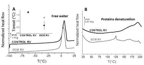
Figure 2. Thermal characterization (DSC) of healthy and dilated pig ventricles (A: hydrated state; B: freeze-dried state).
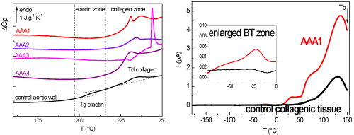
Figure 3. Thermal (A: DSC) and dielectric (B: TSC) characterization of human atheroma plaques.
In this last case biophysical techniques are used to characterize right and left ventricles from patients suffering from cardiomyopathy. This current study involves 16 patients, and the objective of this study is to compare thermal/vibrational indicators according to the localization (right/left ventricles, remote or infarcted zone) and the pathology (control/non-ischemic/ischemic cardiomyopathy). The feasibility of vibrational and thermal techniques to analyse such human cardiac tissues has been demonstrated and relevant markers can be extracted. Multivariate data analysis is now in progress to correlate biochemical and biophysical markers in this clinical study.
The molecular, conformational and physical characterization of the cardio-vascular tissues (aortic wall, pericardium, myocardium) has emerged as a novel approach to study remodeling in diseased tissues. The vibrational FTIR spectra of cardiovascular tissues can be correlated to the vibrational answer of the different components, allowing to quantify them and to discriminate the different secondary structures of proteins. Using calorimetry and dielectric techniques, the thermal transitions and dynamics of free water, elastin, collagens or muscle proteins can be detected in cardiovascular tissues, evidencing a strong correlation between chain architecture and mesophase organization. The evolution of these indicators with pathology (cardiomyopathy, infarction, atherosclerosis) completes biochemical analysis and contributes to a better knowledge of the involved mechanisms. It is also promising to optimize the conception of substitutive biomaterials in the cardiovascular research.
- Kuwahara M, Bannai K, Segawa H, Miyamoto K, Yamato H (2014) Cardiac remodeling associated with protein increase and lipid accumulation in early-stage chronic kidney disease in rats. Biochim. Biophys 1842: 1433-1443. [Crossref]
- Marzec KM, Rygula A, Gasior-Glogowska M, Kochan K, Czamara K, et al. (2015) Vascular diseases investigated ex vivo by using Raman, FT-IR and complementary methods. Pharmacol Rep 67: 744-750. [Crossref]
- Samouillan V, Revuelta-López E, Dandurand J, Nasarre L, Badimon L, et al. (2014) Cardiomyocyte intracellular cholesteryl ester accumulation promotes tropoelastin physical alteration and degradation. Int J Biochem Cell Biol 55: 209-219. [Crossref]
- Aktas N, Tülek Y, Gökalp HY (1997) Determination of freezable water content of beef semimembranous muscle DSC study. J Therm Anal 48: 259-266.
- Samouillan V, Dandurand J, Lacabanne C, Thoma RJ, Adams A, et al. (2003) Comparison of chemical treatments on the chain dynamics and thermal stability of bovine pericardium collagen. J Biomed Mater Res A 64: 330-338. [Crossref]
- Miles CA, Ghelashvili M (1999) Polymer-in-a-box mechanism for the thermal stabilization of collagen molecules in fibers. Biophys J 76: 3243-3252. [Crossref]
- Samouillan V, Dandurand-Lods J, Lamure A, Maurel E, Lacabanne C, et al. (1999) Thermal analysis characterization of aortic tissues for cardiac valve bioprostheses. J Biomed Mater Res 46: 531-538. [Crossref]
- Lee J, Pereira C, Abdulla D, Naimark WA, Crawford I (1995) A multi-sample for collagenous denaturation biomaterials temperature tester. Mol Eng Phys 17: 115-121. [Crossref]
- Samouillan V, Delaunay F, Dandurand J, Merbahi N, Gardou JP, et al. (2011) The use of thermal techniques for the characterization and selection of natural biomaterials. J Funct Biomater 2: 230-248. [Crossref]
- Wright DJ, Wilding P (1984) Differential scanning calorimetric study of muscle and its proteins: myosin and its subfragments. J Sci Food Agric. 35: 357-372. [Crossref]
- Dergez T, Könczöl F, Farkas N (2005) DSC study of glycerol-extracted muscle fibers in intermediate states of ATP hydrolysis. J Therm Anal Calorim 80: 445-449.
- Tamilmani P, Pandey MC (2016) Thermal analysis of meat and meat products. J Therm Anal Calorim 123: 1899-1917.
- Samouillan V, Dandurand J, Tintar D (2011) Dielectric relaxations in biological tissues as a function of physiology and pathology. Proc Int Symp Electrets.
- Samouillan V, Lamure A, Maurel E, Dandurand J, Lacabanne C, et al. (2000) Dielectric characterization of collagen, elastin, and aortic valves in the low temperature range. J Biomater Sci Polym Ed 11: 583-598. [Crossref]
- Samouillan V, Dandurand J, Lacabanne C, Hornebeck W (2002) Molecular mobility of elastin: effect of molecular architecture. Biomacromolecules 3: 531-537. [Crossref]
- Samouillan V, Dandurand J, Nasarre L, Badimon L, Lacabanne C, et al. (2012) Lipid loading of human vascular smooth muscle cells induces changes in tropoelastin protein levels and physical structure. Biophys J 103: 532-40. [Crossref]
- Samouillan V, Revuelta-López E, Soler-Botija C, Dandurand, Benitez-Amaro A, et al. (2017) Conformational and thermal characterization of left ventricle remodeling post-myocardial infarction. Biochim Biophys Acta Mol Basis Dis 1863: 1500-1509. [Crossref]
- Cheheltani R, Rosano JM, Wang B, Sabri AK, Pleshko N, et al. (2012) Fourier transform infrared spectroscopic imaging of cardiac tissue to detect collagen deposition after myocardial infarction. J Biomed Opt 17: 56014. [Crossref]
- Kong J, Yu S (2007) Fourier Transform Infrared Spectroscopic Analysis of Protein Secondary Structures Protein FTIR Data Analysis and Band Assign- ment. Acta Bioch Biophys Sin 39: 549-59. [Crossref]
- Benitez-Amaro A, Samouillan V, Jorge E, Dandurand J, Nasarre L, et al. (2018) Identification of new biophysical markers for pathological ventricular remodelling in tachycardia-induced dilated cardiomyopathy. J Cell Mol Med 22: 4197-4208. [Crossref]
- Samouillan V, Dandurand J, Lacabanne C, Stella A, Gargiulo M, et al. (2010) Characterization of aneurysmal aortas by biochemical, thermal, and dielectric techniques. J Biomed Mater Res A 95: 611-619. [Crossref]
- Gargiulo M, Stella A, Spina M, Faggioli G, Cenacchi G, et al. (1993) Content and turnover of extracellular matrix protein in human "nonspecific" and inflammatory abdominal aortic aneurysms. Eur J Vasc Surg 7: 546-553. [Crossref]
- Palazzuoli A, Alberto P, Gallotta M, Guerrieri G, Quatrini I, et al. (2008) Prevalence of risk factors, coronary and systemic atherosclerosis in abdominal aortic aneurysm: comparison with high cardiovascular risk population. Vasc Health Risk Manag 4: 877-883. [Crossref]