Such cells which retain a chromosomal aberration, for several cell cycles (mitotic divisions) and even may pass them to next generations (by meiosis) are termed as “rogue” cells. Rogue cells, may trigger to result in many variables. Translocations are more predominantly effective and among them “Levanian translocations” have been particularly found to be more frequent than other complex translocations. Levanian translocation is one, in which a chromosome is attached to a nonhomologus chromatid. This rare translocation has been recorded in natural population of plants and animals including man.
rogue cells, rare translocations, natural plant populations and translocation, translocations and malignancy
Chromosomal aberrations have played very significant role in evolutionary events in the biological system for millions of years. Except duplication, translocation and inversion often result after two breaks while deletion may have a minimum of one break. Chromosome translocations refer to transfer of a chromosome or segment or part thereof, to any non-homologous chromosome. Chromosome translocations are very important aberrations and are believed to play a major role in speciation among plants and animals including Man (Homo sapiens). Translocations are chiefly responsible to make a cell ‘different” from being normal, and such cells with breaks and or structurally altered chromosomes have been termed as “Rogue” cells [1]. Translocations are also understood to be the cause or consequences of many pathological diseases, syndromes including malignancy among humans [2-8].
This communication emphasizes on a new category, Levanian translocation along with a very significant aberration, “ Marker Dot” (chromatin dots expelled by some or the other chromosome) which are molecularly installed and maintained within some cells and do persist even after many cell divisions. Marker dots are positive indicators of chromosomal mutagenesis [3-5]. Levanian translocation (transfer of a chromosome on one chromatid of a nonhomologous chromosome [3-6; Figures 1 & 2] often results in “appearance” of one or more chromatin dots within the cells. Since marker dots persist in future generation of many cells (even in natural populations of plants) these supernumerary structures must be capable of replicating thereby clearing meiotic barriers as laggards. This is unknown though, but may be, these could be precursors of B chromosomes (?) arising in some plant and animal populations. Marker dots are also observed in cells which have different categories of translocations and also in those cells which do not display any kind of aberration.
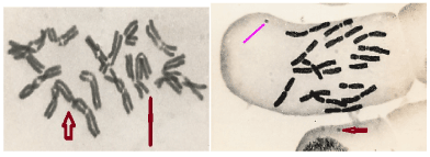
Figure 1A. Allium sativum chromosomes (2n=16) showing Levanian translocation after treatment of Urethane; Arrowed (After Goswami, 1983)
Figure 1B. Allium sativum (2n= 16 chromosomes as revealed in C-metaphase) in root cells show a chromatin dot released after urethane treatment (arrowed)
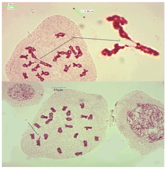
Figure 2A. Rogue cells in natural population of Plants (Courstey: Dr Iruduraj) A. Meiosis I: Diplotene in the spore mother cell of Osmunda hugelina (a fern: 2n=44; n= 22) showing exact transfer of two different chromosomes; each one translocated on different chromosome of another chromosome of the bivalent
Figure 2B. Meiosis I. Early Diakinesis in Osmunda hugelina: A bivalent showing attachment of a chromatid at the terminal end of a chromosome (chromatid end). Courstey: Dr Iruduraj
Chromosome translocations are not uncommon in natural populations of plants and animals and such transfer of a chromosome or a part thereof, on a nonhomologous chromosome in human cells often is associated with some or the other pathological condition. Translocations are of more than a dozen types [2,3,7-9] and the frequency of occurrence is often correlated with lot many diseases among humans. That the chromosomes can break and also translocate often due to some or the other abnormal chemical reaction was best explained by Levan and his colleagues [10-13] by exposing roots of Allium cepa (onion) and A. sativum (garlic) roots and their experiments emphatically correlated the translocations as possible affiliations to malignancy. That chromosome aberrations are always associated with cancer-onset mechanisms was originally propounded by Boveri [14]. Series of such experiments had demonstrated that various chemicals, including carcinogens do result in many chromosomal aberrations which are also encountered in cancer cells naturally occurring among humans. On the basis of similar experiments I could also find that in garlic root cells exposed to, urethane (a carcinogen; kindly sent by Late Professor Albert Levan) many aberrations were seen but the most frequent was regular record of linear transfer-attachment of a chromosome on the chromatid of a chromosome. Such a translocation was never reported earlier which was too peculiar. Since this was repeatable on Allium sativum chromosomes, this rare translocation (Figure 1A) was designated as “ Levanian translocation”. Then alive professor Albert Levan suggested me (personal communication) to postpone this naming of the rare translocation in his honour until, definite chromosome configuration is obtained and figured adequately and also the validity of the translocation should be acceptable only when such a chromatid-chromosome translocation is detected in the naturally occurring populations.
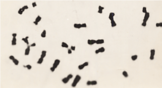
Figure 3. A part of somatic metaphase in a human lymphocyte showing two acrocentric chrmosomes getting attached to the different chromatid of the large acrocentric chromosome (After Goswami; Exhibiting Levaniann translocation with other chromosomal aberrations in a man. This person was seriously exposed to MIC in Dec 1984; died in 1999)
Not only our slides of chromosome preparations based on studies on human lymphocytes but also published old papers and books were searched for such figures. The truth discovered was that such a complex figure of chromosomes was ignored and most workers were assessing on the basis of other aberrations. Exactly same chromosomal configurations were figured by various workers by earlier publications [7-9,15-17] who had reported or incorporated figures from other’s work on chromosome aberrations among human subjects signifying various disorders but such figures were ignored by authors. But now these chromatin structures are termed as small supernumerary marker chromosome (s). Many such chromosomal mentioning positive relationships with “levanian translocations, marker dots and malignancy aberrations have been recently reviewed by Goswami [18,21].
Rogue cells in plant populations
This paper also offers an additional valid documentation that not only human chromosomes but even naturally occurring plant population (Figures 2 A & B) may exhibit such a rare kind of genuine attachment of a chromosome –chromatid translocation, termed as Levanian translocation. This extremely rare chromosome translocation has been observed in a few plants belonging to Osmunda hilsenbergii Grev. & Hook. Such a rare translocation was also observed by Zhang et al [22] in the Royal fern Osmunda banksiifolia (Presl) Kuhn but they did not mention anything in particular about transferring of a chromatid on chromosome. Normally, this is considered to be abnormal meiosis offering explanation for irregular segregation. But, biologically, each chromosome is packed at both the ends and linear attachment of a chromatid with another chromatid of an intact chromosome is an extremely rare phenomenon demanding breaks at the telomeric end. As the natural populations of plants do present sometimes variants exhibiting chromosomal aberrations which persist in subsequent generations, we can assume that rogue cells are also encountered among plants, obviously offering new path for speciation. May be, also the prevalence of rogue cells may cause obstacles in future survival due to aberrant segregations in cell divisions.
A few pertinent observations (Figures 3A & B) have also been discussed in papers published on human chromosomes and the zest of all these observations lies on the fact that while Levanian Translocation is a definite kind of translocation, the net outcome appears to be the appearance of marker dot/ small supernumerary chromosome. The marker dot is also seen in many other translocations and has also been seen in those cells which do not display a typical translocation, no matter the cell belongs to a plant (Figures 1B; 3B) in natural population or under some external stress (so also in human lymphocytes). The appearance of a marker dot may also be an outcome of any intrachromosomal mechanism which enforces release of chromatin dot (Induced heterochromatinization or DNA mehylation; may be both?) from any specific chromosome.
Specific identification
While we have countless information on each category of aberration affecting speciation among plants and animals singly or collectively, translocations have been of vital importance often becoming deleterious for survival or at least, influencing some or the other evolutionary change. In human populations these marker dots [4] have been found to be present among less than 5% seemingly normal persons but often, associated with many pathological conditions including cancers [5,6,18]. Several banding and other methods of identifying chromosomal aberrations [15-19] have been discovered and lately many workers have identified small supernumerary marker chromosomes and most of them are employing various methods of banding techniques [18-21]. Sawyer et al [20] have studied by employing multicolour spectral karyotyping (SKY) on patients with multiple myeloma (MM) that showed complex clonal chromosome aberrations which were not fully characterized by G-banding. Detailed molecular cytogenetic studies are needed so as to discover the mechanism due to which certain specific chromosomes (of plants or animals) release chromatin dots of variable sizes, sometimes appearing like a small chromosome, not only due to an external influence but also, in natural populations.
- Awa, A.A.and Neel, J.V. 1986. Cytogenetic “rogue” cells: what is their frequency, organic, and evolutionary signification? Proc Nati Acad Sci USA 83: 1021-1025.
- Borgaonkar, D S. 1991. chromosomal variations in Man: A Catolog of chromosomal variants and anamolgies .5th Edtn Alan Liss New York.
- Goswami, H. K. 1983. Is chromosomal involvement obligatory to cancer? In, Genetics & Public Health. Catholic Press, Ranchi.
- GoswamiHK .1986. Cytogenetic effects of Methyl Isocyanate exposure in Bhopal. Human Genetics (Berlin) 74: 81-84.
- Goswami H K (2006) Levanian translocation: An entity among translocations. Bionature. 26: 87-89.
- Goswami, H. K., Rangnekar, G. V., Varshney, S., Gandhi, P., Jain, B. and Joshi, A. 1992. Crossed Renal Ectopia with pelvic lipomatosis: A new Syndrome involving Chromosome 1. Human Genetics 89: 666-670 (Springer-Verlag).
- Lewis, R. 1997. Human Genetics: Concepts and applications. WCB, Mc-Graw Hill, New York, USA
- Therman Eva and Susman M. 1993. Human chromosomes: Structure, behaviour and Effects . Springer Verlag 3rd Edin. New York
- Levan A. 1949. The influence on chromosomes and mitosis of chemicals, as studied by the Allium test. Hereditas. 35(1):325–337.
- Levan A,1969. Chromosome abnormalities and carcinogenesis. In Handbook of Molecular cytology. Pp 717-731.Edt A.Lima de Faria, Amsterdam.
- Levan A. 1977. Chromosomes in malignancy. Proc.VI International Chromosome conference. Helsinki.pp 339-343.
- Levan A, Levan G and Mitelman F. 1977. Chromosomes and cancer. Hereditas 86:15-30.
- Boveri, T.1914. Zur Frage der Enstehung maligner Turnoren. Gustav Fischer Jena p. 64.
- Vogel F and Motulsky A G. 1986. Human Genetics: Problems and Approaches (2nd Edn). Springer Verlag, Berlin pp 807.
- Pathak, S. Nemeth, M.A. Mutani, A.S., Thalmann, G.N., Von Eschenbach, A.C. and Chung, L. W.K. 1997. Cancer cells transform normal host cells into malignant cells? British Journal of cancer; 76 (9): 1134-1138.
- Howell, R.T and Taylor, A.M. H. 1992, Chromosome Instability Syndrome, In Human Cytogenetics: A Practical Approach. Edts: D. E. Rooney and B. H. Czepudkowski. Pp 209-234 Oxford Univ. Press.
- Goswami H K. 2020. Many Developmental Errors and or Pathological Conditions May Be Associated with Marker Dots /Small Supernumerary Chromosomes. J Family Med Community Health 7(1): 1169. DOI: 10.13140/RG.2.2.17016.80643
- Ferguson-Smith, M. A., Yang, F. and O’Brien, P.C.M. 1998. Comparative mapping using chromosome sorting and painting. ILAR. J. 39: 200-208.
- Sawyer J R, Lukacs J L, Thomas E L, Swanson M et al. 2001. Multicolour spectral karyotyping identifies new translocations and a recurring pathway for chromosome loss in multiple myeloma. British Journal of Haematology. 112: 167-17
- Goswami, H K. 2021. Genes Chromosomes and Malignancy. Eliva Press. Moldova, Europe.Pp 200.
- Zhang, S. Z., He, Z. C., Fan, C. R. and Yan, B. 2008. A cytogenetic study of five species in the genus Osmunda. J. Syst. & Evol. 46 (4): 490-498.