brain, choroid plexus cysts, xanthogranuloma, magnetic resonance (MR) imaging
Intracranial cysts and cystic-appearing lesions have a broad spectrum in imaging and differential diagnoses and can be challenging for radiologists to assess [1,2]. Choroid plexus cysts (CPC) can be symptomatic, if they are large in size and/or cause obstructive hydrocephalus [1,3]. Histologically, most of these cysts prove to be degenerative xanthogranulomas (XG) [1]. We present a case of a female patient with an incidental finding of bilateral choroid plexus cysts.
A 61-year woman presented to our emergency department with recurrent vertigo, ataxia and paresthesia of the upper and lower left extremity in the last few days. No motor weakness or other neurologic deficits were present. A week before, the patient was examined and treated for a transitoric ischemic attack (TIA) in another institution. Regarding cerebrovascular risk factors, medical history showed a hyperlipidemia, treated with atorvastatin.
Initial non-enhanced computed tomography (NECT) of the cerebrum ruled out intracranial hemorrhage or demarcated ischemia. No hydrocephalus was present. As an additional finding in both atria of the lateral ventricles partially calcified formations with a density of 18 Houndsfield Units (HU), slightly hyperdense to cerebrospinal fluid (CSF, 10 HU) were seen.
Subsequent magnetic resonance (MR) imaging of the cerebrum showed no parenchymal diffusion restriction or contrast enhancing lesions. In an additional MR examination 3D time-of-flight-angiography (3D-TOF) and contrast enhanced MR angiography normal extra- and intracranial vessels were present. On unenhanced T1- and T2-weighted turbo spin-echo (TSE) images, the lesions were almost isointense to CSF. In fluid-attenuated inversion recovery (FLAIR) sequences, the formations in the atria of the lateral ventricles were almost isointense to the cortex and significantly hyperintense compared to CSF. Diffusion weighted imaging (DWI) showed partial diffusion restriction with high signal in diffusion (b = 1000) and low signals in apparent diffusion coefficient (ADC) maps. Following contrast administration, no relevant gadolinium enhancement was observed. On T2-weighted and FLAIR images no tissue edema was found. There were no signs of hydrocephalus.
After completed diagnostic workup the patient was released with the diagnosis of a migraine sans migraine. Since there was no hydrocephalus or tissue edema present, the CPCs were interpreted as an asymptomatic incidental finding.
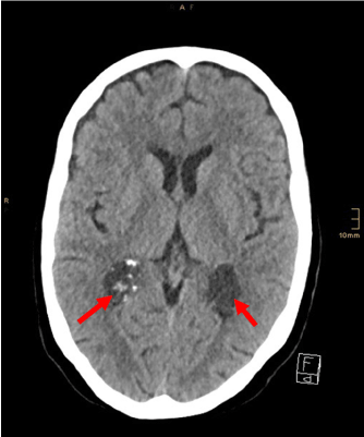
Figure 1. Formations in the atria of the lateral ventricles (arrows), almost isodense to cerebrospinal fluid
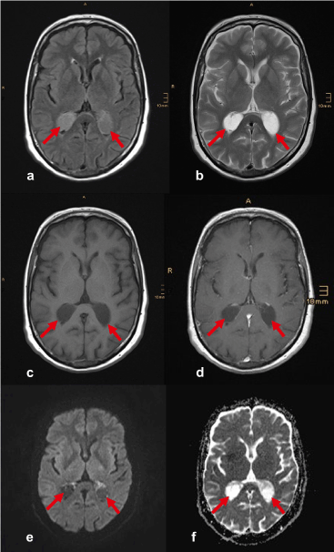
Figure 2. Choroid plexus cysts (arrows) in a) FLAIR, b) T2-weighted TSE, c) T1-weighted SE before, and d) after i.v. administration of contrast agent, e) Diffusion-weighted (b=1000) and f) ADC map images
CPCs are usually benign incidental findings and for the most part asymptomatic [1]. Symptomatic CPCs are mostly close to the foramen of Monro and can cause obstructive hydrocephalus [1,3]. Large CPCs can be symptomatic, if compression of adjoining brain tissue occurs [3]. Histologically, most CPCs are XGs.1 Although the full pathomechanism is not yet fully understood, some authors postulate, that XGs result from lipid accumulation from degenerative changes in the choroid epithelium with cholesterol clefts and inflammatory cells [1,3-5]. Hyperlipidemia, as present in our case, seems to be a risk factor for the development of CPCs [4,5].
On NECT and T1- and T2-weighted MR images, the appearance of CPCs is heterogeneous and depends on the proportions of lipid accumulation, calcification, fibrosis and intralesional hemorrhagic changes [1,4,5]. Thus, it can be challenging for radiologists to make a definitive diagnosis [2,4]. DWI and FLAIR sequences can be helpful in the workup and differentiation of cystic brain lesions [1]. CPCs are usually slightly to moderately hyperintense to CSF on FLAIR sequences, two-thirds show restriction on DWI [1]. Gadolinium enhancement of CPCs is variable and unspecific [4].
Important differential diagnosis are ependymal cysts and villous hyperplasia of the choroid plexus [1]. Other differential diagnosis of intraventricular masses include meningioma, ependymoma, choroid plexus papilloma, arteriovenous malformations (AVM), and metastatic neoplasm [2,4,6]. At the foramen of Monro, colloid cysts are a considerable differential diagnosis [1].
Since CPCs or XGs of the choroid plexus are mostly asymptomatic incidental findings in brain imaging, it is important for radiologists to recognize possible complications such as hydrocephalus or parenchymal edema and exclude treatable conditions such as AVMs. Furthermore, it is important to know the differential diagnoses for cystic and cystic-appearing lesions in the ventricular system to avoid unnecessary imaging and therapy in benign, “leave me alone”-lesions.
No conflicts of interest. No grants or financial supports.
- Osborn AG, Preece MT (2006) Intracranial cysts: radiologic-pathologic correlation and imaging approach. Radiology 239: 650-664.
- Kadota T, Mihara N, Tsuji N, Ishiguro S, Nakagawa H, et al. (1996) MR of Xanthogranuloma of the Choroid Plexus. AJNR 17: 1595-1597.
- Yan C, Zhu S, Sun H, Lee WT, Zhang X, et al. (2019) Neuronavigator-guided ventriculoscopic approach for symptomatic xanthogranuloma of the choroid plexus in the lateral ventricle. Medicine 98: 16(e14718).
- Emon ST, Ozturk E, Meric K, Aker F, Orakdogen M (2017) Symptomatic bilateral xanthogranuloma of the choroid plexus. J Neurosci Rural Pract 8(1): 123-126.
- Chen Y, Hamilton AM, Parkins KM, Wang JX, Rogers KA, et al. (2016) MRI and histopathologic study of a novel cholesterol-fed rabbit model of Xanthogranuloma. J Magn Reson Imaging 44(3): 673-682.
- Shetty J, Pai M (2010) Unusual presentation of xanthogranuloma of the choroid plexus. J Neurosci Rural Pract 1(2): 97-98.