Background: Chronic Anal Fissure (CAF) is a common benign condition that reduces the quality of life. Lateral internal sphincterotomy (LIS) is frequently carried out but leads a significant risk of anal incontinence. Posterior Medline Internal Sphincterotomy (PMIS) is an alternative that in our experience it has involved considerable success. This study is designed to confirm the role of PMIS in the treatment of surgically recurrent chronic anal fissure based on an experience over 30 years considering the complications, costs and hospital stay.
Materials and Methods: A total of 1849 patients was treated with PMIS between January 1982 and December 2018. Chronicity was defined by symptoms persisting more than 4-6 weeks despite medical therapy. All patients had proctoscopy before surgery. The sphincterotomy was generally performed with electrocautery through an incision of 3-4 millimetres after local anaesthesia on posterior plane in 86% of patients, the remaining required a spinal anaesthesia. The follow-up of patients was done at 3-6 and 12 months and the first considerations studied were complications like postoperative pain, infections, healing rate of fissure and incontinence rates. Secondary we considered the costs and hospital stay.
Results: All patients reported the disappearance of pain over a period of 3-4 weeks and only a negligible percentage 1,7% (32pt) required a second operation because persistence pain symptoms, 22 patients (1,1%) reported haemorrhage from the small incision resolved spontaneously. None reported infection, recurrence and incontinence. Most patients returned home about one hour after surgery, while a small number of them required more hours but nobody slept in the hospital.
Conclusion: The Authors retains the PMIS safe and effective performed under local anaesthesia with no increased risk of pain or complications and is best suited for resource-poor surgical setting.
anal fissure, posterior Medline internal sphincterotomy.
The anal fissure is the elongated split in the squamous epithelium of the distal anal canal between the anal verge and the dentate line. These lesions rank second after haemorrhoids as the most frequent affliction in the anal region and their peculiarity is characterised by tendency not to heal. While the aetiology of this condition is unclear [1], numerous investigators have suggested that the internal sphincter plays a major role in the development of the lesion that is the midline ischemia and high resting anal pressure have been proposed aetiologies [2-3]. These theories are still valid today [4].
Anal fissures occur primarily in the median posterior plane. The predominance of this location may be explained by its low anatomical resistance, greater expansibility and elliptical shape (Figure 1) [5]. All anodermal lesions are accompanied by pain and hypertonicity of the internal sphincter. Various conservative treatments have been tested like Glyceryl Trinitrate (GTN) [6], topical nitric oxide (NO) [7], botulin toxin injection (Botox) [8] but without results. Therefore, in case of failure of medical therapy, the patient should be sent to the surgeon.
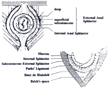
Figure 1. Anatomy of the anus(schematic): the fibres of the external sphincter muscle are attached to the retro anal skin posteriorly along the anal groove to the anococcygeal raphe.
Clinicians have proposed a variety of different techniques of sphincterotomy aimed to reducing this hypertonicity [9-10]. In 1969 Notaras [11] described a simpler method of lateral subcutaneous sphincterotomy which has been widely adopted. In 1978, Oh [12] modified Park’s method of open lateral internal sphincterotomy but this method manifests as incontinence of gas, liquid, or stool. Therefore, the surgeons can be too cautious in its use making ineffective superficial incision or avoiding the operation altogether. We report the results of our personal experience with posterior midline internal sphincterotomy with a clinic follow-up at 3-6-12 months of patients.
This retrospective study was conducted between January 1982 and December 2018. A total of 1849 adult patients with CAF were operated because persistent anal pain after the failure of medical therapy lasting 4-6 weeks. The patients were aged between 28 and 82 years (mean 57) and all underwent preoperative proctoscopy before surgery to document the persistent of the lesion. All patients were referred to surgery after failing conservative treatment more than 6-8 weeks duration. The therapy included the intake of lactulose oil after meals to facilitate the evacuation and the use of anal dilators (Dilatan) by applying xylocaine 2% ointment. All patients were operated upon by the same surgeons (the Authors) using a uniform method with local anaesthesia (LA) in 86% and spinal injection in 14% of the patients. For this second group detailed blood work-up like haemoglobin, blood sugars, renal function test and electrolytes were done before undergoing fitness for anaesthesia. For patients in LA group, only blood tests for coagulation were done as they were not required to undergo detailed anaesthetic work-up.
Operative technique
The patients were asked to assume the lithotomy position and the perianal sphincter area was shaved with iodine solution. The anaesthetist was in stand-by for all operations. The left index finger inserted in the anal canal as a guide and the submucosal plane was infiltrated with 5cc of bupivacaine hydrochloride 0,5% with adrenalin and lidocaine hydro chlorine 2% in equal amounts using a 25G needle. After a dilatation of the anal canal gently which allows the direct vision of the lesion, a 0,5cm. incision is made on the posterior margin of the sphincter at the level of the base of the lesion. After thorough haemostasis, the fibres of the internal sphincter were separated using a baby dissector and affecting also the fissure. This was carried-out with the index finger inserted in the anal canal in order to avoid any laceration of the mucosa. The internal sphincter is then incised under direct vision (Figure 2) with a cautery. A pressure is applied on the mucosa for few minutes for the complete haemostasis and the wound was packed with haemostatic gauze which was removed after the first evacuation. The patients were asked to maintain normal toilet habits and the wound usually healed within 5-7 days.

Figure 2. The internal sphincter incised under direct vision with a cautery.
The symptoms and sites of the fissure are listed in table 1. The results after operative treatment are shown in table 2.
Table 1. Symptoms and sites of the fissure
Principal symptoms |
Number of patients |
% |
Pain |
1849 |
100 |
Haemorrhage |
1568 |
84.8 |
Perianal itching |
423 |
22.8 |
Site of fissure |
|
|
Posterior |
1682 |
89.9 |
Lateral |
141 |
7.6 |
Anterior |
|
|
Table 2. Results after operative treatment
Healing of the fissure |
100% |
Time of healing (week) |
3-4 |
Disappearance of pain (days) |
2-3 |
Hospitalization (days) |
- |
Recurrence (4 pt) |
0.2% |
The majority of patients are cured completely except for 32 of them (1,7%) that after few months continued to complain of anal pain after evacuation. In fact, for them it was necessary a second operation of resphincterotomy performed always with the same modalities. Twenty-two patients (1,1%) had modest bleeding from the small incision resolved spontaneously after a few days. Nine patients (0,4%) complained of a moderate anal incontinence for gas but everything is regressed within 4-6 weeks. For these patients was performed anorectal manometry at 6 months after operation in order to exclude permanent injury of the anal sphincter. The pain was not intentionally included among the complications because it was present in all patients postoperatively but however with lower intensity in comparison with preoperative period. The clinic control with proctoscopy was made after 2-6-12 months in all patients it showed complete healing and epithelisation of the fissure. There has been no recurrence.
The anatomy of the terminal portion of the anal canal together with the defecation mechanism can explain the aetiology of the anal fissure and its frequent location in the posterior wall [13]. Anal fissures are accompanied by spasms of the internal sphincter, a phenomenon amply documented by manometric data [14-15].
The cause of this disease still remains unclear and many studies have suggested various etiopathogenic theories for anal fissure, and so a multifactorial origin for this condition like constipation, diarrhoea, vascular or infection condition is most likely [16-17]. Irrespective of whether this condition is a consequence or a cause of the fissure, it is maintained by the reflex stimulus induced by the fissure itself and by ampullary reflexes resulting from incomplete defecation. This is one explanation for the pain patients experience during and after defecation. A great variety of therapeutic methods for the treatment of chronic fissure have been proposed when conservative treatment fails: non-surgical procedures, as topical treatment such as botulinum toxin [18-19], nitrate preparation [20-21], and nifedipine [22]; and surgical approach such as anal dilatation [23-24], sphincterotomy [25-26], and advanced flap [27].
All these techniques aim at a high rate of healing in association with a low morbidity rate. Historically the LIS has been proven the procedure of choice in various comparative studies and in the literature only two type of LIS have been widely discussed: open and closed sphincterotomy without statistically significant differences between the two methods [28]. We have always preferred to perform the sphincterotomy through a posterior approach on the midline at first because technically easier, secondly a better control of bleeding in case of haemorrhage. In fact, over time the results obtained given us reason for our choice. On such a large number of patients we have reported a remarkably low percentage of complications which, moreover, did not require further operation. In fact, the patients with haemorrhage in immediate postoperative period (1,1%), anyone needed a reoperation. Likewise, for 0,4% of patients who complained mild incontinence spontaneously regressed. Only 1,7% of patients required a surgical review for a poor section of internal sphincter in our opinion because ipertrophy.
If we want to make a self-criticism, we must recognize that the limit of this retroactive study is the follow-up was done only for one year after surgery, which was not enough to reflect any long-term results or recurrence of the symptoms. On the other hand, we consider that over the time the number of the patients has become uncontrollable and the most of them have not accepted a further follow-up because they were symptom free.
This study, based on the experience gained over 30 years and on a large number of patients, showed that PMIS for the treatment of chronic anal fissure has advantages over other procedures both in terms of postoperative morbidity and surgical feasibility. In fact, it can be performed with local anaesthetic in an outpatient facility and with low-cost. The procedure also allows a direct view of the operative field and therefore easy haemostasis in case of need.
treatment offers a high percentage of healing, rapid remission of symptoms and involves no hospitalization.
Above all I had a low incidence of early and late complications and no recurrences.
- Lund JN, Scholefield JH (1996) Aetiology and treatment of anal fissure.Br J Surg83: 1335-1344. [Crossref]
- Abcarian H, Lakshmanan S, Read DR, Roccaforte P (1982) The role of internal sphincter in chronic anal fissures.Dis Colon Rectum25: 525-528. [Crossref]
- Goligher JC (1982) Surgery of the anus and rectum 4th edition Piccin Publisher.180-203.
- Farouk R, Gunn J, Duthie GS (1998) Changing patterns of treatment for chronic anal fissure.Ann R Coll Surg Engl80: 194-196. [Crossref]
- Oh C, Kark AE (1973) Anatomy of the perineal body.Dis Colon Rectum16: 444-454. [Crossref]
- Lund JN, Scholefield JH (1997) A randomized, prospective, double blind, placebo-controlled trial of glyceryl trinitrate ointment in treatment of anal fissure. Lancet 1997; 349-656.
- Gorfine SR (1995) Treatment of benign anal disease with topical nitroglycerin.Dis Colon Rectum38: 453-456. [Crossref]
- Jost WH (1997) One hundred cases of anal fissure treated with botulin toxin: early and long-term results.Dis Colon Rectum40: 1029-1032. [Crossref]
- Bennett RC, Goligher JC (1962) Results of internal sphincterotomy for anal fissure.Br Med J2: 1500-1503. [Crossref]
- Lund JN, Scholenfield JH (1997) Aetiology and treatment of anal fissure. Br J Surg. 84: 279. [Crossref]
- Notaras (1969) MJ Lateral subcutaneous sphincterotomy for anal fissure; a new technique. Proc R Soc Med 62:713-715. [Crossref]
- Oh C (1978) A modified technique for lateral internal sphincterotomy. Surg Gynecol Obstet 146: 623-625. [Crossref]
- Goligher JC (1984) Surgery of the Anus, Rectum and Colon. 5th edition London: Bailliere Tindall
- Sailer M, Bussen D, Debus ES, Fuchs KH, Thiede A (1998) Quality of life in a patient with benign anorectal disorders. Br J Surg 85:1716-179. [Pubmed]
- Xynos E, Tzortzinis A, Chrysos E, Tzovaras G, Vassilakis JS (1993) Anal manometry in patients with fissure-in-ano before and after internal sphincterotomy.Int J Colorectal Dis8: 125-128. [Crossref]
- Sales R, Martinez P, Lobez T, Culell P (2000) Chronic anal fissure surgery: long-term results. Cir Esp 68: 467-70.
- Klosterhalfen B Vogel P, Rixen H, Mittermayer C (1989) Topography of the inferior rectal artery: a possible cause of chronic, primary anal fissure. Dis Colon Rectum 32: 43-52. [Crossref]
- Maria G, Brisinda G, Bentivoglio AR, Cassetta E, Gui D, et al. (1998) Botulinum toxin injections in the internal anal sphincter for the treatment of chronic anal fissure. Long-term results after two different dosage regimens. Ann Surg 228: 664-669. [Crossref]
- Brisinda G, Maria G, Sganga G, Bentivoglio AR, Albanese A, et al. (2002) Effectiveness of higher doses of botulinum toxin to induce healing in patients with chronic anal fissures. Surgery 131: 179-84. [Crossref]
- Evans JE, Luck A, Hewett P (2001) Glyceryl nitrate vs lateral sphincterotomy for chronic anal fissure. Prospective randomized trial. Dis Colon Rectum 44: 93-97. [Crossref]
- Richard CS, Gregoire R, Plewes EA, Silverman R, Burul C, et al. (2000) Internal sphincterotomy is superior to topical nitro-glycerine in the treatment of chronic anal fissure: results of randomized, controlled trial by the Canadian Colorectal Surgical Trials Group. Dis Colon rectum 43: 1084-1057. [Crossref]
- Cook TA, Humphreys MM, McC Mortensen NJ (2000) Oral nifedipine reduces resting anal pressures and heals chronic anal fissure. Br J Surg 87: 251. [Crossref]
- Gaj F, Trecca A, Crispino P (2006) Efficacy of anal dilators in the treatment of acute anal fissure. A controlled clinical trial.Chir Ital58: 761-765. [Crossref]
- Giovaninetti GD, Manzi M, Marchi L, Ricci G, Rodella L, et al. (1986) Use of anal dilators in the conservative therapy of anal rhagades.Chir Ital38: 666-670. [Crossref]
- Abscarian H (1980) Surgical correction of chronic anal fissure: results of lateral internal sphincterotomy vs fissurectomy-midline sphincterotomy. Dis Colon rectum 23: 31-36. [Crossref]
- Higuero T (2015) Update on the management of anal fissure.J Visc Surg152: S37-43. [Crossref]
- Leong AF, Seow-Choen F (1995) Lateral sphincterotomy compared with anal advancement flap for chronic anal fissure. Dis Colon Rectum 38: 69-71. [Crossref]
- Garcia-Anguilar J, Belmonte C, Wong WD, Lowry AC, Madoff RD (1996) Open vs closed sphincterotomy for chronic anal fissure: long term results. Dis Colon Rectum 39: 440-443. [Crossref]