The sustentaculum tali is a bony eminence at the medial surface of the calcaneus that articulates with the middle calcaneal articular surface of the talus and provides an important support for the medial column of the foot. Its upper articular surface is concave, and its lower surface has a groove for the tendon of the flexor hallucis longus. The sustentaculum tali gives attachment to the plantar calcaneonavicular (spring) ligament on its anterior margin and the tibiocalcaneal ligament and the medial talocalcaneal ligament (part of the deltoid ligament) on its medial margin [1-4].
The os sustentaculi (OS) is a rare accessory bone located at the posterior end of the sustentaculum tali which is usually attached to the calcaneus by fibrous or fibrocartilaginous tissue. The first report of this accessory bone was published by Pfitzner in 1896 [5]. Since then a few case reports have appeared discussing the clinical manifestations and radiographic features of this bony variant [6-8]. We report on the case of an unusual medial malleolar fracture in a patient presenting an OS with a concomitant talocalcaneal synchondrosis. Due to subtalar motion restriction an abnormal force may have been applied to the ankle mortise causing the atypical fracture.
An 11-year-old boy was admitted to our hospital after a right ankle sprain. The mechanism of injury was a combination of eversion movement and external rotation. He immediately complained of severe pain over the medial malleolus. Clinical examination determined swelling and bruising on the medial side of his ankle and full weight bearing was impossible. Anteroposterior and lateral radiographs revealed a fracture of the anterior aspect of the medial malleolus. A CT-scan was conducted to accurately evaluate the size and the morphology of the fracture. The exam confirmed the radiographic findings but revealed also an os sustentaculi with a concomitant incomplete talocalcaneal coalition (Figure 1 and 2).
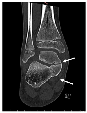
Figure 1. Coronal CT images of the right ankle: coalitions are seen between the sustentaculum tali and the os sustentaculum tali, and between the os sustentaculum tali and the talus (arrows)
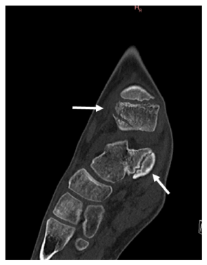
Figure 2. Sagittal CT image of the right ankle: the arrow highlights the avulsion fracture of the anterior tibiotalar and tibionavicular ligaments from the anterior aspect of the medial malleolus
Open reduction and internal fixation of the malleolar fracture was performed under general anaesthesia using an anteromedial approach; the fragment was stabilized with a 2.7 mm screw. The ankle was then immobilized in an under-the-knee cast for 6 weeks, followed by physiotherapy instructed rehabilitation.
Talocalcaneal coalition (TCC) is defined as a union between the talus and the calcaneus, usually associated with an abnormal hypertrophy of the medial aspect of the talus and the sustentaculum tali. TCC is diagnosed in less than 1% of the general population [8]. However, as the bony variation is often asymptomatic the real prevalence is probably much higher [9]. In the paediatric population, talocalcaneal coalition is of congenital origin and results from a failure of mesenchymal segmentation of calcaneus and talus [10]. Usually, this condition becomes symptomatic in the second decade of life [11-12]. A TCC can present itself as a synostosis, as a synchondrosis, or as a syndesmosis [13-16]. Furthermore, it can be classified according to its location, as intraarticular (affecting either anterior, medial, or posterior facets) or extraarticular (usually posteromedial) [17-18]. Recently, a new classification of TCC has been established using multiplanar CT or MRI scans [16]. Surprisingly, the scans showed that an os sustentaculum occurred more often in conjonction with a TCC than previously thought. Furthermore, the coalition was frequently determined between the sustentaculum tali and the os sustentaculum, and between the os sustentaculum and the talus [16] as was the case in our patient.
The mechanical characteristics of the ankle are complex [19-21], due to the two contributing joints. The principal motion of the talocrural joint is in the sagittal plane, whereas the subtalar joint moves in the frontal plane [10]. The subtalar complex is defined as a polyaxial articulation, whose range of motion accounts for 70% to 90% of the overall range of motion of the ankle complex [19]. Therefore, patients suffering from subtalar joint coalitions display a severe restriction of subtalar joint motion. In the absence of subtalar joint motion an excessive rotational stress is exerted on the talocrural joint, especially during inversion movements. This mechanical stress may cause peroneal spastic flatfoot [22-24] or may result in the development of a ball and socket joint at the level of the talocrural joint [10]. It is also known that a motion restriction at the subtalar joint increases the mechanical stress on collateral ligaments of the ankle which can result in frequent ankle sprains [9].
The injury mechanism in our patient was classified as pronation-eversion-external rotation stage I in the Lauge-Hansen classification. The morphologic features of the fracture corresponded to an avulsion fracture of the anterior tibiotalar and tibionavicular ligaments from the anterior aspect of the medial malleolus. It is unanimously recognized that the main cause of an isolated injury of the anterior tibiotalar and tibionavicular ligament is pronation and external rotation of the hind & midfoot. The TCC in our patient restricted the range of motion in the subtalar joint and we therefore believe that the rotational force was not absorbed properly. Thus, the excessive rotational force may have been transmitted to the talocrural joint, leading to an overload of the superficial deltoid ligament, which resulted in an avulsion fracture of the anterior aspect of the medial malleolus.
In our review of the published data, we found only a few studies that described unusual fracture patterns of the ankle in patients presenting with a TCC. Imade et al reported the case of a 16-year-old male who experienced a bimalleolar fracture of the ankle, classified as a supination-adduction type in the Lauge-Hansen classification [25]. Compared to the typical supination-adduction type fracture, the authors considered that the TCC may have caused more severe injury as the medial malleolar fracture was positioned more laterally and had changed to a spiral fracture [25]. Godoy and al reported the case of 53-year-old female who sustained an isolated intraarticular tibial pilon fracture, without lesion of the medial and lateral malleoli, and intact syndesmosis. In this case, computed tomography revealed both talocalcaneal and talonavicular coalitions [10].
In conclusion, we consider that patients with TCC should undergo a full radiographic evaluation as altered biomechanics can cause atypical fractures and increase the risk of more significant ankle injuries.
We have no conflict of interest to declare.
The authors received no specific funding for this work.
- Olexa TA, Ebraheim NA, Haman SP (2000) The sustentaculum tali: anatomic, radiographic, and surgical considerations. Foot Ankle Int 21: 400-403. [Crossref]
- Gitajn IL, Toussaint RJ, Kwon, JY (2013) Assessing accuracy of sustentaculum screw placement during calcaneal fixation. Foot Ankle Int 34: 282-286. [Crossref]
- Gitajn IL, Abousayed M, Toussaint RJ, Ting B, Jin J (2014) Anatomic alignment and integrity of the sustentaculum tali in intra-articular calcaneal fractures: is the sustentaculum tali truly constant? J Bone Joint Surg Am 96: 1000-1005. [Crossref]
- Pazour J, Křivohlávek M, Lukáš R (2016) Positions of sustentacular screw in osteosynthesis of calcaneal fractures: clinical and radiographic study. Acta Chir Orthop Traumatol Cech 83: 182-185. [Crossref]
- Pfitzner W (1896) Beiträge zur Kenntnis des menschlichen Extremitatenskeletts VII. Die Variationen im Aufbau des Fussskeletts. Schwalbes Morphol Arb 6: 245.
- March HC, London RI (1956) The os sustentaculi. AJR 96: 1114-1118.
- Sarrafian SK (1993) Anatomy of the foot and ankle, 2nd edn. Philadelphia: Lippincott. 1-112.
- Tsuruta T, Shiokawa Y, Kato A, Matsumoto T, Yamazoe Y (1981) Radiological study of the accessory skeletal elements in the foot and ankle. Nihon Seikeigeka Gakkai Zasshi 55: 357-70. [Crossref]
- Bellapianta JM, Andrews JR, Ostrander RV (2011) Bilateral Os Subtibiale and Talocalcaneal Coalitions in a College Soccer Player: A Case Report. J Foot Ankle Surg 50: 462-465. [Crossref]
- Godoy HM, Micciche MJ (2017) An Incidental Finding of a Talonavicular and Talocalcaneal Joint Coalition After a Tibial Pilon Fracture: A Case Report. J Foot Ankle Surg 56: 1332-1334. [Crossref]
- Stormont DM, Peterson HA (1983) The Relative Incidence of Tarsal Coalition. Clin Orthop Relat Res 181: 28-36. [Crossref]
- Newman JS, Newberg AH (2000) Congenital Tarsal Coalition: Multimodality Evaluation With Emphasis on CT and MR Imaging. Radiographics 20: 321-32. [Crossref]
- Cowell HR (1972) Talocalcaneal coalition and new causes of peroneal spastic flatfoot. Clin Orthop Relat Res 85: 16-22. [Crossref]
- Scranton PE Jr (1987) Treatment of Symptomatic Talocalcaneal Coalition. J Bone Joint Surg Am 69: 533-539. [Crossref]
- Crim JR, Kjeldsberg KM (2004) Radiographic Diagnosis of Tarsal Coalition. AJR Am J Roentgenol 182: 323-328. [Crossref]
- Yun SJ, Jin W, Kim GY, Lee JH, Ryu KN, et al. (2015) A Different Type of Talocalcaneal Coalition With Os Sustentaculum: The Continued Necessity of Revision of Classification. AJR Am J Roentgenol 205: 612-618. [Crossref]
- Downey MS (1991) Tarsal Coalitions. A Surgical Classification. J Am Podiatr Med Assoc 81: 187-197. [Crossref]
- Lim S, Lee HK, Bae S, Rim NJ, Cho J (2013) A radiological classification system for talocalcaneal coalition based on a multi-planar imaging study using CT and MRI. Insights Imaging 4: 563-567. [Crossref]
- Leardini A, Stagni R, O'Connor JJ (2001) Mobility of the Subtalar Joint in the Intact Ankle Complex. J Biomech 34: 805-809. [Crossref]
- Siegler S, Udupa JK, Ringleb SI, Imhauser CW, Hirsch BE, et al. (2005) Mechanics of the Ankle and Subtalar Joints Revealed Through a 3D Quasi-Static Stress MRI Technique. J Biomech 38: 567-578. [Crossref]
- Stagni R, Leardini A, O'Connor JJ, Giannini S (2003) Role of Passive Structures in the Mobility and Stability of the Human Subtalar Joint: A Literature Review. Foot Ankle Int 24: 402-409. [Crossref]
- Leonard MA (1974) The inheritance of tarsal coalition and its relationship to spastic flat foot. J Bone Joint Surg Br 56B: 520-526. [Crossref]
- Jayakumar S, Cowell HR (1977) Rigid Flatfoot. Clin Orthop Relat Res 122: 77-84. [Crossref]
- Takakura Y, Tanaka Y, Kumai T, Sugimoto K (1999) Development of the Ball-And-Socket Ankle as Assessed by Radiography and Arthrography. A Long-Term Follow-Up Report. J Bone Joint Surg Br 81: 1001-1004. [Crossref]
- Imade S, Takao M, Nishi H, Uchio Y (2007) Unusual malleolar fracture of the ankle with talocalcaneal coalition treated by arthroscopy-assisted reduction and percutaneous fixation. Arch Orthop Trauma Surg 127: 277-80. [Crossref]