Introduction: Specialized studies say that tears Achilles tendon injury is one of the pathologies with the highest incidence in the spectrum of orthopedic pathologies nature. This particular is interest to motor age groups, with or without specific physical training and activity ranging from sedentary to intense physical activity. The management of therapy is the paramount importance, as the decision between surgical and conservative treatment and often is not easy in practice.
Case presentation: A 14-year-old female patient was presented to the ER (emergency room) for pain in the left Achilles tendon, which is associated with functional impotence with plantar flexion.
The patient was carefully examined and investigated, and the surgery was decided to restore functional integrity.
Conclusion: Although, the Achilles tendon (the tendon of the sural triceps muscle), is one of the strongest tendons, with an average length between 9-11 centimeters, having the largest diameter in the human body, can suffer a rupture at an early age.
The particularity of this case consists in solving the rupture of the Achilles tendon by suturing with allograft taken from the tendon of the plantar muscle. The patient's evolution was satisfactory with the resumption of the functionality of the left lower limb.
Achilles tendon rupture, tendon suture, positive Thompson sign, allograft
Specialized studies say that tears Achilles tendon injury is one of the pathologies with the highest incidence in the spectrum of orthopedic pathologies nature. This particular is interest to motor age groups, with or without specific physical training and activity ranging from sedentary to intense physical activity.
The currently available information, in the literature, confirms that the Achilles tendon (tendon of the sural triceps muscle) is one of the strongest tendons, with an average length of 9-11 centimeters, having the largest diameter in the human body.
From an anatomical point of view, it is formed by joining the muscle fibers, coming from the gastrocnemius and solar muscles, corresponding to the posterior face of the talocrural joint.
It is important to remember that in its structure, each of the two muscles mentioned above has various contributions. The distribution of the tendon fibers has a spiral appearance, with a lateral twist at an angle of 90 degrees, while following a downward trajectory, so that the fibers from the gastrocnemius muscle are inserted on the lateral portion of the calcaneus, compared to the medial insertion of the solar muscle [1,2].
The specialized literature confirms that the etiology of traumatic tendon ruptures is stratified. Thus, behind the primary cause, represented in this case by the mechanical force applied post- traumatic which may have this direct consequence, can be found a number of other underlying causes (autoimmune diseases, inflammatory pathologies, or genetic defects). In the case of this tendon, a high degree of variability of insertion sites is observed, depending on the age of the patients, which is why the low incidence of calcaneal tendinopathy in pediatric patients can be explained (Figure 1) [3].
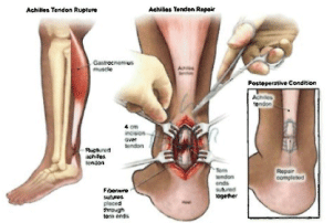
Figure 1. Anatomy details.
One of the main reasons why calcaneal tendon ruptures do not always show clinical expression is defined by the fact that it is not the only plantar flexor tendon of the ankle. The blood supply is characterized by the predominant presence of a recurrent branch of the posterior tibia artery, the area most prone to ruptures being avascular. It is important to remember that in the case of this anatomical unit there is an additional irrigation, from the adjacent part of the tendon. This justifies the presence of one of the intramuscular arterial branches (which irrigate the proximal portion ofthe tendon), while the distal portion is vascularized by a periosteal network on the posterior face of the calcaneus [4,5].
From the anamnestic point of view, the patient admitted to the ER with tendon rupture presents symptoms dominated by the appearance of intense pain, with sudden onset, located in the distal part of the tendon, thus making it impossible to perform plantar flexion. In the vast majority of cases this change is palpable, with a positive Thompson test on clinical examination. Pathognomonic is the impossibility of lifting the affected leg on tiptoes [6].
Complementary radiological examination is useful only if the doctor does not obtain sufficient data from the anamnesis and clinical examination. The protocols in the specialized literature define the ultra-sonographic examination as the first step in the imaging evaluation, due to the possibility of highlighting the focal defect, but also the thin film of fluid existing around this tendon. Ultrasonography can detect, in dorsiflexion, the broken ends of the tendon. MRI is especially useful in sagittal presentation, or for the differential diagnosis of tendinopathy. From the series of pathologies that can be part of the differential diagnosis of tendon ruptures, we mention severity of ruptures (total and partial), fractures accompanied by avulsion of the calcaneus, traumatic hematomas located in the lower 1/3 of the thigh, Achilles tendonitis (called tennis - leg), bursitis, etc. (Figure 2) [7-9].
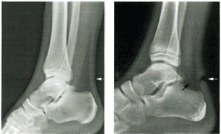
Figure 2. Radiological appearance of Achilles tendon rupture of the patient.
The differential diagnosis is made between total and partial rupture, Achilles tendonitis ("tennis-leg"), traumatic hematoma of the lower 1/3 of the thigh, fracture-avulsion of the heel, bursitis or Achilles tendinopathy [10].
A 14-year-old female patient was admitted to the ER of Children's Emergency Hospital "Sfantul loan" from Galati (where I am working as orthopedic surgeon specialist) on 08/01/2021.
Her anamnesis shows that the patient denies the existence of significant antecedent's collateral inheritance for the reasons of presentation. At the time of clinical examination, a good general condition is noticed, with symmetrical fades, skin and normally colored mucous membranes. Normally implanted, trophic veneers. The patient weighs 85 kg, excessively represented connective-adipose tissue, significant for grade 11-111obesity. The ganglion system is superficially intangible, painless, with the presence of the normal status, normal trophicity, normal kinetics muscular system. Balanced cardio-pulmonary and digestive. Urination is present spontaneously, bilateral negative Giordano. From a neurological point of view, the patient is stable, without signs of meningeal irritation, with bilateral symmetrical pupils.
The clinical examination, from a strictly visual point of view, expresses the skin integrity, with the lack of damage to the structures in the vascular-nervous bundle. Palpation of the affected area revealed sensitivity and even pain in the tendon insertion, with discrete irradiation in the adjacent area. Bruising was also observed in the posterior area of the calcaneus. The patient presented gait disorders with the difficulty of trying to support herself on the tips.
Clinical examination detects the existence of signs that support the definite diagnosis of Achilles tendon rupture, these being:
• Pain detected in the Achilles tendon
• Functional impotence when the patient tries to perform plantar flexion
• Interruption of the continuity of the Achilles tendon ("pencil mark")
• Positive Thompson sign with the association of Celsius signs (local pain, edema and bruising).
Imaging examination indicates the lack of degenerative elements at the tendon level, thus confirming the presumptive diagnosis mentioned above.
The patient underwent immobilization in the anterior plaster cast femur - foot splint in the "equine" position in the ER, the surgery being timed for 11.01.202
In the surgery room she was positioned in a prone position, being constantly monitored under spinal anesthesia. Once the posterior-medial incision was made, the adjacent part of the tendon was highlighted. These steps allowed the rupture heads to be easily detected and subsequently dissected both proximally and distally. First of all, the evacuation of the post-traumatic hematoma and the cleaning of the rupture ends were performed. Thus, Kessler-type sutures were used using allograft taken from the tendon of the plantar muscle. It is important to keep in mind that this therapeutic alternative was chosen because it was desired to obtain the best result in terms of suture firmness. Postoperatively, an immobilization in the anterior equatorial femoral plaster splint is established (with an average duration of 6 weeks). Subsequently, the progressive reduction of the equine will be performed by repeated immobilizations of the legs (Figure 3).
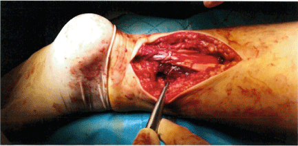
Figure 3. Surgical appearance of Achilles tendon rupture of the patient during the surgery.
The specialized literature confirms the fact that the minimally invasive and percutaneous surgical techniques follow the possibility of early mobilization and implicitly simplifying the healing process of the patients, creating the possibility of a fast healing, but also of an evolution without complications. The first attestation of a percussive intervention technique was described by Ma and Griffiths [11]. They hypothesized that two combined suturing techniques could be used to resolve the Achilles tendon rupture as follows: Bunnell for the proximal abutment and Kessler for the distal abutment. Later, another technique was used, this time a little more elaborate, developed by Kaluchi. This technique involves the use of a combination of the percutaneous approach and open surgery: it is a mini-incision that is performed at the level of the tendon defect (highlighted by palpation), thus allowing the abutments of the rupture to be highlighted. With the help of patterned Kirschner brooches that are inserted under the adjacent part of the tendon, percutaneous suturing will be performed [12,13].
It is important to remember that post-surgery, the treatment of patients consists in performing plaster immobilization. The immobilization that is progressively modified, progressively, to bring the forefoot into a neutral plantar position. All this treatment is performed for 12 weeks (with multiple immobilizations - femur-foot in equine position, followed by successive immobilizations with progressive reduction of the equine) (Figure 4 and Figure 5) [14].
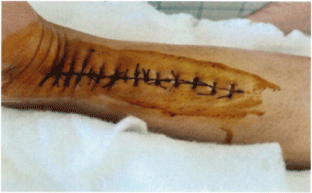
Figure 4. Surgical suture of Achilles tendon rupture of the patient.
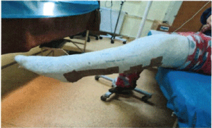
Figure 5. Post-surgery - plaster immobilization of the patient.
In the case of these patient, the post-surgery drug treatment consisted in the concomitant administration of double antibiotic therapy (with Oxacillin lg/ 6h and Gentamycin - a vial at 12 hours), in combination with anticoagulant medication (anticoagulant drug named Clexane 0.4 ml), analgesics (Algocalmin 1 ampoule as needed), as well as protective medication (Omeprazole 40 mg, respectively Eubiotic). Under this treatment, associated with the application of local ice to reduce inflammation, the patient showed favorable developments, with good general condition, without fever. For this reason, it was decided to discharge her on 15.01.2021.
The patient received the following recommendations for the post-discharge period:
1. To maintain the femur-pedios-plaster cast in equine for 6 weeks, with the indication to avoid support in the affected limb.
2. The mobilization will be performed with crutches without support on the left pelvic limb.
3. Suppression of sutures every 14 days.
4. Re-evaluation in the office of the polyclinic with a referral note from the family doctor (29.01.2021), or if necessary.
5. Medical prescription: anticoagulant Clexane 0.4 ml XXI ampoules, for 21 days; Oxacillin 500 mg/tablet XX tablets (1 tablet/ 6 h), 5 days; Omeprazole 20 mg; 5tablets - administration 1 tablet/ day for 5 days; Eu biotic tablet X, with the administration of 2 tablets/ day, for 5 days.
The specialized literature confirms that the process of repair and post-surgery time healing of tendon ruptures is a complex one. The involvement of inflammation mediators, simultaneously with neurotransmitters and mechanical stimuli ensures this phenomenon. Cellular activation is performed by mediators of inflammation, which is followed by a phenomenon of invasion of tendon tissue (normally hypo or even avascular) by vessels of new-formation and nerve branches. This creates a vital neurovascular structure for the healing process [15,16].
It is subdivided into three structural phases as follows:
- The inflammatory response
- Collagen formation phase
- Completed by the callus remodeling phase
From the point of view of post-surgery complications, it is known that minimally invasive or percussive interventions are associated with low incidences of infections, compared to interventions performed by open surgery (with an infection rate of 19.6%). Another complication mentioned is the tendon rupture again, relatively, low compared to percutaneous interventions (Figure 6) [17,18].
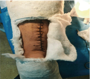
Figure 6. Post-surgery evolution of the patient: before removing the sutures.
Conservative treatment of traumatic rupture of the Achilles tendon is an alternative to surgical treatment considered viable, although the management of the entire therapeutic act is relatively difficult, especially in the case of young adults with active life. Traditionally, conservative management of traumatic rupture of the Achilles tendon is especially addressed to elderly patients, in whom operative stress is not an easy risk to assume, to patients with a minimal retraction of the ends of the rupture imaged, to patients who do not live a life. active or whose associated diseases may slow healing [19].
To conclude and make a sentence:
l. Systematic evidence is required for a correct assessment of the healing process. It is also important to follow the characteristic elements for making the correct diagnosis of Achilles tendon rupture, by corroborating the information obtained from the detailed anamnesis and para-clinical examinations.
2. Although, the Achilles tendon (the tendon of the sural triceps muscle), is one of the strongest tendons, with an average length between 9-11 centimeters, having the largest diameter in the human body, can suffer rupture at an early age.
3. The particularity of the present case consists in solving the rupture of the Achilles tendon by
suturing with allograft taken from the tendon of the plantar muscle.
4. The patient's evolution was satisfactory with the resumption of the functionality of the left lower limb.
Sources of Funding: No funding for the research
Ethical approval: This is not a research study. No ethical approval was necessary.
Consent: Consent for publication was obtained from the parents of the patient being a minor.
ANGHELE Aurelian -Dumitrache took the pictures; collected the data; made the surgery and wrote part of surgery.
MARINA Virginia wrote the article, processed the pictures.
Registration of research studies: This is a case report. Is not a research study.
Guarantor: MARINA Virginia
Provenance and peer review: Not commissioned externally peer- reviewed.
Declaration of Competing Interest: The author declarate that there is not conflict of interest regarding the publication of this article.
- Ion Albu (2006) Victor papilian Human Anatomy. The locomotor system - 11th Edition.
- Iagnov Z, Repciuc E, Russu IG, Simionescu N, Simu M (1962) Anatomia omului: Aparatul locomotor. Editura Medicala Bucure ti.
- Viorel Ranga (2002) Anatomia omului, vol. 2-Membrele, Editura CERMA.
- Mihail tefanet (2007) Anatomia omului. Volumul I, Chi inau, Centrul Editorial-Poligrafic Medicina.
- Susan Standring (2016) Gray's Anatomy: The Anatomical Basis of Clinical Practice. Expert Consult- Online and Print. 41st edn. Elsevier.
- Snow SW, Bohne WH, DiCarlo E, Chang VK (1995) Anatomy of the Achilles tendon and plantar fascia in relation to the calcaneus in various age groups. Foot Ankle Int 16: 418-421. [Crossref]
- Kim PJ, Martin E, Ballehr L, Richey J-M, Steinberg JS (2011) Variability of insertion of the Achilles tendon on the calcaneus: an MRI study of younger subjects. J Foot Ankle Surg 50: 41-43. [Crossref]
- Kim PJ, Richey JM, Wissman LR, Steinberg JS (2010) The variability of the Achilles tendon insertion: a cadaveric examination. J Foot Ankle Surg 49: 417-420. [Crossref]
- Lohrer H, Arentz S, Nauck T, Dorn-Lange NV, Konerding MA (2008) The Achilles tendon insertion is crescent-shaped: an in vitro anatomic investigation. Clin Orthop Relat Res 466: 2230-2237. [Crossref]
- Cummins EJ, Anson BJ (1946) The structure of the calcaneal tendon (of Achilles) in relation to orthopedic surgery, with additional observations on the plantaris muscle. Surg Gynecol Obstet 83: 107-116. [Crossref]
- Yamada H (1970) Strength of Biological Materials. Huntington, NY: Robert E. Krieger.
- Takigawa M (1953) Study upon strength of human and animal tendons. J Kyoto Prof Med Univ 53: 915-933.
- Salmon M, Taylor GI, Razaboni RM (1994) Anatomic Studies: Arteries of the Muscles of the Extremities and the Trunk: Arterial Anastomotic Pathways of the Extremities. St Louis: Quality Medical Publishers.
- Carr AJ, Norris SH (1989) The blood supply of the calcaneal tendon. J Bone Joint Surg Br 71: 100-101. [Crossref]
- Chen TM, Rozen WM, Pan WR, Ashton MW, Richardson MD, et al. (2009) The arterial anatomy of the Achilles tendon: anatomical study and clinical implications. Clin Anat 22: 377-385. [Crossref]
- Ahmed IM, Lagopoulos M, McConnell P, Soames RW, Sefton GK (1998) Blood supply of the Achilles tendon. J Orthop Res 16: 591-596. [Crossref]
- Zantop T, Tillmann B, Petersen W (2003) Quantitative assessment of blood vessels of the human Achilles tendon: an immunohistochemical cadaver study. Arch Orthop Trauma Surg 123: 501-504. [Crossref]
- Yepes H, Tang M, Geddes C, Glazebrook M, Morris SF, et al. (2010) Digital vascular mapping of the integument about the Achilles tendon. J Bone Joint Surg Am 92: 1215-220. [Crossref]
- E. Proca (1988) Tratat de patologie chirurgicala. Vol. III-Ortopedia (Coordonator: Prof. dr. A. Denischi). Editura Medicala, Bucure ti.