Lip cancers are common tumors of the oral-maxillary region. The etiologic causes of lip cancers include; smoking, exposure to ultraviolet (UV) rays, alcohol use. In this article, we discussed a case of lower lip squamous cell carcinoma in a male patient with active tobacco use and a history of direct contact with UV rays. This case report is important because of recurrent axillary metastases occured after subsequent treatment.
lower lip cancer, recurrent, axillary metastases
Head and neck cancer is the sixth most common cancer in the world [1]. Squamous cell carcinoma of the lower lip (SCC) is 25% of oral cancer [2].
Persons with a greater likelihood of lip SCC are indicated below; male patients over 45 years of age, with a history of chronic sun exposure, tobacco, and alcohol drinking habits [2,3]. According to a previous comprehensive study, SCC; It is a dominant type of lip cancer among men aged over 53 and exposed to UV rays [4].
Distant metastases play a critical role in the treatment and prognosis of oral cancer patients [5]. With regard to distant metastases of oral cancers, hypopharynx is the most common primary site (60%), followed by the base of the tongue (53%) and the anterior tongue [6].
In this case report, we report the metastases of the lower lip squamous cell carcinoma to the axillary lymph nodes, a rare metastases site. The purpose of this publication is to contribute to the literature.
A 64-year-old male patient was admitted to the hospital with a lesion on the lower lip. A biopsy was planned after the head and neck examination.
The patient's lower lip incisional biopsy revealed SCC. The patient was scheduled for surgery, but he did not accept it. 10 months after the development of swelling on the neck re-admitted to the hospital. The patient's lower lip mass was excised and neck dissection was performed. Six of the 50 lymph nodes were metastatic. The largest metastatic lymph node was a 5 cm sized conglomerated mass and had an extracapsular extension. Postoperative chemoradiotherapy was administered to the patient. He received 30 sessions of radiotherapy with 50 mg of cisplatin per week. The patient was invited to follow-up for 3 months after treatment.
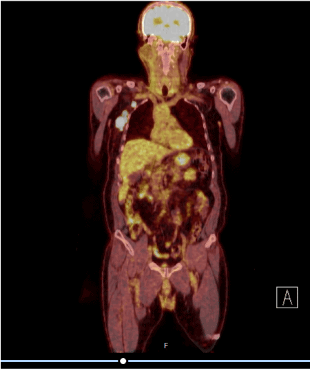
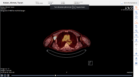
The patient's Positron emission tomography (PET/CT) showed multiple lymphadenopathies with a diameter of 3.6 cm with pathological glucose metabolism increases in the right axillary region Standardized Uptake Value (SUVmax: 42.6). Pathologic glucose metabolism increases were observed in 2 adjacent lymph nodes (SUVmax: 31.9) in the right infraclavicular region (Figures 1 and 2).

Figure 1. Image of first relapse after curative treatment

Figure 2. Image of first relapse after curative treatment
Lymph node biopsy was performed from the axillary region. The pathology result was reported as squamous cell carcinoma. DCF (Daxotere, cisplatin, fluorouracil) treatment was given to the patient as metastatic head and neck cancer. PET/CT was evaluated as a complete response according to RECIST 1.1 criteria after 4 cycles of DCF. Two cycles were recommended to the patient. But the patient did not accept.
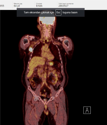
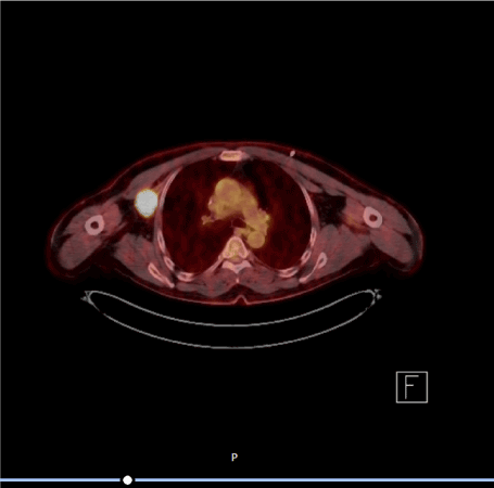
After 3 months of PET/CT, lymphadenopathy of the right axillary region was approximately 4 cm (SUV max. 39.2) (Figures 3 and 4).

Figure 3. Relaps after DCF treatment

Figure 4. Relaps after DCF treatment
Cisplatin, cetuximab, 5-FU treatment was planned as second-line chemotherapy as metastatic head and neck cancer. Radiological response after the completion of 12 cycles of treatment was reported as stable disease. As maintenance therapy setuximab is ongoing. Stable disease persists at 10 months.
Head and neck cancer is the sixth most common cancer in the world [1].
In a previous study; the rate of male patients was higher in 107 patients with head and neck cancer. The most common soft tissue metastases occurred in the lung in 23 cases, followed by heart in 10 cases [7]. Distant metastase is not very common in oral cancer and it is problem Associated with advanced stages of oral cancer [8]. Another previous study showed that; 186 of 415 patients who presented with lip lesions were diagnosed as carcinoma. Bone metastases were present in 4 of these patients and axillary lymph node involvement was observed in 1 patient [9]. In lip cancers; axillary lymph node metastases are extremely rare. Oral cavity malignancies have a significant recurrence rate and usually metastases to cervical lymph nodes [10].
In our literature search, it was found that lip cancers were less likely to have distant metastases. In addition, we were unable to detect a case report with an isole axillary lymph node metastases that recurred in the same region after the treatment. The patient we presented; he received DCF treatment in the first step of the metastatic disease and relapsed in the same region 3 months after the end of chemotherapy. After recurrence, cisplatin, cetuximab, 5-FU treatment was planned as second-line treatment. Radiological response after the completion of 12 cycles of treatment was reported as stable disease. As maintenance therapy setuximab is ongoing. Stable disease persists at 10 months.
- Parkin DM, Bray F, Ferlay J, Pisani P (2005) Global cancer statistics, 2002.CA Cancer J Clin55: 74-108. [Crossref]
- Han AY, Kuan EC, Mallen-St Clair J, Alonso JE, Arshi A, et al. (2016) Epidemiology of squamous cell carcinoma of the lip in the United States: A population-based cohort analysis. JAMA Otolaryngol Head Neck Surg 142: 1216-1223. [Crossref]
- Maruccia M, Onesti MG, Parisi P, Cigna E, Troccola A, et al. (2012) Lip cancer: A 10-year retrospective epidemiological study. Anticancer Res 32: 1543-1546. [Crossref]
- Czerninski R, Zini A, Sgan-Cohen HD (2010) Lip cancer: incidence, trends, histology and survival: 1970-2006.Br J Dermatol162: 1103-1109. [Crossref]
- Takes RP, Rinaldo A, Silver CE, Haigentz M Jr, Woolgar JA, et al. (2012) Distant metastases from head and neck squamous cell carcinoma. Part I. Basic aspects. Oral Oncol 48: 775-779. [Crossref]
- Kotwall C, Sako K, Razack MS, Rao U, Bakamjian V, et al. (1987) Metastatic patterns in squamous cell cancer of the head and neck.Am J Surg154: 439-442. [Crossref]
- Irani S (2016) Distant metastasis from oral cancer: A review and molecular biologic aspects. J Int Soc Prev Community Dent 6: 265-271. [Crossref]
- Betka J (2001) Distant metastases from lip and oral cavity cancer.ORL J Otorhinolaryngol Relat Spec63: 217-221. [Crossref]
- Luna-Ortiz K, Güemes-Meza A, Villavicencio-Valencia V, Mosqueda-Taylor A (2004) Lip cancer experience in Mexico. An 11-year retrospective study. Oral Oncol 40: 992-999. [Crossref]
- Okura M, Aikawa T, Sawai NY, Iida S, Kogo M (2009) Decision analysis and treatment threshold in a management for the N0 neck of the oral cavity carcinoma. Oral Oncol 45: 908-911. [Crossref]


