Key words
99mTc-MDP, liver metastases, breast cancer
Case report
We report the case of a fifty-eight years old woman with history of left breast cancer diagnosed in November 2010 and treated with radical mastectomy, chemotherapy and radiation therapy, followed by hormone therapy. From August 2011 to November 2017 the patient was in imaging follow up, free from recurrences or metastases.
In December 2017, due to the onset of intense dorsal-lumbar spine pain, the patient underwent 99mTc-methylene-diphosphonate (MDP) scintigraphy to evaluate possible skeletal involvement. The whole-body scan showed multiple bone metastases, more evident in skull and axial skeleton, and inhomogeneous 99mTc-MDP uptake in the right upper quadrant of the abdomen, suspect for hepatic localization (Figure 1). Bone scan was completed with single photon emission computed tomography/computed tomography (SPECT/CT) confirming the focal 99mTc-MDP uptake areas in liver, more evident in the sixth and eighth segments. At the CT images these areas showed slight inhomogeneity without macroscopic calcifications. Moreover SPECT/CT accurately highlighted metastases of the VIII and IX right ribs and confirmed the other sites of bone metastases (Figure 2).
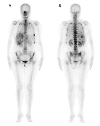
Figure 1: (A) anterior and (B) posterior whole body 99mTc-MDP scintigraphy shows multiple bone metastases of skull and axial skeleton and inhomogeneous uptake in the right upper quadrant of the abdomen.
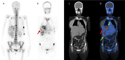
Figure 2: (A) MIP and (B) SPECT, (C) CT, (D) SPECT/CT coronal images of 99mTc-MDP scintigraphy show focal uptake areas in the 6th and 8th liver segments (green arrows) and VIII and IX right ribs metastases (red arrow) in addition to the multiple skeletal metastases detected at the whole-body scan.
Various conditions can influence MDP accumulation in tissues, such as serum calcium increase, PO4 ion, tissue pH and factors which alter cell integrity like infection, radiotherapy and expansion of the extracellular fluid [1].
Hepatic uptake of 99mTc-MDP is an specific finding and its pattern can be focal or diffuse, with different meaning. Literature reports some cases of focal faint uptake in various malignancies, in particular in liver metastases from breast, colon or lung cancer. Also, primary breast cancer could uptake 99mTc-MDP [2-4].
Liver 99mTc-MDP uptake pathogenesis is firstly related to calcific regions in necrotic or proliferating fibrous tissue. The necrotic metastatic mass produces passive congestion and liver enlargement and then hypoxia with hemosiderin and calcium deposits with 99mTc-MDP affinity growing. Furthermore, ischemia damages cell integrity, calcium precipitates within mitochondria and on denatured proteins. So 99mTc-MDP is evidenced in irreversibly damaged cells in presence of residual blood flow [1].
Otherwise diffuse liver 99mTc-MDP uptake can be related to amyloidosis, hepatitis or recent intravenous injection of gadolinium contrast for Magnetic Resonance. These causes of diffuse hepatic uptake have to be considered after technical problems exclusion, due to 99mTc-MDP dose degradation by excess allumina chemical impurity, with colloid formation [2,5].
Incidental findings of extraosseous uptake can have an important impact on cancer patient management; for this reason the role of hybrid SPECT/CT imaging appears particularly relevant because fused technology provides the great opportunity to combine nuclear medicine examinations sensitivity with CT anatomical information. The role of SPECT/CT in management of breast cancer patients have been already investigated in other procedures such as lymphoscintigraphy for sentinel lymph node radioguided surgery [6-8].
Currently bone scan is the gold standard in breast cancer follow up for skeletal metastases. Occasionally incidental extraosseous findings are possible and the knowledge of their meaning is important for a correct diagnostic and therapeutic classification and a better patient’s management.
The most modern multimodal SPECT/CT technology have a decisive role in improving the accuracy of 99mTc-MDP bone scintigraphy.
Disclosure
No conflict of interest.
No grants of financial supports.
References
- Üstün F, 2021 Copyright OAT. All rights reservver metastases of lung cancer detected with similar uptake pattern on bone scintigraphy and fluorine-18 fluorodeoxyglucose positron emission tomography/computed tomography: What's the pathophysiologic mechanism? World J Nucl Med 16: 163-165. [crossref]
- Chen P, Marentis T, Brown RK (2014) Diffuse liver uptake on (99m) Tc-MDP bone scan secondary to severe hepatic failure. Clin Nucl Med 39: 658-659. [crossref]
- Baumert JE, Lantieri RL, Horning S, McDougall IR (1980) Liver metastases of breast carcinoma detected on 99mTc-methylene diphosphonate bone scan. AJR 134: 389-391.
- Piccolo S, Lastoria S, Mainoffi C, Muto P, Bazzicalupo L, Salvatore M (1995) Technetium-99m-methylene diphosphonate scintimammography to image primary breast cancer. J Nucl Med 36: 718-724. [crossref]
- Al-katib S, Al-faham Z, Balon H (2015) Liver uptake on bone scanning: A diagnostic algorithm. J Nucl Med Technol 43: 135-136. [crossref]
- Loutfi I, Collier BD, Mohammed AM Nonosseous (2003) Nonosseous abnormalities on bone scans. J Nucl Med Technol 31: 149-153. [crossref]
- Waldman LE, Scharf SC (2017) Bone SPECT/CT of the spine, foot, and ankle: Evaluation of surgical patients. Semin Nucl Med 47: 639-646. [crossref]
- Frusciante V, Niccoli Asabella A, Castriotta G, Guerra M, Murgo R, et al. (2016) Added value of SPECT/CT over planar imaging in improving sentinel node detection in breast cancer patients. Recenti Prog Med 107: 444-449. [crossref]