Purpose: The aim of this study was to identify changes in swallowing in patients who underwent supracricoid laryngectomy with cricohyoidoepiglottopexy (SCL-CHEP) for laryngeal cancer.
Methods: Retrospective analysis of dynamic videofluoroscopic swallowing examinations was performed in 45 patients who received SCL-CHEP as a treatment for laryngeal cancer. The examinations were completed by protocol and then digitally analyzed. The analysis focused on measuring the displacement and timing related to swallowing sequence. The results were compared to published normative data.
Results: All the patients were male (mean age, 71 years; age range, 66–79 years). The examinations were performed around five years after surgery. The findings were then compared with normative data, and the duration of opening of the pharyngoesophageal segment (PES) and airway closure were found to be prolonged (p <0.001, p < 0.001) with an accentuated hyolaryngeal elevation of increased duration (p < 0.001). There was no significant deterioration in the displacements’ measurements in critical swallowing events.
Conclusions: Swallowing characteristics can be identified in patients who have undergone SCL-CHEP, suggesting swallowing safety through increased duration of airway closure and PES opening with enhanced hyolaryngeal elevation. These results suggest that safe and effective deglutition without aspiration can be achieved after a SCL-CHEP treatment for laryngeal cancer.
supracricoid laryngectomy, deglutition, deglutition disorders, vfss, pharyngoesophageal segment, hyoid bone, rehabilitation
Supracricoid laryngectomy with cricohyoidoepiglottopexy (SCL-CHEP) is a function preservation surgery as effective as near total laryngectomy for laryngeal cancer. This procedure was popularized by Piquet [1] and Laccourreye et al. [2]. SCL-CHEP can be applied for T2 and well-selected T3 and T4 patients and can also be utilized to salvage radiation failures [3]. SCL-CHEP fulfills three major functions: (1) respiration via a natural airway, (2) rough yet acceptable speech and (3) adequate swallowing ability [4-6]. It was introduced in 1997 at our institute [3]. We have previously reported a correlation between surgical variation and swallowing acquisition [7]. Once swallowing is regained, in our experience, most of the patients are able to maintain an adequate swallowing function for years. Swallowing functions after SCL-CHEP have also been investigated in earlier studies [8,9]. However, there is very limited information about the key factors that determine swallowing compensation after SCL-CHEP, which is a surgery that resects the bilateral vocal cords and the entire thyroid cartilage. Quantitative physiological analyses such as videofluoroscopic swallowing study (VFSS) or functional endoscopic evaluation of swallowing (FEES) are essential. Utilizing dynamic VFSS, we quantitatively evaluated characteristics of swallowing compensation after SCL-CHEP. The aims of this study were to accurately identify the compensatory swallowing mechanisms developed in this cohort by means of comparison with reliable normal data, published by Leonard [10-12].
This study was approved by our Institutional Review Board (B ethics 11-107). Clinical data of patients who had undergone SCL-CHEP at the Department of otolaryngology head and neck surgery, Kitasato University Hospital (ORL-KUH) between October 2003 and April 2013 were included for comparison. Patients who received VFSS at ORL-KUH to evaluate post-surgical deglutition five years after SCL-CHEP were sorted. Patients under the age of 64 years at VFSS and two female patients were excluded for comparison with normal data of men >65 years old, as published by Leonard and Kendall [10-12]. Four PEG-dependent patients were also excluded to comply with the study aim. Measurement protocols of the VFSS method and statistical analyses are presented below.
VFSS method
VFSS was performed around five years after surgery. Radiographic studies were conducted on a 1.2 mA-type output for the full field of view (9 in. input phosphor diameter). Fluoroscopy study results were recorded on a Multi-drive DVD/HDD Recorder “RD-XS31” (Toshiba Co., Ltd.[A]) for further analysis. The swallowing studies involved recording at 30 frames/s, where patients were asked to swallow 3 mL of Omnipaque® (iohexol) (Daiichi Sankyo Co., Ltd.[B]). A ruler was displayed in the image field adjacent to the patient prior to fluoroscopic examinations. The VFSS method has been unchanged since 2003, when we first started this study.
Measurement protocols
Fluoroscopy study videos were transferred to MacBook Air computers (Apple Inc.[C]). All the quantitative measures were performed from lateral views. We obtained timing measures by observing the video studies in slow motion and frame-by-frame advance (forward and reverse) modes using a time counter embedded in QuickTime Player 7 ver.7.6.6 (Apple Inc.[C]).
The timing of both selected swallowing gestures and bolus transit was determined (Figure 1). Duration was also measured with this timer. Values included (1) Hmax: the duration of hyoid bone remaining in maximum superior/anterior displacement; (2) PESstart: the duration of initial opening of the PES to the time of first entrance of the bolus through the PES; (3) PESopen: the duration of pharyngoesophageal sphincter (PES) opening; (4) AEstart: the duration from onset of swallowing to airway closure start, which was detected by the first anterior movement of the arytenoid, to an end by firm contact of the arytenoid to the epiglottis (tongue base); (5) AEclose: the duration from first firm contact of arytenoid-epiglottis to close the neoglottis to the point of airway re-opening. (6) Oropharyngeal transit time (OPT): bolus transit time through oral - pharyngeal section, that is the duration from first movement of the bolus head past the posterior nasal spine that represents swallowing transit onset to first exit of the bolus from the valleculae; and (7) Hypopharyngeal transit time (HPT): bolus transit time through the hypopharynx, that is the duration from first exit of bolus from the valleculae to clearance of the bolus tail from the PES.
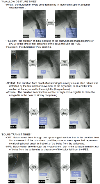
Figure 1. Timing measures for bolus movements and swallow gestures: definitions
Displacement was measured on the ImageJ (ver.1.45[D]) software program that was utilized for the analysis of fluoroscopic images. Area and distance measurements were performed after calibrating the digitized image to a ruler shown in front of the face. Displacement measures included (1) Hmax: distance between hyoid position at “hold” i.e., to the bolus held in the oral cavity, and at its point of maximal anterior and superior excursion during the swallow; (2) PESmax: narrowest point of opening between C3 and C6 during maximal distention for bolus passage; (3) PAhold: pharyngeal area holding the bolus in the mouth at rest; (4) PAmax: pharyngeal area at maximum constriction; (5) pharyngeal constriction ratio (PCR): the ratio of PAmax divided by PAhold to assess pharyngeal constriction; (6) HLhold: distance from inferior hyoid bone to inferior cricoid cartilage/top of air column anteriorly, that represents larynx–hyoid distance; and (7) HLmax: distance from inferior cricoid (top of tracheal column) to inferior border of hyoid at their point of maximum approximation during swallow, that represents maximum larynx–hyoid approximation (Figure 2).
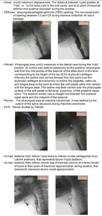
Figure 2. Displacement measures: definitions
Statistical analysis
All data are presented as mean ± standard deviation (SD). The mean values for each variable were then compared between groups, applying Student’s t tests, if standard variations between the groups did not differ, or by applying Welch’s correction if they did. PASW Statistics 18 was used for statistical analyses. For all comparisons, a two-tailed p value of 0.05 was considered statistically significant.
Patients
Between October 2003 and April 2013, 79 patients underwent SCL-CHEP in our institute. Forty-five patients were selected for VFSS’s analysis. All the participants were male (mean age, 71 ± 3.8 years; age range, 66–79 years). One or both arytenoids were preserved in 27 (60%) and 18 (40%) patients, respectively. Twenty (44%) of the selected patients underwent radiotherapy before SCL-CHEP (Table 1). No patient had tracheostoma, nasogastric tube, dietary restrictions, recurrent pulmonary infections, or uncontrolled systemic diseases at the time of VFSS.
Table 1. Patient population after SCL-CHEP (*SCL=supracricoid laryngectomy; CHEP=cricohyoidoepiglottopexy)
Characteristics |
No. of patients, (%) |
Male gender, N (%) |
45 (100%) |
Age (mean±SD, min/max) |
71±3.8, 66/79 years old |
Preserved Arytenoid |
|
Bilateral |
18(40%) |
Unilateral |
27(60%) |
Radiation history before surgery |
20 (44%) |
Swallow gesture times
Hyoid bone elevation: After SCL, the patients’ duration of hyoid elevation was significantly longer than the controls’ (p < 0.001) (Table 2).
Table 2. Means and standard deviations for the duration of hyoid movement, and the duration between the opening of PES and three bolus transit points
Duration |
SCL |
norm |
p-value |
Hmax (sec) |
0.34 (0.22) |
0.12 (0.10) |
P<0.001 |
PESstart (sec) |
0.07 (0.04) |
0.03 (0.03) |
P<0.001 |
PESopen (sec) |
0.59 (0.20) |
0.41 (0.07) |
P<0.001 |
Pharyngoesophageal sphincter opening: In the post-SCL group, opening of the PES was relative to the arrival of the bolus at the PES (p < 0.001). The duration of PES opening was also significantly longer in the post-SCL group than in controls (p < 0.001) (Table 2).
Aryepiglottic fold closure (airway closure): Despite the fact that SCL involved resectioning the entire thyroid cartilage, including both vocal folds and often one arytenoid, the arytenoid and aryepiglottic fold could be identified in all patients in the lateral plane on VFSS. The aryepiglottic fold closure in the post-SCL group was achieved much earlier than in the control group prior to the onset of swallowing (p < 0.001). The closure of the airway in the post-SCL group was significantly prolonged when compared with that in the control group (p < 0.001) (Table 3).
Table 3. Means and standard deviations for the duration of airway closure, the duration between airway closure and the related swallow onset, and for the bolus transit times
Duration |
SCL |
norm |
p-value |
AEstart (sec) |
0.12 (0.41) |
0.64 (0.29) |
P<0.001 |
AEclose (sec) |
0.49 (0.31) |
0.34 (0.25) |
P<0.001 |
OPT (Sec) |
0.45 (0.26) |
0.49 (0.23) |
P=0.386 |
HPT (Sec) |
0.72 (0.27) |
0.63 (0.23) |
P=0.06 |
Bolus transit times: The longer durations of the post-SCL group were relatively uniform throughout the swallow, being 0.09 s longer than the control group in HPT. However, no statistically significant differences in both OPT and HPT were observed (Table 3).
Swallow gesture displacements
Laryngeal elevation: The degree of hyoid displacement was not significantly different when the post-SCL group was compared to the control group. Therefore, the hyoid bone in the post-SCL patients was elevated to the same extent as those in the control group. Both HLhold and HLmax were significantly shortened in post-SCL group (p < 0.001) (4).
Pharyngoesophageal sphincter opening: The maximum extent of PES opening was statistically enhanced in the study group as compared to the control group (p = 0.05) (Table 4).
Table 4. Means and standard deviations for the distance of maximal hyoid movement, maximal PES opening, the ratio of pharyngeal area, and the distance from the hyoid bone to the larynx in a hold position or maximal approximation
Displacement |
SCL |
norm |
p-value |
Hmax (cm) |
2.19 (0.48) |
2.08(0.81) |
P=0.18 |
HL hold (cm) |
0.87 (0.04) |
4.20 (0.37) |
P<0.001 |
HLmax (cm) |
1.03 (0.05) |
1.71 (0.72) |
P<0.001 |
PESmax (cm) |
0.62 (0.21) |
0.50(0.19) |
P=0.05 |
PCR (PAmax/PAhold) |
0.04 (0.03) |
0.03 (0.02) |
P=0.04 |
PAmax (cm2) |
0.38 (0.26) |
0.35 (0.23) |
P=0.50 |
PAhold (cm2) |
8.81 (0.44) |
11.4 (3.0) |
P<0.001 |
Pharyngeal constriction: The pharyngeal constriction ratio (PCR), which is the ratio of PAmax: PAhold, in post-SCL subjects was deteriorated (p = 0.04) (Table 4). The PAhold in the post-SCL group was smaller than in the control group (p < 0.001) (Table 4). There was no significant difference between the groups in PAmax.
SCL-CHEP has been recognized as an effective technique for the treatment of selected glottic and supraglottic cancers, as it avoids total laryngectomy while maintaining physiologic speech and swallowing function. Some articles have reported that SCL-CHEP produces excellent functional results [13] if the patients receive appropriate swallowing rehabilitation [14].
VFSS is useful for identifying patients with postoperative anatomical and physiological (dynamic) information on swallowing. Previous studies suggest that the key parameters to evaluate swallowing on VFSS include tongue base-arytenoid contact, epiglottic function, maximal PES opening, and movement of the hyoid bone [15]. Woisard et al. studied the mechanisms of post-SCL aspiration and reported that faulty synchronization between the posterior motion of the tongue base and neoglottic closure led to aspiration [16]. To obtain evidence supporting the above-mentioned theory, we deemed that swallowing function after SCL-CHEP should be comprehensively analyzed by instrumental examination. The method of VFSS analysis published by Leonard and Kendall includes crucial elements likely to shed light on the mechanisms of post-SCL dysphagia. Furthermore, the obtained values are quantitative and reproducible, and published normative data are available for comparison. We selected pharyngeal transit time, airway closure, hyoid bone elevation, PES opening, PCR, and laryngeal elevation as our main measured variables. Applying a standardized approach to the analysis of swallowing allowed us to identify differences in the VFSS between subjects following SCL and normal subjects. To our best knowledge, this type of study has not been previously conducted to evaluate swallowing after SCL-CHEP.
Swallowing gesture timing and duration
It is well known that prolonged transit times correlate with an increased risk of aspiration [17]. Oropharyngeal transit time (OPT) and Hypopharyngeal transit time (HPT) was not prolonged in post-SCL group compared with that in control group. In normal subjects, PES opening and the arrival of the bolus at the PES occurred almost simultaneously. In contrast, in post-SCL patients, PES opening started far earlier and stayed open for significantly longer to prepare for entry of the bolus than that in normal subjects. This evidence of early and prolonged opening of the PES suggests functional advantage for deglutition after SCL-CHEP.
The timing of other selected swallow gestures also differed between the two groups. The start time of aryepiglottic approximation to the completion of airway closure in the post-SCL group was significantly earlier than in the normal group, by around 0.5 s. Moreover, the duration of airway closure was around 0.15 s longer in post-SCL subjects than in controls. In terms of airway protection, therefore, this result suggests compensation of the post-SCL neoglottis to prevent bolus invasion into the airway.
From the viewpoint of the neoglottis morphology, prior to swallowing initiation, the arytenoid cartilages sit on the cricoid cartilage and are already located in an elevated position due to the pexis procedure. Furthermore, in the case of post-SCL swallow, we frequently found that the valleculae were not easily identifiable due to epiglottic approximation to the root of the tongue. These features appeared to make it easier for the arytenoid to make contact with the tongue base (Figure 3). Thus, by the time the bolus has arrived at the PES, the tongue base (attached to the epiglottis) is seen to approximate the arytenoids effectively and rapidly to close the supraglottic passage (Figure 4). Moreover, the supraglottic airway remains closed until after the PES has opened (Figure 5). These findings, which are consistent with the report by Woisard et al. [17], were demonstrated by the prolonged duration of airway closure and PES opening.
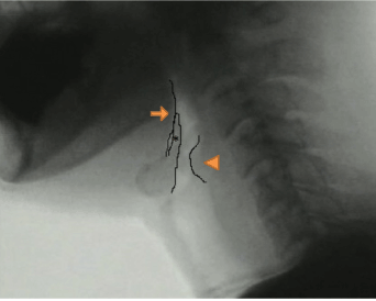
Figure 3. Retrofluorographic lateral view at bolus-hold position, showing an open airway. The arrow, arrowhead, and asterisk show the tongue base, arytenoid, and epiglottis, respectively
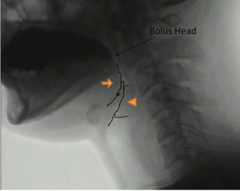
Figure 4. Retrofluorographic lateral view at initiation of neoglottic closure from contacting the epiglottis petiole and mucosa of the arytenoid, showing a closed airway
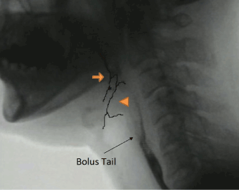
Figure 5. Retrofluorographic lateral view at the passage of the bolus tail at the PES, showing that the airway is still closed
The duration of maximum hyoid elevation in the post-SCL group was longer. This fact likely has dual meanings: (1) increasing the duration of PES opening and (2) ensuring supra-neoglottic occlusion with firm posterior tongue base movement and epiglottic folding [16].
Swallowing gesture displacement
It seems to be of particular value that no displacement measures deteriorated following SCL-CHEP. In fact, post-SCL subjects demonstrated supranormal displacements in some cases. Maximal PES opening was enhanced compared with that in normal subjects. Despite post-surgical effects, namely, fibrosis, hyoid elevation remained the same. During SCL-CHEP, the infrahyoid muscles were resected and the preserved laryngeal structures including the anterior wall of the trachea were separated from surrounding tissue including ligation of the inferior thyroid vein. We believe that this contributed to appropriate hyoid elevation during pexis.
The pharyngeal constriction ratio (PCR; PAmax/PAhold), which is a surrogate for pharyngeal strength, increased (thereby, deteriorated) following surgery. Yip et al. reported that increased PCR was associated with the presence of aspiration on fluoroscopy [18]. In post-SCL patients, PCR was increased; however, this ratio was actually calculated with PAmax and PAhold. PAhold in the post-SCL group was much smaller than in the control group and hence PCR was superficially increased. On the contrary, these results suggest the great compensation that favors to facilitate sufficient pharyngeal constriction.
The results of distance from the hyoid bone to the larynx represented by the top of air column were characteristic of post-SCL patients, because, HLhold was far shortened due to the pexis procedure, which directly led to shorter distance of HLmax in post-SCL group. This finding was consistent with good PES opening and minimal post-swallowing residue despite longer distance between the mandible and the hyoid bone in post-SCL patients than in control ones as post-surgical effects.
Residue and pharyngeal function are crucially linked to the risk of aspiration, and we believe that maintenance of pharyngeal function is critical for the success of post-surgical deglutition. SCL-CHEP, by its nature, enhances displacement measures as viewed on VFSS. These surgical maneuvers are designed to ameliorate post-surgical swallowing risk and assist in swallowing rehabilitation. In contrast, modification of swallowing gesture timing as seen on VFSS in this study is most likely a compensatory change linked to airway protection. Consequently, alterations of both displacement and timing values in patients following SCL are likely to constitute the functional benefits of this surgery. By thorough swallowing rehabilitation, it is possible to instruct SCL patients on these compensatory modifications and thus facilitate the swallowing recovery process.
Swallowing rehabilitation
According to the present study, swallowing rehabilitation should focus on airway protection and PES opening via an increase in the anterior to posterior tongue movement and in the hyoid elevation, respectively. We consider that supraglottic swallow and effortful swallow targeting the two features mentioned above are crucial in post-SCL patients. Chin tuck swallows and Shaker exercises can also be utilized.
Research providing evidence from rehabilitation are very scarce, as most studies focused on the functional outcomes after SCL-CHEP, but not on possible rehabilitation options. The tradition of evidence-based rehabilitation after organ-sacrificing therapies such as total laryngectomy is now well established; hence, it is perhaps time to invest in evidence-based rehabilitation after SCL-CHEP.
SCL-CHEP offers an alternative to total laryngectomy for the control of advanced-stage laryngeal cancers. Analysis of postoperative function, particularly the function of deglutition, is equally crucial. To our best knowledge, this study is the first to report direct comparison of swallowing between normal subjects and SCL-CHEP by VFSS. We noted compensatory changes in timing, such as an earlier onset of PES opening, in post-SCL patients compared with that in normal subjects, relative to bolus arrival at the PES. Moreover, the duration of critical events, such as PES opening, hyoid elevation, and airway closure, was prolonged in patients compared to normal subjects, which was seen to involve direct compensatory strategies following surgery. We also showed an increased ability of post-SCL patients to close the neoglottis. None of the critical displacements such as hyoid elevation, PES opening, and PCR deteriorated after SCL-CHEP. This confirms that SCL-CHEP is an ideal function-retaining surgical option.
[A]. Toshiba Medical Systems Corporation, 1385 Shimoishigami, Otawara-shi, Tochigi, Japan
[B]. Daiichi Sankyo Co., Ltd., 3-5-1, Nihonbashi Honcho, Chuo-ku, Tokyo, Japan
[C]. Apple Computers, Inc., One Infinite Loop, Cupertino, CA 95014
[D]. Rasband WS. Image J, available at: http://rsbweb.nih.gov/ij/, accessed May 21, 2012
Dr. Y. Seino takes responsibility for the integrity of the content of this paper. We express our deep thanks to Jacqui E. Allen, MBChB, FRACS, and Anna Miles, CCC-SLT at the University of Auckland for her elaborated advice and invaluable discussion.
The authors declare that they have no conflict of interest.
- Piquet JJ, Chevalier D (1991) Subtotal laryngectomy with crico-hyoido-epiglotto-pexy for the treatment of extended glottic carcinomas. Am J Surg 162: 357-361.
- Laccourreye H, Laccourreye O, Weinstein G, Menard M, Brasnu D, et al. (1990) Supracricoid laryngectomy with cricohyoidoepiglottopexy: A partial laryngeal procedure for glottic carcinoma. Ann Otol Rhinol Laryngol 99: 421-426.
- Nakayama M, Okamoto M, Miyamoto S, Takeda M, Yokobori S, et al. (2008) Supracricoid laryngectomy with cricohyoidoepiglotto-pexy or cricohyoido-pexy: Experience on 32 patients. Auris Nasus Larynx 35: 77-82.
- Weinstein GS, El-Sawy MM, Ruiz C, Dooley P, Chalian A, et al. (2001) Laryngeal preservation with supracricoid partial laryngectomy results in improved quality of life when compared with total laryngectomy. Laryngoscope 111: 191-199.
- Naudo P, Laccourreye O, Weinstein G, Hans S, Laccourreye H, et al. (1997) Functional outcome and prognosis factors after supracricoid partial laryngectomy with cricohyoidopexy. Ann Otol Rhinol Laryngol 106: 291-296.
- Farrag TY, Koch WM, Cummings CW, Goldenberg D, Abou-Jaoude PM, et al. (2007) Supracricoid laryngectomy outcomes: The johns hopkins experience. Laryngoscope 117: 129-132.
- Nakayama M, Yao K, Nishiyama K, Nagai H, Ito A, et al. (2002) Swallowing function after near-total laryngectomy, cricohyoidoepiglottopexy (CHEP), and cricohyoidopexy (CHP). Nihon Jibiinkoka Gakkai Kaiho 105: 8-13.
- Benito J, Holsinger FC, Perez-Martin A, Garcia D, Weinstein GS, et al. (2011) Aspiration after supracricoid partial laryngectomy: Incidence, risk factors, management, and outcomes. Head Neck 33: 679-685.
- Chevalier D, Piquet JJ (1994) Subtotal laryngectomy with cricohyoidopexy for supraglottic carcinoma: Review of 61 cases. Am J Surg 168: 472-473.
- Kendall KA, Leonard RJ (2007) Dysphagia Assessment and Treatment Planning: A Team Approach. In Kendall KA, McKenzie S, Leonard RJ, Dynamic Swallow Study: Objective Measures and Normative Data in Adults (pp. 233-294). San Diego, USA: Plural Publishing INC.
- Leonard RJ, Kendall KA, McKenzie S, Goncalves MI, Walker A, et al. (2000) Structural displacements in normal swallowing: A videofluoroscopic study. Dysphagia 15: 146-152.
- Kendall KA, McKenzie S, Leonard RJ, Goncalves MI, Walker A, et al. (2000) Timing of events in normal swallowing: A videofluoroscopic study. Dysphagia 15: 74-83.
- Dworkin JP, Meleca RJ, Zacharek MA, Stachler RJ, Pasha R, et al. (2003) Voice and deglutition functions after the supracricoid and total laryngectomy procedures for advanced stage laryngeal carcinoma. Otolaryngol Head Neck Surg 129: 311-320.
- Clayburgh DR, Graville DJ, Palmer AD, Schindler JS (2012) Factors associated with supracricoid laryngectomy functional outcomes. Head Neck 2012 Oct 5.
- Yuceturk AV, Tarhan S, Gunhan K, Pabuscu Y (2005) Videofluoroscopic evaluation of the swallowing function after supracricoid laryngectomy. Eur Arch Otorhinolaryngol 262: 198-203.
- Woisard V, Puech M, Yardeni E, Serrano E, Pessey JJ, et al. (1996) Deglutition after supracricoid laryngectomy: Compensatory mechanisms and sequelae. Dysphagia 11: 265-269.
- Leonard R, McKenzie S (2006) Hyoid-bolus transit latencies in normal swallow. Dysphagia 21: 183-190.
- Yip H, Leonard R, Belafsky PC (2006) Can a fluoroscopic estimation of pharyngeal constriction predict aspiration? Otolaryngol Head Neck Surg 135: 215-217.
Editorial Information
Editor-in-Chief
Yi-Hwa Liu
Senior Research Scientist
Department of Internal Medicine
Yale University
New Haven, USA
Article Type
Research Article
Publication history
Received: April 13, 2020
Accepted: April 27, 2020
Published: April 30, 2020
Copyright
©2020 Seino Y. This is an open-access article distributed under the terms of the Creative Commons Attribution License, which permits unrestricted use, distribution, and reproduction in any medium, provided the original author and source are credited.
Citation
Yutomo Seino, Shunsuke Miayamoto, Meijin Nakayama and Taku Yamashita (2020) A kinematic analysis of swallow gestures and timing using video fluoroscopic study after supracricoid laryngectomy. Radiol Diagn Imaging,1: doi: 10.15761/RDI.1000168

Figure 1. Timing measures for bolus movements and swallow gestures: definitions

Figure 2. Displacement measures: definitions

Figure 3. Retrofluorographic lateral view at bolus-hold position, showing an open airway. The arrow, arrowhead, and asterisk show the tongue base, arytenoid, and epiglottis, respectively

Figure 4. Retrofluorographic lateral view at initiation of neoglottic closure from contacting the epiglottis petiole and mucosa of the arytenoid, showing a closed airway

Figure 5. Retrofluorographic lateral view at the passage of the bolus tail at the PES, showing that the airway is still closed
Table 1. Patient population after SCL-CHEP (*SCL=supracricoid laryngectomy; CHEP=cricohyoidoepiglottopexy)
Characteristics |
No. of patients, (%) |
Male gender, N (%) |
45 (100%) |
Age (mean±SD, min/max) |
71±3.8, 66/79 years old |
Preserved Arytenoid |
|
Bilateral |
18(40%) |
Unilateral |
27(60%) |
Radiation history before surgery |
20 (44%) |
Table 2. Means and standard deviations for the duration of hyoid movement, and the duration between the opening of PES and three bolus transit points
Duration |
SCL |
norm |
p-value |
Hmax (sec) |
0.34 (0.22) |
0.12 (0.10) |
P<0.001 |
PESstart (sec) |
0.07 (0.04) |
0.03 (0.03) |
P<0.001 |
PESopen (sec) |
0.59 (0.20) |
0.41 (0.07) |
P<0.001 |
Table 3. Means and standard deviations for the duration of airway closure, the duration between airway closure and the related swallow onset, and for the bolus transit times
Duration |
SCL |
norm |
p-value |
AEstart (sec) |
0.12 (0.41) |
0.64 (0.29) |
P<0.001 |
AEclose (sec) |
0.49 (0.31) |
0.34 (0.25) |
P<0.001 |
OPT (Sec) |
0.45 (0.26) |
0.49 (0.23) |
P=0.386 |
HPT (Sec) |
0.72 (0.27) |
0.63 (0.23) |
P=0.06 |
Table 4. Means and standard deviations for the distance of maximal hyoid movement, maximal PES opening, the ratio of pharyngeal area, and the distance from the hyoid bone to the larynx in a hold position or maximal approximation
Displacement |
SCL |
norm |
p-value |
Hmax (cm) |
2.19 (0.48) |
2.08(0.81) |
P=0.18 |
HL hold (cm) |
0.87 (0.04) |
4.20 (0.37) |
P<0.001 |
HLmax (cm) |
1.03 (0.05) |
1.71 (0.72) |
P<0.001 |
PESmax (cm) |
0.62 (0.21) |
0.50(0.19) |
P=0.05 |
PCR (PAmax/PAhold) |
0.04 (0.03) |
0.03 (0.02) |
P=0.04 |
PAmax (cm2) |
0.38 (0.26) |
0.35 (0.23) |
P=0.50 |
PAhold (cm2) |
8.81 (0.44) |
11.4 (3.0) |
P<0.001 |


