Keywords
Total Wrist Arthroplasty; Cementation
Abbreviations
TWA: Total Wrist Arthroplasty; WRS: Wrist Reconstructive System; FDA: US Food and Drug Administration; PE: Polyethylene; P/TWF: Partial/Total Wrist Fusion; WJOA: Wrist Joint OsteoArthritis; THA: Total Hip Arthroplasty; DRUJ: Distal RadioUlnar Joint; UHR: Ulnar Head Replacement; PPO: PeriProsthetic Osteolysis.
Note by the author: 77.6% of 98 Scientific References related to TWA are not older than 10 years, and 80.3% of them are not older than 5 years.
Case presentation
This is to inform the community of all Orthopaedic and Hand Surgeons around the world who are interested in Total Wrist Arthroplasty (TWA) about the official decision by the European and German Branches (Winterthur, Switzerland and Freiburg i. Breisgau, Germany) of the company for distribution (Zimmer Biomet Holdings Inc., Warsaw, Indiana/USA) for the MaestroTM Wrist Reconstructive System (WRS, insertion of the carpal plate with variable or fixed locking screws) (Figure 1A-C). Recently, the company informed the author on February 26., 2018 that they have definitively withdrawn the MaestroTM WRS from the marketplace here in Germany, and this decision will be extended in 2018 for all other countries worldwide if the CE certification is finished in these countries in 2018 as well. The company officially wrote that the cause for its withdrawal is that as follows: The MaestroTM WRS is approved only for its cemented use by the US Food and Drug Administration (FDA) and there are no publications in the literature worldwide on favorable results with its cemented use, and so, it cannot be guaranteed the surveillance of this implant by the company if it inserted in a non-cemented manner ( i.e. "off-label" use).
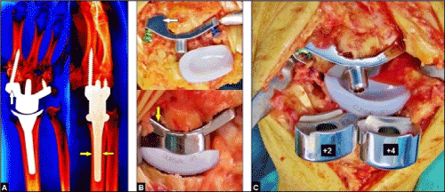
Figure 1. (A) Radiographs of a patient's right wrist in both planes demonstrating correctly positioned MaestroTM WRS utilizing a carpal plate with a scaphoid augment after removal of the entire scaphoid bone (white arrow). The carpal plate is fixed with a variable locking titanium screw into the 2nd metacarpal bone and a fixed locking titanium screw into the hamate bone. Note that the polished titanium alloy radial stem is press-fit inserted (yellow arrows) that does not require cementation. (B) Clinical photographs demonstrating the MaestroTM WRS utilizing capal plates with (white arrow) and without (yellow arrow) scaphoid augments. The undersurface is coated with a thick titanium spray (Macrobond). Note the green colored variable locking screw and the blue colored fixed locking screw that is the first change in comparison to the primary MaestroTM Total Wrist which had non-locking screws only. (C) Clinical photograph demonstrating the MaestroTM WRS after insertion of its carpal plate and radial component. Note that the intercalated carpal heads are set up externally onto the cone of the previously inserted carpal plate that is the second change in comparison to the primary MaestroTM Total Wrist (here the carpal heads are screwed onto the carpal plate over the capitate peg before its insertion that does not more allow its exchange in case of a dislocation without revision of the entire carpal plate) [40].
Generally, TWA with the use of the third generation types (biaxial-anatomical, unconstrained, ellipsoid metal-on-polyethylene (PE) surface articulation) is the recognized motion-preserving treatment option to a Partial or Total Wrist Fusion (PTW or TWA) in cases of rheumatic or non-rheumatic Wrist Joint Osteo Arthritis (WJOA) worldwide [1-13]. Wrist joint motion modulates the synergistic and antagonistic extrinsic / intrinsic motion at the finger joints which is an important prerequisite to realize all powerful functional grips of the hand. PTWs crossing the midcarpal or radiocarpal joints always result in loss of the overall wrist joint motion of approximately 50% [14,15]. Adams et al. demonstrated impressively in 2003 that in healthy subjects limited wrist joint motion inevitably leads to a significant worsening of their ratings in activities of daily living (perineal hygiene, buttoning a blouse or shirt, threading a needle, turning over the pages of a newspaper, opening a door with the door handle, picking up small objects, and so on), and Nydick et al. reported in 2013 that patients who received primarily a TWA for treatment of post-traumatic WJOA rated their functional outcome in Patient-rated wrist evaluation (PRWE) statistically significant better than patients who received primarily a TWF [16,17]. Adey et al. reported in 2005 that nearly all patients who received primarily a TWF would elect to have a procedure that could make their wrist move again if one were available, and Ekroth et al. reported in 2012 that their patients with failed TWAs utilizing older generation types would have a TWA again despite that their long-term results being poor and many of them being revised to a TWF [18,19]. It is still widely known from the literature that for the wrist 5° to 40° of flexion, 30° to 40° of extension, and 10°/15° of radial/ulnar deviation to 40° of combined radial-ulnar deviation are required to realize the most essential activities of daily living [20,21]. When patients are unsatisfied with their primarily performed motion-restricting TWF, then a secondary conversion to a motion-preserving TWA with a third-generation type, also utilizing the MaestroTM WRS, can be an useful option to improve the patient's disability [22,23]. Additionally, the MaestroTM WRS has proven to be useful as well as salvage procedure of a failed previously performed PTW, and in single cases as early treatment option for highly comminuted distal radius fracture in selected older and elderly patients [24,25]. Furthermore, recent evidence suggests that the complication rate of TWA does not differ statistically significant in comparison to patients undergoing a TWF [26,27].
Till 2014, implant survival with the third TWA generation types was reported to be 90 to 100% at 5 years in most series, but it declines from 5 to 8 years [9]. These results are completely comparable with those after Total Shoulder / Elbow / Ankle Arthroplasties which are much more less debate in the literature than TWA. Recently, rheumatoid arthritis remains the most common indication for TWA with a relative portion ranging from 51 to 71%, and followed by 14% for treatment of post-traumatic WJOA of all TWAs performed by surgeons who have published their experiences with this procedure [9,26]. Two questions are not clearly answered currently: first, how can patients load their wrists with a TWA; and second, are young patients suitable receiving a TWA [28]. In the past, TWA was almost exclusively used in older patients with rheumatoid arthritis because of their low-demand lifestyle, whereas patients with post-traumatic WJOA are typically young and active [22-32]. However, there is a trend in the literature towards the increasing expansion of the indication also for selected younger patients aged 31 years or younger with post-traumatic WJOA associated with good bone stock (including the MaestroTM WRS), and high claims in their activities of daily living are not always considered as contraindication [6,17,23,33,35-40]. One major complication with the use of older TWA generation types was instability or dislocation in up to 8% of cases [9,41]. It has been noted by Sterling Bunnel (†1957): “ A painless stable wrist is the key to hand function ” [3]. This problem seems to be solved with the use of the new third generation types. The rates of instability or dislocation for the ReMotionTM Total Wrist is reported to be 0,7% only (1 of 144 cases), and for the MaestroTM WRS 0,6% only (1 of 157 cases) [9,22,31-34].
The non-locking MaestroTM Total Wrist, developed in 2002 by Strickland / Palmer / Graham , available in Europe since 2005 and first published with encouraging results by Dellacqua in 2009, is one of the third generation TWA that is currently in use worldwide [10,17,22,24,25,31-34,37,39,40,42-49]. The design of the implant has 3 essential differences as compared to the other third generation types (ReMotionTM, Universal 2): first: the design resembles a Total Hip Arthroplasty (THA) in which a metal head articulates in a PE cup; second: the titanium alloy radial stem is to be inserted press-fit into the radial diaphysis; and third: the carpal plates are available without or with various scaphoid augments that provides a sufficient support of the carpal plate with it titanium alloy capitate peg onto the base of the trapez/trapezoid bones even in cases when the scaphoid bone is removed that is associated with the advantage that bone grafting between the surrounding carpal bones is exclusively not required (Figure 1A-C). The MaestroTM WRS is the further development of the implant, it has 2 changes as compared to the MaestroTM Total Wrist: first, the carpal plate is inserted with fixed or variable locking screws that should reduce the shear forces at the implant-bone interface (Figure 1B); and second: the intercalated carpal heads are set up externally onto the cone of the previously inserted carpal plate whereas the carpal heads of the MaestroTM Total Wrist are screwed onto the carpal plate over the capitate peg before its insertion. This new feature allows an exchange of the carpal heads such as required in case of instability without the need for revision of the entire carpal plate (Figure 1C). Noted that the FDA in 2009 reported 1 case of backing out of a non-locking screw of the first MaestroTM Total Wrist type (inserted in this case in 2005) that required a conversion to a TWF [50].
Dellacqua noted with the first scientific paper on the MaestroTM Total Wrist in 2009 that the titanium alloy radial stem should be inserted press-fit without the need of cementation [31]. It is still widely known from literature that cementation of a titanium alloy stem in THA does not confer any benefits in survivorship over its insertion in a non-cemented manner (Figure 12) [51]. Moreover, the high elasticity of titanium and the excessive stresses in the mantle of the cement could lead to micromotion and debonding of the stem, and these micromovements at the cement-stem interface may be responsible for cement mantle breakage and generation of titanium debris inducing necrosis and osteolysis [51,52]. The failure risk could be higher for smaller stems due to their greater elasticity, and in men who are physically active, while titanium stems with a larger diameter may be more successful [53]. Furthermore, Thomas et al. noted that in THA the combination of a polished titanium stem (such as with the radial stems of the MaestroTM Total Wrist/WRS) and cement appears to facilitate crevice corrosion [54]. Additionally, cementation of implants can be associated with another disadvantages such as prolonged operation time, fat embolism, and venous thromboembolic disease [55-58]. In the literature, there are no evident scientific data with the use of all third generation TWA types (including the MaestroTM Total Wrist/WRS!) that support the necessity of cementation at time of its primary insertion (Figure 1A-C, Figure 5A-B, Figure 11A-E), cementation is only recommended primarily in patients with poor bone stock (rheumatoid arthritis, advanced stage of osteoporosis) (Figure 2A-B) or in cases of revision TWA, and cementation does not decrease the risk of intra- or postoperative periprosthetic fracture [2-13,16,17,22-49,59-63]. Noted that for metacarpophalangeal implants it has been observed a higher risk of intraoperative periprosthetic fractures when using cementation than implants which are inserted in a non-cemented manner [64]. Moreover, the most important disadvantage of both MaestroTM types is that the PE insert is fixed to the radial body. Hence, when PE wear or fracture occurs then a revision of the entire radial component becomes necessary. If the radial component was inserted with cementation therefore bony windowing of the distal radius shaft along the entire radial component becomes inevitable that could be associated with further complications. Both MaestroTM do not alter the Distal RadioUlnar Joint (DRUJ), hence, it can be combined with an Ulnar Head Replacement (UHR) (Figure 2B, Figure 4) [43-45,47].

Figure 2. (A) A 84-year-old female sustained a right highly comminuted distal radius fracture that was accompanied with a pre-existing scapholunate diastasis, and treated primarily with a TWA (MaestroTM WRS) utilizing a carpal plate with scaphoid augment combined with an ulnar head resection. Due to the pronounced osteoporotic bone stock, the titanium alloy radial stem was inserted with cementation (yellow arrows). (B) A 79-year-old obese female presented with a left highly comminuted distal radius fracture with marked radial shortening that was treated primarily with a TWA (MaestroTM WRS) utilizing a carpal plate with scaphoid augment combined with ulnar shortening and an UHR. Note the severely destroyed articular surface of distal radius (white oval circle). Due to the pronounced osteoporotic bone stock, the titanium alloy radial stem was inserted with cementation (yellow arrows).
An essential advantage of both MaestroTM types is the high degree of modularity that provides the combination of all components among themselves (Figure 3). Intraoperatively, the alignment and stability of implant is to be well documented by fluoroscopy especially under dynamic conditions as well (Figure 4, Figure 5A-B). An essential prerequisite in order to avoid complications such as dislocation or breakout of the radial component and/or carpal plate is that both components should be exactly aligned to the axis of the radial shaft and to the axis of the capitate-3rd metacarpal bones (Figure 6A-B), and there is a consensus in the literature for all third generation TWA types that the carpal plates should be fixed with a first screw radialwards into the 2nd metacarpal bone, and a second screw ulnarwards into the hamate bone (Figure 6C). The main complication with the use of the BIAX was breakout of the metacarpal peg in dorsal direction subsequently leading to instability of its articulation and followed by PE wear and/or fracture [65]. Technical errors at time of insertion of the BIAX led to a further exacerbation of this complication rate, Takwale et al. found that for each degree a carpal component was failed placed in dorsal direction increased the risk of breakout of the tip of the metacarpal peg in dorsal direction in up to 17% respectively [66]. Hence, the success for an acceptable survivorship of a TWA also depends on every surgeon's experience. Ocampos et al. reported on a their retrospective results with the use of the ReMotionTM Total Wrist (N=14), all performed by unexperienced surgeons, none of the implants were correctly aligned, and mostly observed with averaged 10,1° (range: 2°-21°) for failed positioning of the capitate peg in dorsal direction [67]. Noted that with both MaestroTM types combinations with a thumb carpometacarpal total joint replacement or a Total Elbow Arthroplasty are possible [43,47,68]. Contraindications for a TWA including both MaestroTM types are persistent osteomyelitis, irreparable nerve palsies, longstanding posttraumatic/postoperative wrist contracture with or without malalignment, concomitant suspicious intraosseous lytic lesions without its diagnostic clarification, wrist hyperlaxity, untreated extrinsic ( i.e. wrist-bridging) tendon disruptions, unstable soft tissue with or without infection, concomitant soft tissue tumors without its diagnostic clarification, persistent posttraumatic/postoperative lymphedema and/or chronic regional pain syndrome with stiffness of multiple joints in the hand, distinctive substantial bone loss without a possibility for sufficient bony reconstruction, suspicious compliance of the patient, and for young patients the denervation of the wrist can be successful to decrease pain over a time before TWA (Figure 10A-B) [37,69].
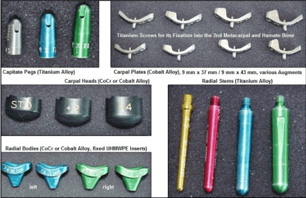
Figure 3. All components of both MaestroTM types demonstrating with the trials. Note the high modularity that allows the combination of all components among themselves.
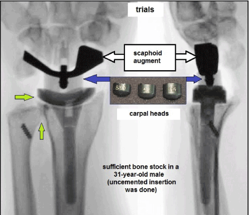
Figure 4. Intraoperative fluoroscopy demonstrating well aligned trials of the MaestroTM WRS, stability of implant can be achieved is to be proofed utilizing the carpal heads with its various heights. Note that the DRUJ is not altered by the implant (green arrows).

Figure 5. (A) Dynamic assessment of the MaestroTM WRS intraoperatively with terminal ranges of radial and ulnar deviation (fluoroscopy) showing correct aligned implant. Note that there are no instabilities nor impingements (white arrows). (B) Dynamic assessment of the MaestroTM WRS intraoperatively with terminal ranges of flexion and extension (fluoroscopy) showing correct aligned implant in the absence of instability. Note that in this case in the presence of good bone stock the titanium alloy radial stem was inserted press-fit without the need of cementation (white arrows).
Recently, in a single-center study with the use of the MaestroTM Total Wrist (published in 2015), the cumulative implant survival after 8 years (N=68) is reported to be 95%, and at the 5-year follow-up radiographic loosening was present in 2% of all cases only that is slightly superior over the other third generation types as published in 2014 [9,33]. Furthermore, the MaestroTM Total Wrist achieves the most favorable functional outcome as compared to other third-generation types, and it may be justified in preserving resection-related carpal height due to its 3 various carpal heads in combination with its design of ellipsoid surface articulation especially in order to avoid radial impingement that is mainly observed with the use of the ReMotionTM Total Wrist [32,46,70]. However, none of all third generation TWAs are able to restore completely motion in the anatomic axes of a normal wrist including the "dart-throwing" motion arc from extension-radial deviation to flexion-ulnar deviation [71-73]. This motion arc that is performed in an oblique axis at the midcarpal joint is particularly prevalent in most occupational, recreational and avocational activities such as hammering, clubbing or fly fishing; whereas the so-called "reversed dart-throwing" motion arc from extension-ulnar deviation to flexion-radial deviation takes place almost exclusively at the radiocarpal joint [15,74]. However, a recent human cadaveric study revealed that the "dart-throwing" motion arc can also takes place at the radiocarpal joint as a compensatory mechanism when a pancarpal fusion was done [75]. In order to obtain the "dart-throwing" motion arc, a newer approach in wrist arthroplasty is midcarpal hemiarthroplasty ( i.e. preserving midcarpal motion by obtaining the proximal pole of capitate bone) with the use of a radial implant alone [76,77]. However, whether this newer approach is superior to a TWA cannot be assessed currently. Additionally, another approach to obtain normal wrist circumduction with a new TWA that has some similarities with MaestroTM Total Wrist is recently in proof with a human cadaveric study [78]. Noted that postoperative deterioration of function in patients receiving a TWA can also be caused by technical errors or early soft tissue complications such as insufficiency of the extensor retinaculum (Figure 7).

Figure 6. (A) Intraoperative clinical photograph and fluoroscopy (MaestroTM WRS) in both planes demonstrating correct central positioning of the K-wire into the radial radial shaft before it's reaming. Note that for correct positioning of the K-wire in the "free hand technique" (white arrow) the axes of radial shaft are equally located in the dorsal-radial quadrant of the articular surface (green circle). (B) Intraoperative fluoroscopy (MaestroTM WRS) in both planes demonstrating reaming of the capitate bone after correct central positioning of the K-wire along the axis of the capitate-3rd metacarpal bones. (C) Intraoperative fluoroscopy (MaestroTM WRS) demonstrating placement of the 2 K-wires for fixation of the carpal plate with the first crew into 2nd metacarpal bone and the second screw into the hamate bone.
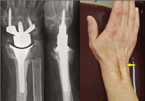
Figure 7. Same patient as in Figure 2A. The extensor retinaculum was primarily reconstructed intraoperatively, but it has disrupted 2 years after surgery again that led to marked bowstring of the extrinsic extensor tendons (yellow), and resulted in a loss of active extension at the wrist of 10° as compared with the 1-year follow up [25, 48].
Both MaestroTM types are approved for carpal hemiarthroplasty creating a "metal-on-cartilage/bone articulation" by the FDA. However, first results with a small number of patients are not encouraging. Gaspar et al. [34] reported in 2016 on 6 patients who were evaluated in mean follow-up of 35 months, and there were 1 failure, 1 instability, 3 contractures, 2 soft tissue imbalances, 2 erosions into the opposite bone, and 2 cases with DRUJ diseases. Huish et al. reported in 2017 on 11 patients who were evaluated at an average follow-up of 4 years, there was a high failure in 45% of cases that required in 2 patients a conversion to a TWA and in 3 patients a conversion in TWF, and they concluded that is probably based on a mismatch between the lunate facet of distal radius articulating with the metal head of implant [79]. Both MaestroTM types are not approved for radial hemiarthroplasty creating a "PE-on-cartilage/bone articulation" by the FDA, and the reported first results were disappointing, and it has been noted by Cooney in 2013 that care must be taken when using this implant in such an "off-label" manner [80]. The background of this procedure is similar to midcarpal hemiarthroplasty by obtaining the proximal pole of the capitate bone that allows midcarpal motion. Culp et al. reported in 2012 on 10 patients who were evaluated at mean follow-up of 19% months, and there was an unacceptable high complication rate mostly based on aseptic loosening and/or tenosynovitis ( i.e. polyethylene disease) [81].
What are the causes for failed TWAs? It is still widely accepted that for older TWA types utilizing a metal-on-metal articulation that metal wear subsequently resulting in metallosis to the surrounding soft tissue, observed in nearly 100% of cases with the APH prosthesis, was the main cause for its failure [82]. Since introducing the newer TWA types utilizing a metal-on-PE articulation, metal wear appears not to be responsible for its failure. It is reported with the Universal 2 in up to 16,7% only, and common initially presented as carpal tunnel syndrome, pisiform impingement, or pseudotumor [83-86]. With the use of the MaestroTM Total Wrist, only 1 publication could be found in which metallosis led to capsular destruction with subsequent attenuation of the DRUJ leading to dislocation and destruction of the extensor mechanism, however, there was an impingement between the TWA and an UHR with a metal head [87]. However, the FDA reported in 2010 another case of metallosis with the MaestroTM Total Wrist due to fretting of a loosened non-locking titanium screw and the carpal plate, but a revision was required due to an infection and not due to implant issues [88]. In the literature there is a consensus with the use of the newer TWA types utilizing a metal-on-PE articulation that its failure is primarily caused by mechanical imbalance especially by the natural course of rheumatoid arthritis (ulnar carpal translocation, progressive deterioration of bone stock, muscular imbalance, ligamentous insuffiency), and secondarily followed by PE or metal wear, and noted as well that was found for the ReMotionTM Total Wrist that there are no statistically significant differences in the amount of metal or PE wear particles (evaluated in histological specimens) in patients with or without loosening of their implants [65,66,89,90]. The natural course of rheumatoid arthritis regarding progressive ulnar carpal translocation especially when an ulnar head resection was performed is impressively described for dislocation of a TWA in ulnar direction, and for lunate migration onto the distal ulnar stump in patients who are not received a TWA [82,91,92]. Furthermore, the natural course of rheumatoid arthritis can also be associated with translocation of the wrist in volar direction that must be considered as contraindication for TWA as well when it leads to wrist contracture accompanied with a longstanding disruption of the volar extrinsic radioscaphocapitate ligament which is the most important stabilizer of the wrist (Figure 8A-B). However, younger patients receiving a TWA with a high mobility and functional needs may be more risky for occurrence of wear [86]. When a TWA should become necessary, correction of post-traumatic deformities at the distal radius is required in every instance if there are no post-traumatic contractures at the wrist, and pre-existent or post-traumatic WJOA is not present as well (Figure 9A-G). Furthermore, when elderly patients with advanced stage of post-traumatic WJOA are not able receiving TWA or TWF for private reasons, then the denervation of the wrist can be "last option" to improve the patient's disability (Figure 10A-B).

Figure 8. (A) A 73-year-old active female presented with advanced stage of rheumatoid arthritis in her right wrist. There is a marked carpal volar translocation (lines) that led to wrist contracture accompanied with longstandig disruption of the radioscaphocapitate ligament (red star). Independently that the patient is well pain-adopted with her rheumatoid-related drugs, the amount of fixed translocation of the entire carpus and the additional loss of ligamentous stabilization of the wrist are to be considered generally as contraindication for TWA. (B) Same patient with her left wrist equally to her right wrist.
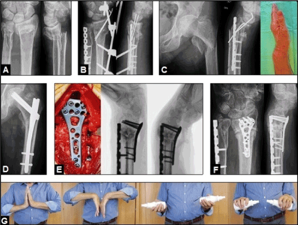
Figure 9. (A) A 72-year-old active male sustained a left highly comminuted distal forearm fracture after a fall in his garden, note that there was no pre-existent WJOA. (B) Same patient, primarily surgical reduction by external fixation and pinning of the dorsally displaced extra-articular distal radius fracture, and locked plating of the distal ulna (the multiple bony fragments distal to the DRUJ were circularly sutured at the plate). The external fixation was removed 3 weeks after surgery, and the wrist was immobilized with a cast for another 3 weeks. (C) Same patient, 7 weeks after the primary injury the patient sustained a second fall-related injury associated with a left subtrochanteric femoral fracture and a re-fracture of the left distal radius with pronounced displacement in volar direction. (D) The left subtrochanteric femoral fracture was surgically reducted by a load-stable Friedl-nail. (E) Same patient, the left distal radius re-fracture was surgically treated by corrective osteotomy utilizing a locking distal volar radius titanium plate and a corticocancellous bone graft from the left iliac crest. Intraoperative dynamic fluoroscopy demonstrating a stable wrist at terminal ranges of passive extension and flexion. (F) Same patient 3 months later, radiographs demonstrating uneventful bony healing of the left distal radius corrective osteotomy. (G) Same patient 6 months later, clinical photographs showing complete restoration of the active extension-flexion and supination-pronation motion arcs in comparison to the right unaffected wrist.
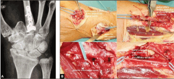
Figure 10. (A) A 72-year-old active female presented with advanced stage of right post-traumatic WJOA that allows both a TWA and a TWF. Unfortunately, she has to take care of her ill and bedridden husband after a stroke at home, and so, she was unable to endure the prolonged time for rehabilitation after TWA or TWF. Hence, the denervation of the wrist was indicated by us at last option to decrease pain in her unfortunate situation. (B) Same patient intra-operatively, the posterior and anterior interosseus nerves and the retrograde branch of the superficial radial were transected and ligated, and the neurovascular structures surrounding the radial artery were dissected carefully as well. Two months after surgery, the patient was happy that she was able to perform her activities of daily living without pain at home again.
What role do PeriProsthetic Osteolyses (PPO) around a TWA play in assessment of its clinical manifestation of loosening? Boeckstyns and Herzberg reported in 2014 on 44 patients received a TWA with the ReMotionTM Total Wrist and evaluated at a mean follow-up of 3,7 years, significant periprosthetic radiolucency (more than 2 mm in width) were found juxta-articularly around the prosthetic components in 36,4% at the radial component and in 15,9% at the carpal component, whereas clinical manifestation of loosening (defined as progressive angulation or subsidence) was present in 14% of all cases only; and radiolucency seemed to be stabilize within 3 years after surgery [93]. These observations were confirmed with the use of the Motec type in which PPO juxta-articularly around the radial component seemed to be stabilize within 8 years after surgery without any clinical signs of loosening [38]. This discrepancy between the amounts of occurrence of PPO and its real clinical manifestation of loosening is also observed in patients receiving an UHR in which PPO around the collar of implant are observed in nearly all cases and followed by its stabilization within 3 years after surgery, and that phenomenon is discussed as a result of "stress-shielding" [92]. Hence, PPOs in the absence of any clinical signs of loosening do not require surgical intervention (Figure 11A-E). That phenomenon is probably not new. Julius Wolff, a german orthopaedic surgeon (†1902), first described in 1892 that cortical bones primarily became atrophic in lesser loaded regions whereas it became hyperthrophic in higher loaded regions, and resulted secondarily in a steady-state over time ("Wolff's law") [95]. That phenomenon is especially observed in THA when utilizing a non-cemented titanium femoral stem which is press-fit inserted into the femoral diaphysis and followed by its sufficient osseointegration distal to the radiolucency (Figure 12). Hence, PPOs juxta-articularly around non-cemented prosthetic components can be probably discussed as well as result of sufficient osseointegration of its titanium alloy stems distal to the PPOs.
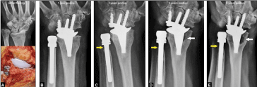
Figure 11. (A) A 59-year-old female presented with advanced stage of WJOA accompanied with pronounced radial shortening as result of a longstanding distal radius physeal arrest and treated by a non-cemented TWA (ReMotionTM Total Wrist) primarily combined with a non-cemented UHR (metal head), formerly published by the author in 2014 at a 1-year follow-up [69]. (B) Same patient 1 year after surgery, there are no signs of PPO. (C) Same patient 2 years after surgery, first signs of PPO around the collar of UHR (yellow arrow). (D) Same patient 3 years after surgery, discrete progression of PPO around the collar of UHR (yellow arrow), and first signs of PPO at the radial component of TWA (white arrow). (E) Same patient 4 years after surgery, stabilization of PPO around the collar of UHR (yellow arrow), and progression of PPO at the radial component of TWA (white arrow) in the absence of any clinical signs of loosening, hence, revision surgery is not required at this time.
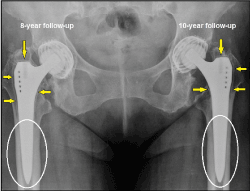
Figure 12. Long-term follow-up of THA at both sides in a 77-year-old active female who is very satisfied with her outcome. Non-cemented femoral titanium stems were inserted press-fit to the diaphyseal cortical bones (Zweymüller stems). There is a steady-state ("Wolff's law") between unchanged insignificant radiolucencies at the lesser loaded trochanteric regions (yellow arrows) whereas the titanium stems distal to the radiolucencies at the higher loaded regions are well osseointegrated with the cortical bones of diaphyses (white ovals).
For a failed third generation TWA there are 2 options for surgical treatment: revision TWA with or without bone grafting or cementation, and conversion to a TWF [29,60,96-100]. However, patients undergoing treatment with TWA must be prepared that it might end with TWF; therefore limited bone resection is a common feature of all contemporary wrist. Both MaestroTM implants have demonstrated uncomplicated conversion to TWF, and a recent study revealed that the outcome of TWF after a failed TWA is comparable to those after primary TWF [22,32-34,101].
Conclusion
TWA using all third generation types is an established motion-preserving alternative to P/TWF for treatment of WJOA. Herzberg and Adams noted in 2012: “The future appears positive for greater adoption of total wrist arthroplasty for patients who are willing to accept somewhat greater risks for the benefit of improved function” [5]. Both MaestroTM types are slightly superior in terms of modularity, survivorship at a 5 to 8-years follow-up, and functional outcome in comparison to the other types. Polished titanium alloy femoral stems (such as in both MaestroTM wrist implants) have proven to be useful and reliable in THA. In the literature, there are no evident scientific data which support the cemented use of all third generation TWAs (including both MaestroTM types!), and cementation is only recommended in the presence of poor bone stock or for revision TWA that is absolutely comparable with THA (Figure 13). Even Dr. Strickland, one of the developers of the MaestroTM Total Wrist in 2002 (!), and honoured as "Pioneer of Hand Surgery" in 2010 by the International Federation of Societies for Surgery of the Hand (IFSSH) at its 11th International Congress in Seoul (South Korea), has been noted in 2007 in original as follows: "With improved implant designs and osseointegrated stems, surgeons can achieve bone ingrowth and stability that couldn’t be achieved in the past, minimizing the need for bone cement ... In unique circumstances, however, surgeons might improve the implants’ ability by filling the medullary canal with bone cement before inserting the implant stems ... Those indications may be very poor bone stock, severe osteoporosis, previous erosive processes, or other circumstances where the surgeon feels that the stability and long-term performance of the components … may be improved by the use of supplementary bone cement ..." [102,103]. The disadvantage of both MaestroTM types is that the PE insert is fixed to the radial body, hence, when PE wear or fracture occurs then a revision of the entire radial component becomes necessary. When the long radial stem of both MaestroTM types is primarily inserted into the radial diaphysis with cementation, then (in case of failure) an extended bony windowing along the entire radial component is needed for its removal that can be followed by another complications in the further course. Hence, the decision by the company to withdraw both MaestroTM types from the market place because it was inserted only in a non-cemented manner in the past, is doubtful. Interestingly, Zimmer Biomet distributes 3 femoral stems (Mayo, Taperloc, M/L Taper) for THA which have similarities in surface structure (polished distally, titanium alloy) with the stem of radial component of both MaestroTM types, and all of them are approved for THAs in a non-cemented manner [104-106]. Finally, all surgeons who are performed TWAs utilizing both MaestroTM implants with encouraging results in the past and willing to perform it in future, now must communicate their operated and very satisfied patients as well as their new patients present with WJOA that the decision by the company Zimmer Biomet is incomprehensible and contradicts any scientific knowledge recently.
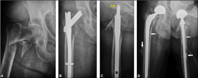
Figure 13. (A) A 76-year-old female presented with an unstable subtrochanteric femoral shaft fracture associated with poor osteoporotic bone stock. (B) Same patient, primarily treated by a non-cemented load-stable Friedl-nail (yellow arrow) introduced in 1996 [107, 108]. (C) Same patient, cut-out of the blade into the hip joint 6 weeks after surgery. (D) Same patient, a conversion procedure was done utilizing a bipolar hip arthroplasty with a long cemented femoral stem (white arrows) which needed crossing the hole of the removed distal interlocking screw of the nail. Note the partial outlet of cement through the hole of the removed interlocking screw at the femoral shaft.
Conflict of interest
The author confirms that this article content has no conflict of interest.
Acknowledgements
The author would like to thank Michel E.H. Boeckstyns (MD DMSc (PhD), Denmark) and Ole Reigstad (MD PhD EBHS Fellow, Norway) for their excellent scientific support especially in understanding the complexity of TWA for many years. The author would also like to thank Prof. Dr. med. Abdul-Kader Martini (Heidelberg / Germany, President of the German Society of Hand Surgery (DGH) in 2006 and Honorary Member of the DGH) for the jointly performed anatomical courses under his scientific leadership at the Anatomical Institute of the Ruprecht-Karls-University Heidelberg from 2010 to 2012 that exclusively included communication for recent indications of TWA and presentation of the operation technique of the MaestroTM Total Wrist at human cadaveric hands.
References
- Menon J (1998) Universal Total Wrist Implant: experience with a carpal component fixed with three screws. J Arthroplasty 13: 515-523.
- Adams BD (2004) Total wrist arthroplasty. Orthopedics 27: 278-284. [Crossref]
- Gupta A1 (2008) Total wrist arthroplasty. Am J Orthop (Belle Mead NJ) 37: 12-16. [Crossref]
- Herzberg G (2011) Prospective study of a new total wrist arthroplasty: short term results. Chir Main 30: 20-25. [Crossref]
- Herzberg G, Adams BD (2012) Future of wrist arthroplasty. J Wrist Surg 1: 5-6. [Crossref]
- Herzberg G, Boeckstyns M, Sorensen AI, Axelsson P, Kroener K, et al. (2012) "Remotion" total wrist arthroplasty: preliminary results of a prospective international multicenter study of 215 cases. J Wrist Surg 1: 17-22.
- Boeckstyns ME, Herzberg G, Merser S (2013) Favorable results after total wrist arthroplasty: 65 wrists in 60 patients followed for 5–9 years. Acta Orthop 84: 415-419.
- Adams BD (2013) Wrist arthroplasty: partial and total. Hand Clin 29: 79-89. [Crossref]
- Boeckstyns ME (2014) Wrist arthroplasty--a systematic review. Dan Med J 61: A4834. [Crossref]
- Reigstad O, Røkkum M (2014) Wrist arthroplasty: where do we stand today? A review of historic and contemporary designs. Hand Surg 19: 311-322. [Crossref]
- Reigstad O (2014) Wrist arthroplasty: bone fixation, clinical development and mid to long term results. Acta Orthop Suppl 85: 1-53.
- Halim A, Weiss AC (2017) Total Wrist Arthroplasty. J Hand Surg Am 42: 198-209.
- Boeckstyns MEH, Herzberg G (2017) Current European Practice in Wrist Arthroplasty. Hand Clin 33: 521-528. [Crossref]
- Meyerdierks EM, Mosher JF, Werner FW (1987) Limited wrist arthrodesis: a laboratory study. J Hand Surg Am 12: 526-529. [Crossref]
- Leventhal EL, Moore DC, Akelman E, Wolfe SW, Crisco JJ (2010) Carpal and forearm kinematics during a simulated hammering task. J Hand Surg Am 35:1097-1104.
- Adams BD, Grosland NM, Murphy DM, McCullough M (2003) Impact of impaired wrist motion on hand and upper-extremity performance(1). J Hand Surg Am 28: 898-903. [Crossref]
- Nydick JA, Watt JF, Garcia MJ, Williams BD, Hess AV (2013) Clinical outcomes of arthrodesis and arthroplasty for the treatment of posttraumatic wrist arthritis. J Hand Surg Am 38: 899-903.
- Adey L, Ring D, Jupiter JB (2005) Health status after total wrist arthrodesis for posttraumatic arthritis. J Hand Surg Am 30: 932-936.
- Ekroth SR, Werner FW, Palmer AK (2012) Case report of long-term results of biaxial and volz total wrist arthroplasty. J Wrist Surg 1: 177-178. [Crossref]
- Palmer AK, Werner FW, Murphy D, Glisson R (1985) Functional wrist motion: a biomechanical study. J Hand Surg Am 10: 39-46. [Crossref]
- Ryu JY, Cooney WP 3rd, Askew LJ, An KN, Chao EY (1991) Functional ranges of motion of the wrist joint. J Hand Surg Am 16: 409-419.
- Nydick JA, Greenberg SM, Stone JD, Williams B, Polikandriotis JA, et al. (2012) Clinical outcomes of total wrist arthroplasty. J Hand Surg Am 37: 1580-1584. [Crossref]
- Nicoloff M (2015) Total wrist arthroplasty--indications and state of the art. Z Orthop Unfall 153: 38-45. [Crossref]
- Schmidt I (2016) An Unusual and Complicated Course of a Giant Cell Tumor of the Capitate Bone. Case Rep Orthop 2016: 3705808. [Crossref]
- Schmidt I (2015) Can Total Wrist Arthroplasty Be an Option for Treatment of Highly Comminuted Distal Radius Fracture in Selected Patients? Preliminary Experience with Two Cases. Case Rep Orthop 2015:380935. doi: 10.1155/2015/380935. Epub 2015 Sep 29.
- Melamed E, Marascalchi B, Hinds RM, Rizzo M, Capo JT (2016) Trends in the utilization of total wrist arthroplasty versus wrist fusion for treatment of advanced wrist arthritis. J Wrist Surg 5: 211-216.
- Hinds RM, Capo JT, Rizzo M, Roberson JR, Gottschalk MB (2017) Total Wrist Arthroplasty Versus Wrist Fusion: Utilization and Complication Rates as Reported by ABOS Part II Candidates. Hand (NY) 12: 376-381.
- Weiss AP, Akelman E (2012) Total wrist replacement. Med Health R I 95: 117-119. [Crossref]
- Adams BD (2010) Complications of wrist arthroplasty. Hand Clin 26: 213-220. [Crossref]
- Kamal R, Weiss AP (2011) Total wrist arthroplasty for the patient with non-rheumatoid arthritis. J Hand Surg Am 36: 1071-1072.
- Dellacqua D (2009) Total wrist arthroplasty. Tech Orthop 24: 49-57.
- Sagerfors M, Gupta A, Brus O, Rizzo M, Pettersson K (2015) Patient related functional outcome after total wrist arthroplasty: A single center study of 206 cases. Hand Surg 20: 81-87.
- Sagerfors M, Gupta A, Brus O, Pettersson K (2015) Total Wrist Arthroplasty: A Single-Center Study of 219 Cases With 5-Year Follow-up. J Hand Surg Am 40: 2380-2387.
- Gaspar MP, Lou J, Kane PM, Jacoby SM, Osterman AL, Culp RW (2016) Complications following partial and total wrist arthroplasty: A single center retrospective review. J Hand Surg Am 41: 47-53.e4.
- Cooney W, Manuel J, Froelich J, Rizzo M (2012) Total wrist replacement: a retrospective comparative study. J Wrist Surg 1: 165-172.
- Boeckstyns ME, Herzberg G, Sørensen AI, Axelsson P, Krøner K, et al. (2013) Can total wrist arthroplasty be an option in the treatment of the severely destroyed posttraumatic wrist? J Wrist Surg 2: 24-329.
- Schmidt I (2017) Does Total Wrist Arthroplasty for Treatment of Posttraumatic Wrist Joint Osteoarthritis in Young Patients Always Lead to Restriction of High-demand Activities of Daily Living? Case Report and Brief Review of Recent Literature. Open Orthop J 11: 439-446.
- Reigstad O, Holm-Glad T, Bolstad B, Grimsgaard C, Thorkildsen R, et al. (2017) Five- to 10-Year Prospective Follow-Up of Wrist Arthroplasty in 56 Nonrheumatoid Patients. J Hand Surg Am 42: 788-796. [Crossref]
- Schmidt I (2017) Kienböck's disease mimicing gouty monoarthritis of the wrist. Int J Case Rep Images 8: 423-426.
- Schmidt I (2017) The MaestroTM Wrist Reconstructive System for treatment of posttraumatic wrist joint osteoarthritis after a devasting course of a distal radius fracture: Case presentation and technical note to the implant. Trauma Emerg Care 2: doi: 10.15761/TEC.1000124.
- Ma JX, Xu YQ (2016) The instability of wrist joint and total wrist replacement. Chin J Traumatol 19: 49-51. [Crossref]
- Nair R (2015) Past, Present, and Future in Total Wrist Arthroplasty: A Perspective. Curr Orthop Pract 26: 318-319.
- Schmidt I (2015): Surgical treatment options in thumb carpometacarpal osteoarthritis: a recent literature overview searching for practice pattern with special focus on total joint replacement. Curr Rheumatol Rev 11: 39-46.
- Schmidt I (2015): Combined replacements using the Maestro total wrist and uHead ulnar head implants. J Hand Surg Eur Vol 40: 754-755.
- Schmidt I (2017): Distal radioulnar synostosis after primary combined replacements for treatment of highly comminuted distal radius fracture in an elderly patient. J Hand Surg Eur Vol 42: 97 - 98.
- Schmidt I (2017): RE-MOTIONTM total wrist arthroplasty for treatment of advanced stage of scaphoid non-union advanced collapse. Does excision of the entire scaphoid bone prevent impingement at terminal range of radial deviation? Trauma Emerg Care 2: doi: 10.15761/TEC.1000127.
- Schmidt I (2017) Combined replacements of the wrist, ulnar head, and thumb carpometacarpal joint. Case report, technical note and recent evidence to the ArpeTM prosthesis. Trauma Emerg Care 2: doi: 10.15761/TEC.1000130.
- Schmidt I, Nennstiel I, Muradyan M, Schmieder A, Kaisidis A, et al. (2017) The rare injury of a closed digital extensor tendon avulsion of the third finger at its musculotendinous junction: Case presentation and brief overview of literature with regard to general, specific and practicable aspects to extensor tendon surgery involving tendon transfer procedures. Trauma Emerg Care 2: doi: 10.15761/TEC.1000155.
- Schmidt I, Schmitz H, Muradyan M, Kaisidis A, Großmann D (2017) Who introduced the "quadrangular-construct" modification of the Sauvé- Kapandji procedure first? Case presentation and brief overview of literature with regard to general, specific and practicable aspects to primary trauma-related surgical treatment including possible salvage options at the distal radioulnar joint. Trauma Emerg Care 3: doi: 10.15761/TEC.1000157.
- MAUDE Adverse Event Report: BIOMET ORTHOPEDICS MAESTRO TOTAL WRIST CARPAL PLATE 9MM X 37MM W/6MM AUGMENT PROSTHESIS, WRIST.
- Head WC, Bauk DJ, Emerson RH Jr (1995) Titanium as the material of choice for cementless femoral components in total hip arthroplasty. Clin Orthop Relat Res 311: 85-90.
- Mcgrath LR, Shardlow DL, Ingham E, Andrews M, Ivory J, et al. (2001) A retrieval study of capital hip prostheses with titanium alloy femoral stems. J Bone Joint Surg Br 83:1195-1201.
- Jergesen HE, Karlen JW (2002) Clinical outcome in total hip arthroplasty using a cemented titanium femoral prosthesis. J Arthroplasty 17:592-599.
- Thomas SR, Shukla D, Latham PD (2004) Corrosion of cemented titanium femoral stems. J Bone Joint Surg Br 86: 974-978.
- Engh CA Jr, Claus AM, Hopper RH Jr, Engh CA (2001) Long-term results using the anatomic medullary locking hip prosthesis. Clin Orthop Relat Res 393: 137-146.
- Francis CW, Marder VJ, Evarts CM (1986) Lower risk of thromboembolic disease after total hip replacement with non-cemented than with cemented prostheses. Lancet 1: 769-771.
- Kim YH, Suh JS (1982021 Copyright OAT. All rights reservsis after cementless total hip replacement. J Bone Joint Surg Am 70: 878-882.
- Ries MD, Lynch F, Rauscher LA, Richman J, Mick C, et al. (1993) Pulmonary function during and after total hip replacement: findings in patients who have insertion of a femoral component with and without cement. J Bone Joint Surg Am 75: 581-587.
- Boeckstyns ME, Merser S (2014) Psychometric properties of two questionnaires in the context of total wrist arthroplasty. Dan Med J 61: A4939.
- Fischer P, Sagerfors M, Brus O, Pettersson K (2017) Revision Arthroplasty of the Wrist in Patients with Rheumatoid Arthritis, Mean Follow-Up 6.6 Years. J Hand Surg Am 7. pii: S0363-5023(17)30123-5.
- Gil JA, Kamal RN, Cone E, Weiss AC (2017) High Survivorship and Few Complications With Cementless Total Wrist Arthroplasty at a Mean Follow up of 9 Years. Clin Orthop Relat Res 475: 3082-3087.
- Wagner ER, Srnec JJ, Mehrotra K, Rizzo M (2017) What Are the Risk Factors and Complications Associated With Intraoperative and Postoperative Fractures in Total Wrist Arthroplasty? Clin Orthop Relat Res 475: 2694-2700.
- Schmidt I, Schmieder A (2017) Necessity of secondary Syme amputation of the foot after severely destroyed hindfoot injury due to suicidal jump from height in a psychiatric patient: Case presentation and brief review of literature. Trauma Emerg Care 2: doi: 10.15761/TEC.1000153.
- Wagner ER, Demark RV 3rd, Wilson GA, Kor DJ, Moran SL, et al. (2015) Intraoperative periprosthetic fractures associated with metacarpophalangeal joint arthroplasty. J Hand Surg Am 40: 945-950.
- Kretschmer F, Fansa H (2007) BIAX total wrist arthroplasty: management and results after 42 patients. Handchir Mikrochir Plast Chir 39: 238-248.
- Takwale VJ, Nuttall D, Trail IA, Stanley JK (2002) Biaxial total wrist replacement in patients with rheumatoid arthritis. Clinical review, survivorship and radiological analysis. J Bone Joint Surg Br 84: 692-699.
- Ocampos M, Corella F, del Campo B, Carnicer M (2014) Component alignment in total wrist arthroplasty: success rate of surgeons in their first cases. Acta Orthop Traumatol Turc 48: 259-261.
- Kane PM, Stull JD, Culp RW (2016) Concomitant Total Wrist and Total Elbow Arthroplasty in a Rheumatoid Patient. J Wrist Surg 5: 137-142.
- Schmidt I (2017) Posttraumatic ulnar carpal translocation type I accompanied with disruption of the lunotriquetral ligament caused by a severe radiocarpal fracture-dislocation injury type II accompanied with complete luxation of the distal radioulnar joint. What are the salvage options with its special features in indication when patients develop posttraumatic painful wrist joint osteoarthritis? Trauma Emerg Care 2: doi: 10.15761/TEC.1000138.
- Schmidt I (2014) Primary combined replacements for treatment of distal radius physeal arrest. J Wrist Surg 3: 203-205.
- Leonard L, Sirkett D, Mullineux G, Giddins GE, Miles AW (2005) Development of an in-vivo method of wrist joint motion analysis. Clin Biomech (Bristol, Avon) 20: 166-171.
- Rizzo M, Hooke A, An KN, Cooney WP (2015) Wrist Kinematics and Function: Comparison of Normal and Wrist Joint Replacement. J Wrist Surg 04 - A030: DOI: 10.1055/s-0035-1567922.
- Singh HP, Bhattacharjee D, Dias JJ, Trail I (2017) Dynamic assessment of the wrist after total wrist arthroplasty. J Hand Surg Eur Vol 42: 573-579.
- Moritomo H, Apergis EP, Garcia-Elias M, Werner FW, Wolfe SW (2014) International Federation of Societies for Surgery of the Hand 2013 Committee's report on wrist dart-throwing motion. J Hand Surg Am 39: 1433-1439.
- Kane PM, Vopat BG, Mansuripur PK, Gaspar MP, Wolfe SW, et al. (2018) Relative Contributions of the Midcarpal and Radiocarpal Joints to Dart-Thrower’s Motion at the Wrist. J Hand Surg Am 43: 234-240.
- Vance MC, Packer G, Tan D, Crisco JJ, Wolfe SW (2012) Midcarpal hemiarthroplasty for wrist arthritis: rationale and early results. J Wrist Surg 1: 61-68.
- Anneberg M, Packer G, Crisco JJ, Wolfe S (2017) Four-Year Outcomes of Midcarpal Hemiarthroplasty for Wrist Arthritis. J Hand Surg Am 42: 894-903.
- Hooke AW, Pettersson K, Sagerfors M, An KN, Rizzo M (2015) An anatomic and kinematic analysis of a new total wrist arthroplasty design. J Wrist Surg 4: 121-127.
- Huish EG Jr, Lum Z, Bamberger HB, Trzeciak MA (2017) Failure of Wrist Hemiarthroplasty. Hand (NY) 12: 369-375.
- Cooney WP 3rd (2013) “Off-label” use of orthopedic implants in the wrist. J Hand Surg Am 38: 416-417.
- Culp RW, Bachoura A, Gelman SE, Jacoby SM (2012) Proximal row carpectomy combined with wrist hemiarthroplasty. J Wrist Surg 1: 39-46.
- Radmer S, Andresen R, Sparmann M (2003) Total wrist arthroplasty in patients with rheumatoid arthritis. J Hand Surg Am 28: 789-794.
- Johnson ST, Patel A, Calfee RP, Weiss AP (2007) Pisiform impingement after total wrist arthroplasty. J Hand Surg Am 32: 334-336.
- Day CS, Lee AH, Ahmed I (2013) Acute carpal tunnel secondary to metallosis after total wrist arthroplasty. J Hand Surg Eur Vol 38: 80-81.
- Taha R, Roushdi I, Williams C (2015) Pseudotumor secondary to metallosis following total wrist arthroplasty. J Hand Surg Eur Vol 40: 995-996.
- Heyes GJ, Julian HS, Mawhinney I (2017) Metallosis and carpal tunnel syndrome following total wrist arthroplasty. J Hand Surg Eur Vol Jan 1:1753193417748243.
- Marinello PG, Peers S, Bafus BT, Evans PJ (2015) Modified brachioradialis wrap for stabilizing the distal radioulnar joint: case report. Hand (NY) 10: 802-806.
- MAUDE Adverse Event Report: BIOMET ORTHOPEDICS MAESTRO TOTAL WRIST DISTAL RADIAL BODY 7.5MM LEFT PROSTHESIS, WRIST.
- Boeckstyns ME, Toxvaerd A, Bansal M, Vadstrup LS (2014) Wear particles and osteolysis in patients with total wrist arthroplasty. J Hand Surg Am 39: 2396-2404.
- Pfanner S, Munz G, Guidi G, Ceruso M (2017) Universal 2 Wrist Arthroplasty in Rheumatoid Arthritis. J Wrist Surg 6: 206-215.
- Herren DB, Simmen BR (2002) Limited and complete fusion of the rheumatoid wrist. J Am Soc Surg Hand 2: 21-32.
- de Roeck NJ, Packer GJ (2003) Lunate migration following Darrach’s procedure: a case report. J Orthop Surg (Hong Kong) 11: 213-216.
- Boeckstyns ME, Herzberg G (2014) Periprosthetic osteolysis after total wrist arthroplasty. J Wrist Surg 3: 101-106.
- Herzberg G (2010) Periprosthetic bone resorption and sigmoid notch erosion around ulnar head implants: a concern? Hand Clin 573-577.
- Wolff J (1862) Das Gesetz von der Transformation der Knochen (August Hirschwald, Berlin).
- Reigstad O, Røkkum M (2014) Conversion of total wrist arthroplasty to arthrodesis with a custom-made PEG. J Wrist Surg 3: 211-215.
- Boeckstyns MEH, Herzberg G (2015) Revision surgery for failed total wrist arthroplasty. Chir Main 34: 355.
- Adams BD, Kleinhenz BP, Guan JJ (2016) Wrist Arthrodesis for Failed Total Wrist Arthroplasty. J Hand Surg Am 41: 673-679.
- Sargazi N, Philpott M, Malik A, Waseem M (2017) Ulna Autograft for Wrist Arthrodesis: A Novel Approach in Failed Wrist Arthoplasty. Open Orthop J 11: 768-776.
- Pinder EM, Chee KG, Hayton M, Murali SR, Talwalkar SC, et al. (2018) Survivorship of Revision Wrist Replacement. J Wrist Surg 7: 18-23.
- Reigstad O, Holm-Glad T, Thorkildsen R, Grimsgaard C, Røkkum M (2016) Successful conversion of wrist prosthesis to arthrodesis in 11 patients. J Hand Surg Eur Vol 42: 84-89.
- Bone cement use decreasing in hand, wrist applications, rising in unique cases. Orthopedics Today March 2007 (Healio).
- Pioneers of Hand Surgery (IFSSH). http://www.ifssh.info/pioneers.html
- http://www.zimmerbewegt.de/templates/t3.php?id=1317&PHPSESSID=b3d8e39a47c662e4ac5d4d3325ddca36
- http://www.biomet.de/de-patients/de-patients-hip/de-patientship-surgeryoptions
- http://www.zimmergermany.de/medical-professionals/products/hip/ml-taper.html
- Friedl W (1996) The gliding nail. A new implant for complication-free primary load-bearing management of per- and subtrochanteric femoral fractures. Langenbecks Arch Chir Suppl Kongressbd 113: 970-973.
- Friedl W, Göhring U, Fritz T, Krieglstein C (1998) Gliding nail osteosynthesis. A new universally applicable implant for management of per- and subtrochanteric femoral fractures. Chirurg 69: 191-197.


