Abstract
Background: The aim of this study is to compare two established methods of securing the chest drain (‘Tape’ and ‘Suture’) against a novel 3D printed device.
Methods: The fixation methods were used on a standard 36F chest drain. Each method of fixation was tested 100 times, or until it failed. Failure was defined as migration of more than 25mm from original position. The design of the experiment included pretension of 0.2N to simulate the weight of the tubing and the chest drain receptacle, maximal pull was 30N.
Results: Suturing- in place: The suturing securement method exhibited 0% failure rate. Across the testing cycles the tube initially slipped approximately 10-15mm.
Tape: The tape securement method remained intact for an average of 39.9 cycles. There was an initial slip in the tube of approximately 10-15mm, and with each subsequent cycle.
Prototype 3D printed securement device: The 3D printed device remained in-situ for 55 cycles on average. Notably, there was minimal initial slippage of approximately 1mm, significantly less than that of the suture or tape. With each cycle the tube slipped between 0.5mm and 1mm.
Discussion: The results demonstrate that the suture was the superior method, with no failure noted in suture method experiments. Interestingly, the 3D device averaged 55 cycles prior to failure compared to 39.9 cycles in the tape-only method, which represents a 38% increase in the number of successful attempts.
Of note, the 3D printed securing device was superior in relation to initial slippage. This method showed slippage of initially 1-2 mm, whereas both tape and suture involved initial slippage of 10-15 mm. Further potential advantages were displayed by the 3D device i.e., reduced damage to underlying tissue and easy repositioning of the drain. These would necessitate the need for further research.
Background
Chest tube drainage is a common procedure performed by Emergency Physicians. The indication for such procedures in the Emergency Department is to remove air, blood, purulent material, or fluid from pleural spaces and to restore the mechanical function of the lung [1]. A rarer indication would include a cohort of patients with small pneumothorax who are receiving positive pressure ventilation [2]. Therefore, the dislodgement of a drain prematurely, or the retraction of the drain into the wound, can have significant adverse outcomes for the patient (including repeat drain insertion) with resulting morbidity, delay in further procedures such as pleurodesis, and prolonged hospital stay [3].
Factors that influence the security of chest drains include: the seal of the wound around the drain, the method(s) of fixation applied by the doctor performing the procedure and the force of external pull on the drain [4]. The latter is predominantly related to the weight of the collection device, lack of additional adhesives around the site, level of patient mobility or patient compliance with the drain [4].
Varying techniques of securing chest drains have been described in the literature, but limited data are available on which method is optimal, including with the British Thoracic Society guidelines [5]. The purpose of this study is to compare common types of drain fixation techniques used in clinical practice including sutures, tape to a novel 3D printed device to secure chest drains. The device was designed to address the possibility of some complications of the suture securement methods such as: i) risk of possible infection at the securement site, ii) damage/scarring to the skin, iii) difficulty in repositioning of the chest drains after confirming the position on the chest x-ray.
Methods
Study design
This represents an experimental comparative study.
The Method
Method 1: suturing-in-place
Suturing-in-place is widely regarded as the gold standard [5] of securing chest drains. In this study the Perma-Hand Silk suture size 0 was used in all cycles. The suture was attached to a metal hook in order to test the suture material and pull forces without the added confounding of the quality of the material the suture was attached to (i.e. skin). The setup is illustrated in Figure 1.

Figure 1: Suture securement method.
Method 2: sleek tape
This method was a test of simple adhesive skin tape - sleek. The tape used was a pink, sleek surgical tape easily accesable in all acute hospital settings. The method used was a simple bridge technique, where a single piece of the sleek tape was attached to approximately 50% of the external diameter of a 36F chest drain and then fixed to either side of the simulated ‘chest wall’. This method of securement is demonstrated in Figure 2. This method of securement was chosen after a smaller unpublished experiment demonstrated that of six tested methods (bridge, short and long mesentry method, a wrap around and wrap 75% and 90% of the tube methods), the bridge method proved to be the strongest and most successful.
Method 3: Novel 3D printed securement device
The device is a 3D printed prototype of a mechanical fixation device. The device was originally designed as a Central Venous Catheter securement device for use in Dialysis, but the design was modified slightly to fit the size of the 36F chest drain. It functions by exerting a radial force to the tube to secure it. This feature of design allows for mutiple tightenings and re- tightening of the device. This potentially allows for the repositioning of the chest drain. The novel device has an adhesive side that attaches it to the simulated chest wall ensuring close securement. The securement method demonstrated in Figure 3 and Figure 4 shows a close up view of the device.

Figure 2: Tape securement method.
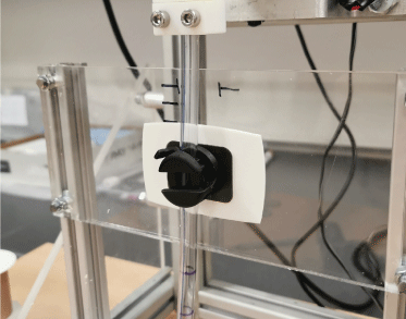
Figure 3: Novel 3D printed device method.
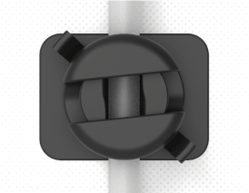
Figure 4: The novel device.
Procedure
Each method of fixation was subject to repeated tension forces to the chest drain to simulate line tugging, in keeping with previous studies of this nature [4]. The maximal number of cycles of tension was 100, or until failure. All fixation methods were applied by the same experienced Emergency Physician. The measurements taken included the number of cycles to failure. The failure of the cycle was defined as the displacement of the chest drain by 25mm, when slipped reached this level the experiment was ceased.
The design of the experiment included a pretension of the 0.2N to simulate the weight of the tubing and the chest drain receptacle. The drain pull in each instance was 30N.
Ethical approval
Ethics approval was not required for this study, as the study did not reach ethical requirement threshold.
Results
Suture
All sutures survived the 100 cycles of testing (Table 1). None of the chest drains secured with this method failed. Throughout all experimental cycles, all tests of the suture material securement method initially slipped approximately 10-15mm from its original position (Table 2). Following this initial movement, the testing reached an equilibrium where the cyclical force of 30N was not sufficient to make the chest drain migrate any further or to break the sutures.
Table 1: Number of successful cycles prior to failure per method of securement.
Method |
Sleek Tape |
3D device |
Suture |
Experiment number |
|
|
|
1 |
28 |
51 |
100 |
2 |
55 |
72 |
100 |
3 |
38 |
76 |
100 |
4 |
55 |
59 |
100 |
5 |
28 |
42 |
100 |
6 |
42 |
45 |
100 |
7 |
27 |
46 |
100 |
8 |
33 |
68 |
100 |
9 |
48 |
48 |
100 |
10 |
45 |
43 |
100 |
Average |
39.9 |
55 |
100 |
Table 2: Initial slippage vs each subsequent cycle per method.
Method |
Sleek tape |
3D device |
Suture |
Initial slip |
10-15mm |
1-2mm |
10-15 |
Slipping with each subsequent cycle |
1-2 mm |
0.5-1 mm |
0 mm |
Adhesive skin tape (Sleek)
The sleek tape method failed on average at 39.9 cycles of loading (Table 1) making it the least successful method of securement of chest drain. There was initial slippage of the tube of approximately 10-15mm (Table 2) and with each subsequent 30N cycle, there was a small amount of further slippage until the tube migrated in total 25mm from its original starting point. The specific failure point of the tape was the adhesive.
Novel 3D Printed Device
The novel printed device on average survived to 55 cycles (Table 1). There was minimal initial slippage of approximately 1 – 2 mm (Table 2), significantly less that the suture or tape. With each cycle the tube slipped between 0.5mm and 1mm. Each test was stopped after the tube migrated 25mm. However, unlike the tape, it maintained a consistent level of securement throughout the test. There was no single failure point, rather consistent slippage during each pull cycle
Discussion
Statement of principal findings
The results of the experiment indicate that for the methods tested, the suture is the preferred standard method for securement of chest drains. However, the novel device showed, on average, 55 cycles prior to failure with a 38% increase on the tape-only method (average = 39.9 cycles). It could be argued that this number could be sufficient as, on average, the chest drains tended to remain in-situ for 24 to 48 hours [6]. There are no data available as to how many daily ‘tugs’ devices are likely to encounter.
The 3D printed device was designed to address three major issues that the suture securement method exhibits: i) risk of possible infection, ii) damage to the skin, iii) difficulty in repositioning of the chest drains after confirming the position on the chest x-ray. To expand on this, the suture device is embedded in the skin with the use of a curved needle and thread (suture). As such it has the potential to become a source of infection [7]. Thirdly, issues that are commonly experienced by physicians include the position of the drain within the chest cavity which is deemed unacceptable (this could be due to failure of reaching the desired position). If this occurs, the suture must be removed, the drain repositioned, and the device secured for a second time with the suture. There is a lack of research into the area of chest drain misplacement. However, the conservative reports suggest that approximately 10% of drain insertions are ectopic (outside the chest cavity) [8]. This number includes a wide range of positions from soft tissues around the chest cavity to very dangerous and often requiring a surgical management intrabdominal insertions. There are however no data available on how many chest drains need to be repositioned if inserted into the chest cavity but in less than satisfactory position (i.e. too medial or too superior). Drains inserted in this fashion would not be seen as a complication/adverse event. As this is not considered a complication, there is no database to report it, likely explaining the lack of research in the area. The chest radiograph is repeated post every manipulation to assess the position. Occasionally this process must be repeated on several occasions until the position is deemed satisfactory. This is obviously an issue for both the physician and their patient who is put through an unpleasant procedure, often more than once.
Therefore, in designing the device, the researchers focused on an adhesive that could be placed on patients’ skin to avoid disrupting the barrier of the skin in securing the drain. We hypothesised that the use of the adhesive could reduce the possibility of skin damage from tearing through with a suture and therefore reduce the risk of possible source(s) of infection at the site of suture bed. Additionally, the mechanism of the drain securement was designed in such a way that the device could be re-opened and the drain repositioned without changing of the device or causing discomfort to the patient. Also, the time required to open and then tighten the device is seconds rather than minutes, as is the case with the suture method.
Our findings indicate that both the tape method showed initial slippage of 10-15 mm. It was clear that the securement dropped with each subsequent cycle as the tape lost its adhesion to the tube and the test bed. This is a noteworthy finding as, if a chest drain was placed in a shallow position, this could lead to movement of the drain outside of the chest cavity but would not be visible externally, leading to the possibility of causing surgical emphysema even at the initial pull [9]. If this occurred, it could potentially lead to an extended hospital stay and patient morbidity [3]. The 3D printed device showed only 1-2 mm of initial slippage; however, the device continued to slip 0.5-1 mm with each subsequent force applied. The suture method showed similar initial slippage of 10-15 mm when compared to that of the tape method. It was clear that the suture held the tube securely and the 30N force caused the soft PVC tube to elongate, acting as a shock absorber for the force. Following the initial elongation of the suture, the drain remained firmly in-situ with no subsequent slippage when force was repeatedly reapplied.
Strengths and Weaknesses
This study applied cyclical mechanical testing of three securement methods of chest drain fixation based on clinical practice. As this was a concept testing method, the device was printed on the 3D printer (3D Systems – Figure 4® Standalone) with a use of rigid polycarbonate like material (Figure 4® PRO-BLK 10 Resin). The actual device would be factory manufactured from a significantly stronger medically safe polymer capable of withstanding stronger forces than the relatively simpler concept device.
The suturing method showed the strongest performance however it is also a procedure with the highest risk to the patients, inc. use or local anaesthetic, invasive procedure (breaking of the skin barrier). Giving an indication that a less invasive method might be safer and beneficial.
Another possible advantage of a novel 3D device could be a temporising measure there the device is being used while the position of the drain is being established and once its confirmed additional securement method might be used.
Lastly, this study was designed to experiment with each securement method in isolation. However, clinical setting it would be impossible not to mention that often methods are combined for example suture could be used in conjunction with the tape. It might be of value to in the future test a performance of the 3D device with tape to assess whether it continues to perform superiorly to suture in the area of slippage.
In conclusion, the suture method was preferred based on both the cycles to failure and slippage metrics as used in this study design. However, the 3D printed design was superior regarding minimisation of initial slippage. If the 3D printed design could prevent the consistent slippage during repeated loading, then it may offer an important alternative to chest drain securement. These changes may yield an improvement in slippage observed while minimising the issue of difficult repositioning, which is the main criticism of the gold standard method.
Declaration Section
- Ethics approval and consent to participate
- This research did not require ethics approval. There was also no participants as it was an experiment therefore no consent was required.
- Consent for publication
- No human or animal participants were utilised therefore no consent for publication was required.
- Availability of data and materials
- All collected data is included in the results section of the manuscript.
- Competing interests
- There are no financial or non-financial competing interest to disclose.
- Funding
- There was no funding for any part of this research.
- Authors' contributions
- Dr Aidan O'Sullivan and Prof. Fergal Cummins designed and conducted the experiment. Dr Katarzyna Domanska has conducted the literature review and wrote the paper. Professors Ryan, O'Sullivan and Galvin reviewed the manuscript and provided advice on necessary changes.
References
- Kamio T, Iizuka Y, Koyama H, Fukaguchi K (2021) Adverse events related to thoracentesis and chest tube insertion: evaluation of the national collection of subject safety incidents in Japan. Eur J Trauma and Emerg Surg 1-8. [Crossref]
- Kirmani B, Zacharias J (2016) Insertion of a chest drain for pneumothorax. Anaesthesia & Intensive Care Medicine 17: 196-198.
- Al-Ameri A, Hicken L, Thomas S (2012) Chest drain fixation: How can we prevent drains from falling out? Lessons from an audit. European Respiratory Journal 40 (Suppl 56).
- Heskin L, Cahill V, Filobbos G, Regan P, O'Sullivan ST, et al. (2019) A new adaptation for a secure surgical drain placement and a comparison with four common drain fixation methods. Ann R Coll Surg Engl 101: 60-68. [Crossref]
- Laws D, Neville E, Duffy J (no date) BTS guidelines for the insertion of a chest drain.
- Utter GH (2013) The Rate of Pleural Fluid Drainage as a Criterion for the Timing of Chest Tube Removal: Theoretical and Practical Considerations. Ann Thorac Surg 96: 2262-2267. [Crossref]
- Urschel JD, Takita H, Antkowiak JG (1997) Necrotizing soft tissue infections of the chest wall. Ann Thorac Surg 64: 276-279. [Crossref]
- Osinowo O, Softah A, lateef L, Eid Zahrani M (2002) Ectopic chest tube insertions: diagnosis and strategies for prevention. Afr J Med Med Sci 31: 67-70. [Crossref]
- Jones PM, Hewer RD, Wolfenden HD, Thomas PS (2001) Subcutaneous emphysema associated with chest tube drainage. Respirology 6: 87-89. [Crossref]