Purpose: To present a novel case of central serous chorioretinopathy (CSCR) diagnosed in a woman three weeks after implantation of Nexplanon, a progestin-only contraceptive device.
Methods: Case report.
Results and Discussion: A 35-year-old healthy woman presented with one-week duration of headache and photopsias in the right eye, three weeks after implantation of a progestin-only contraceptive device. Imaging demonstrated subretinal fluid in the right eye suggestive of central serous chorioretinopathy. Removal of the device was associated with a rapid restoration of visual acuity as well as resolution and sustained absence of CSCR on imaging.
Central serous chorioretinopathy, etonogestrel implant, Nexplanon, progestin-only contraceptive device
Central serous chorioretinopathy (CSCR), characterized by focal serous detachments of the neurosensory retina, is often associated with conditions of increased levels of corticosteroids. Other well-described associations include “Type A” personality patterns, Cushing’s syndrome, gastroesophageal reflux, and pregnancy [1-3]. No association between CSCR and implantable contraceptive devices has been previously reported.
A 35-year-old healthy woman presented for symptoms of headache and photopsias in the right eye for one week. Her history was notable for poor vision in the eye since childhood and recent implantation of a subdermal contraceptive, Nexplanon® (etonogestrel implant) (Merck & Co., Inc., Whitehouse Station, NJ), 3 weeks prior to presentation.
On examination, best corrected visual acuity in the right eye was 20/60 while the left eye was 20/600. The anterior segment exam was unremarkable, without signs of inflammation. There was no vitreous cell noted. Fundus exam of the right eye was significant for subretinal fluid extending over the entire macula but no other lesions; the left eye revealed a large macular scar (Figure 1). Optical coherence tomography (OCT) of the right eye demonstrated a large collection of subretinal fluid; in the left eye, atrophy of all retinal layers and RPE (Figure 2). The choroid was of normal thickness, and no pigment epithelial detachments were noted in either eye. Of noted, OCT-assisted enhanced depth imaging was not available at the time. Fluorescein angiography was also performed and confirmed a multifocal expansile dot pattern with pooling of dye in the macula extending from arcade to arcade (Figure 3). A diagnosis of central serous chorioretinopathy (CSCR) was determined. Given the temporal relationship between the device implantation and the development of symptoms, our service recommended removal of the device.
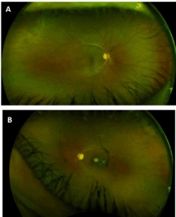
Figure 1. Fundus photo of (a) the right eye, demonstrating subretinal fluid extending superiorly toward the equator between 12 and 1 o’clock, and (b) the left eye, demonstrating a large chorioretinal scar
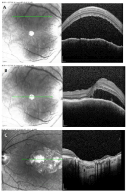
Figure 2. OCT imaging of the macula at presentation in (a, b) the right eye, demonstrating a large serous retinal detachment, and (c) the left eye, demonstrating chorioretinal scarring and atrophy.
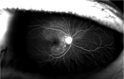
Figure 3. Fluorescein angiography of the right eye demonstrating pooling that extended beyond the macula toward the equator superiorly.
The patient had the etonogestrel implant removed one day after presentation and returned for follow-up three weeks after the initial visit. Visual acuity improved to 20/20 in the right eye. Follow up OCT demonstrated resolution of the subretinal fluid in the right eye (Figure 4a and 4b). At 3 months follow up, visual acuity still measured 20/20 and the OCT showed sustained resolution of the subretinal fluid. (Figure 4c).
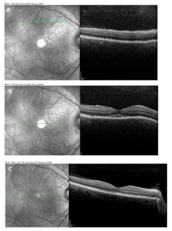
Figure 4. OCT imaging of (a) the right superior macula after Nexplanon removal, 3 weeks after initial presentation and (b) the central macula where a residual amount of subretinal fluid persisted. (c) OCT imaging of the right macula 3 months after initial presentation showed complete and sustained resolution of the fluid.
The etonogestrel implant is a progestin-only radiopaque contraceptive device that is implanted sub dermally into the arm. Progestins are synthetic agonists of the progesterone receptor and are either structurally derived from progesterone or testosterone.
Both the glucocorticoid receptor and the mineralocorticoid receptor have been implicated in the pathogenesis of CSCR. Activation of choroidal mineralocorticoid receptors by aldosterone or glucocorticoids leads to vasodilation and smooth muscle relaxation of the choroid vessels [4-5]. This is one possible mechanism by which etonogestrel could cause CSCR. Progestins interact with not only progesterone receptors, but other steroid receptors as well, including androgen, estrogen, glucocorticoid, and mineralocorticoid receptors [6-8]. It is possible that etonogestrel, or one of its metabolites, interacts with either glucocorticoid or mineralocorticoid receptors in the choroid, leading to vasodilation and increased permeability.
The time course from device implantation and disease, to device removal and rapid resolution, strongly suggests an association between this patient’s CSCR and the etonogestrel implant. The patient’s symptoms began roughly one and half weeks after implantation of the device; her symptoms and clinical findings abated promptly after removal of the device. While the natural course of the disease often results in spontaneous resolution, this typically occurs over the course of 2-3 months [1,4,9]. Fortunately, in our patient’s case the resolution was strikingly brisk.
There were several atypical features in this patient’s presentation with CSCR. The patient recalls a history of poor vision in the left eye since childhood, a history clinically implying ocular toxoplasmosis. However, the scarring may also have been due to recurrent attacks of CSCR. The left eye had evidence of macular scarring and retinal atrophy, possibly from childhood Toxoplasmosis. Alternatively, it may have been the consequence of previous attacks of CSCR. Fluorescein angiography demonstrated a multifocal expansile dot pattern clustered towards the nasal macula, suggesting a higher degree of virulence. One hypothesis is that this patient may have had an underlying propensity for developing CSCR, and the progestin implant was the proverbial tipping point that led to her presentation.
Although this is the first case of CSR presumed due to progestin therapy. It is also consistent with other existing reports of CSR linked to exogenous hormone therapy [10-12].
Since contraceptives are ubiquitous, why has this phenomenon not been observed more frequently? Nexplanon consists of progestin alone. Most contraceptives consist of a combination of estrogen and progestin, and estrogen may counterbalance the vasodilatory effects of progestin. Furthermore, estrogen is also thought to be protective against development of CSCR. Evidence from a report of CSCR after initiation of tamoxifen, which blocks estrogen receptor activation, further supports this idea [13]. More importantly, a myriad of different progestins exist that differ widely in structure, pharmacokinetics, and biochemistry [8]. Etonogestrel is a highly potent synthetic derivative of testosterone, which may lend itself a greater binding affinity for these receptors compared to other progestins [6-8].
In summary, we present a case of a woman who developed CSCR shortly after implantation of the etonogestrel implant. Device removal was associated with rapid restoration of visual acuity and return of anatomical findings to baseline with sustained absence of CSCR on follow-up.
Written informed consent for publication of their clinical details and/or clinical images was obtained from the patient/parent/guardian/ relative of the patient. A copy of the consent form is available for review by the Editor of this journal.
The authors acknowledge the departmental support from an RPB unrestricted grant.
None of the authors had any proprietary interest.
None of the authors had any relevant conflicts of interest.
- Chatziralli I, Kabanarou SA, Parikakis E (2017) Risk factors for central serous chorioretinopathy: Multivariate approach in a case-control study. Curr Eye Res 42: 1069-1073. [Crossref]
- Liew G, Quin G, Gillies M (2013) Central serous chorioretinopathy: A review of epidemiology and pathophysiology. Clin Exp Ophthalmol 41: 201-214. [Crossref]
- Liu B, Deng T, Zhang J (2016) RISK FACTORS FOR CENTRAL SEROUS CHORIORETINOPATHY: A systematic review and meta-analysis. Retina 36: 9-19. [Crossref]
- Daruich A, Matet A, Dirani A (2015) Central serous chorioretinopathy: Recent findings and new physiopathology hypothesis. Prog Retin Eye Res 48: 82-118. [Crossref]
- Zhao M, Celerier I, Bousquet E (2012) Mineralocorticoid receptor is involved in rat and human ocular chorioretinopathy. J Clin Invest 122: 2672-2679. [Crossref]
- Sitruk-Ware R (2004) Pharmacological profile of progestins. Maturitas 47: 277-283. [Crossref]
- Sitruk-Ware R (2005) Pharmacology of different progestogens: The special case of drospirenone. Climacteric 8: 34-12. [Crossref]
- Stanczyk FZ (2003) All progestins are not created equal. Steroids 68: 879-890. [Crossref]
- Daruich A, Matet A, Marchionno L (2017) Acute central serous chorioretinopathy: Factors influencing episode duration. Retina 37: 1905-1915. [Crossref]
- Bussel II, Lally DR, Waheed NK (2014) Bilateral central serous chorioretinopathy associated with estrogen modulator diindolylmethane. Ophthalmic Surg Lasers Imaging Retina 45: 589-591. [Crossref]
- Grieshaber MC, Staub JJ, Flammer J (2007) The potential role of testosterone in central serous chorioretinopathy. Br J Ophthalmol 91: 118-119. [Crossref]
- Nudleman E, Witmer MT, Kiss S (2014) Central serous chorioretinopathy in patients receiving exogenous testosterone therapy. Retina 34: 2128-2132. [Crossref]
- Koulisis N, Moysidis SN, de Koo, Lisa C Olmos (2016) The tipping point: Tamoxifen toxicity, central serous chorioretinopathy, and the role of estrogen and its receptors. Am J Ophthalmol Case Rep 38: 13. [Crossref]