Abstract
Uterine lipoleiomyoma is a rare type of leiomyoma consisting of smooth muscles and fat tissue. It is considered as the most common uterine tumor in the reproductive years. We report a 52-year-old multipara postmenopausal woman who presented an acute exacerbation of chronic pelvic pain. Ultrasound scan showed a 10 cm heterogenic lateral uterine mass. A primary diagnosis of teratoma was made and laparotomy was performed showing an anterior uterine wall mass. Histopathology examination showed mature lipoleiomyoma.
Key words
Lipoleiomyoma (LL), uterus, postmenopausal
Introduction
Uterine lipoleiomyoma (LL) is an unusual uterine fatty tumor wich most frequently occurs in asymptomatic obese perimenopausal or postmenopausal women [1]. Theses tumors are histologically composed of variable amounts of smooth muscle, fat cells, and fibrous tissue. LL is considered as a benign variant of leiomyoma predominantly located in intramural uterine fundus [2-4]. The incidence of this neoplasm varies from 0.03% to 0.2% [2,5,6]. Most frequently clinical manifestations include pelvic painful mass, micturiton problem, menstrual disturbances or infertility. In the majority of cases, patients report no symptoms. The treatment options vary from expectant management to surgical therapy.
Case report
A 54-year-old postmenopausal woman gravida 4, para 4 presented with a two month of pelvic pain and increased frequency of micturition. She experienced the last menstruation at the age of 49 years. Her past medical history was significant for a nervous depression and a pelvic mass diagnosed as a uterine fibroma and treated as such by the progesterone. A physical examination did not reveal any abnormalities. A pelvic examination detected a palpably solid and tender mass in right midline area. Blood laboratory test showed anaemia that would be in relation with breakthrough bleeding minimal and recidivantes; without any others abnormalities. Tumor markers were within normal limits. Uterine cervix presented no visible abnormalities. Cervical cytology was normal. Pelvic ultrasonography revealed a 10 cm heterogeneous pelvic hypoechoic mass, exerting mass effect on the uterine. Bilateral ovaries and adnexa were unremarkable. Computed tomography showed a heterogenic right lateral-uterine mass of 9×8 cm evocating in primary ovarian mature teratoma (Figure 1a-b). The patient underwent laparotomy for excision of the mass. On exploration, we found out that the mass was independent of ovaries and was developed from the right anterior uterine wall. It was pale yellow with a softer consistency on its cut surface (Figure 2). Both ovaries and fallopian tubes appeared normal. The mass was mobilized and freed intact. Pathology revealed that the tumor consists of mature lipocytes and smooth muscle evocating a LL (Figure 3a-b). The patient was discharged from the hospital without complications 5 days following surgery and was healing well without evidence of recurrence one year post-surgery.
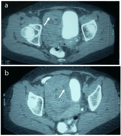
Figure 1a-b. Cross sectional view: shows a heterogenic latero uterine mass.
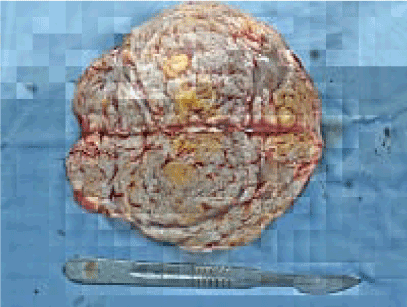
Figure 2. Cut section of the tumor shows yellowish-white areas.
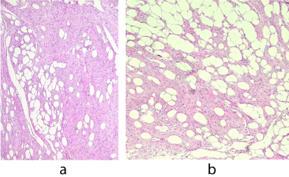
Figure 3a-b. Smooth muscle cell associated with lobules of mature fat cells (a: HE X 100 ; b: HE X 200).
Discussion
LL of the uterus is a rare variant of uterine leiomyoma in gynaecological practices. Obese menopausal women ranging from 50 to 70 years old are the most vulnerable group for uterine LL and 90% of the patients are older than 40 years of age [7]. Histologically, LL is composed of benign smooth muscle cells and mature adipose tissue as was observed in our case (Figure 3). Adipocytes may be regularly positioned within the tumor or may exhibit a focal location [8].
Although the cause is unclear, fatty metamorphogenesis of the smooth muscle cells of leiomyomas is the most likely cause for the development of LL [2]. Fukunaga considered that some LL can result from lipomatous metaplasia of leiomyomas, a hypothesis supported by immunohistochemical findings [9]. Hormonal stimulation especially estrogen, growth hormone and progesterone, has been suggested as a possible cause [2]. In fact, a retrospective study analyzing 76 cases of LL in Turkey between January 2000 and April 2013 suggested that estrogenic manifestations may be an important factor in the development of the tumor [10]. This study found that 75.7% of LL had different types of lesions associated with hyper-estrogenic status such as adenomyosis, endometriosis, endometrial hyperplasia, polyps, complex atypical endometrial hyperplasia, and gynecologic carcinomas. The current case showed a history of fibroma treated by progesterone.
Uterine LL may be associated with metabolic diseases such as hyperlipidemia, hypothyroidism, and diabetes [10]. In the present case, only a history of uterine fibroma and nervous depression was reported. Uterine LL have a spectrum of clinical manifestations including pelvic pain, increased or abnormal menstrual bleeding periods, infertility problem, or pressure effects on surrounding organs like bladder or rectum. The differential diagnosis includes mature ovarian teratoma, benign pelvic lipoma, liposarcoma, extra-adrenal myelolipoma, lipoblastic lymphadenopathy and retroperitoneal cystic hamartoma. However, uterine LL are often diagnosed preoperatively as uterine leiomyomas or mature ovarian teratomas [11].
Radio imaging techniques such as MRI and CT can play an important role in determining the intra-uterine location and fatty nature of lipoleiomyomas but most of these are incidental findings postoperatively on histopathology. The sonographic appearance of leiomyomas is that of a heterogeneous mass containing hyperechoic areas due to the presence of fat. Similarly, on CT images it shows a fatty mass with areas of non-fat soft-tissue density arising from the uterus. MRI including fat suppression sequence is a useful technique to diagnose uterine LL with its high sensitivity and specificity to fat. It reveals a heterogeneous mass with high signal intensity on T1-weighted images in the lesion [6]. Although, it is not often possible to differentiate uterine LL with other lipomatous tumours [12].
LL when asymptomatic require no treatment and are clinically similar to leiomyomas. Surgical management of these tumors is regularly by hysterectomy. Nevertheless, uterine artery embolization and myomectomy can be performed depending on patients’ symptoms, fertility desire, the site of the mass and associated uterine fibroids. It is important to differentiate these LL from ovarian teratoma, which requires surgical excision [13]. Lipoleiomyomas seem to have a benign clinical course. However, lipoleiomyosarcomas arising in uterine lipoleiomyomas had been reported in literature [14].
Some associations are also reported such as: lipoleiomyoma and primary ovarian leiomyoma [7], lipoleiomyoma of uterus and lipoma of broad ligament [8]. In our case, lipoleiomyoma happened in a postmenopausal woman. The main symptom was a frequent mixture due to bladder compression by the tumor. The primary diagnosis based on CT was a mature ovarian teratoma. Histological examination after surgical removal of the tumor revealed a lipoleiomyoma.
Conclusion
Uterine LL is a rare and specific type of leiomyoma with a considerable amount of adipocytes. These benign tumors of the uterus do not affect mortality. Although the final diagnosis of LL is established based on a histopathological test involving the tissue specimen, imaging studies play an important role especially in preoperatively demonstrating the fatty nature and exact location.
References
- Ding DC, Chu TY, Hsu1 YH (2010) Lipoleiomyoma of the uterus. Taiwan J Obstet Gynecol 49: 94-96.
- Karaman E, Çim N, Bulut G, Elçi G, Andiç E, et al. (2015) A Case of Giant Uterine Lipoleiomyoma Simulating Malignancy. Case Rep Obstet Gynecol: 926961.
2021 Copyright OAT. All rights reserv
- Oh SR, Cho YJ, Han M, Bae JW, Park JW, et al. (2015) Uterine Lipoleiomyoma in Peri or Postmenopausal Women. J Menopausal Med 21: 165-170. [Crossref]
- Bajaj P, Kumar G, Agarwal K (2004) Lipoleiomyoma of broad ligament: A case report. Indian J Pathol Microbiol 3: 457-458. [Crossref]
- Avritzcher R, Iyer BB, Ro J (2001) Lipoleiomyoma of the uterus. Am J Roentgenol 177: 856.
- Abdussamet Batur AE, Muhammed Alpaslan B, Ilyas Dundar F, Mesut Ozgokce F, Alpaslan Yavuz A (2015) Uterine Lipoleiomyoma: MR Findings. Pol J Radiol 80: 433-434.
- Kelekci S, Eris S, Demirel E, Aydogmus S, Ekinci N (2015) Lipoleiomyoma of the Uterus and Primary Ovarian Leiomyoma in a Postmenopausal Woman: Two Rare Entities in the Same Individual. Case Rep Pathol 2015: 564846.
- Wahal SP, Mardi K (2014) Lipoleiomyoma of uterus and lipoma of broad ligament-a rare entity. J Can Res Ther 10: 434-436. [Crossref]
- Fukunaga M (2003) Benign “metastasizing” lipoleiomyoma of the Uterus. Int J Gynecol Path 22: 202-204. [Crossref]
- Akbulut M, Gündoğan M, Yörükoğlu A (2014) Clinical and Pathological Features of Lipoleiomyoma of the Uterine Corpus: A Review of 76 Cases. Balkan Med J 31: 224-229. [Crossref]
- Kiyokoba R, Yagi H, Yahata H, Kawano Y, Kaneki E, et al. (2015) Tumor-To-Tumor Metastasis of Poorly Differentiated Gastric Carcinoma to Uterine Lipoleiomyoma. Case Rep Obstet Gynecol 2015: 352369. [Crossref]
- Kitajima K, Kaji Y, Imanaka K, Sugihara R, Sugimura K (2007) MRI findings of uterine lipoleiomyoma correlated with pathological findings. AJR Am J Roentgenol 189: 100-104. [Crossref]
- Manjunatha HK, Ramaswamy AS, Kumar BS, Kumar SP, Krishna L (2010) Lipoleiomyoma of uterus in a postmenopausal woman. J Midlife Health 2: 86-88. [Crossref]
- Mc Donald AG, Dal Cin P, Ganguly A, Campbell S, Imai Y, et al. (2011) Liposarcoma arising in uterine lipoleiomyoma: a report of 3 cases and review of the literature. Am J Surg Pathol 35: 221-227. [Crossref]