Craniostenosis is rare but important clinical condition characterized by early fusion of skull suture causing restriction of growth of developing brain and ultimately arrest of brain growth. It is a cause of acquired mental retardation, which can be avoided and prevented by early diagnosis and early surgical intervention as delay causes irreversible loss of vision and permanent cognitive impairment. So all physicians should be made aware of this rare congenital deformity. Authors report a neglected case of craniostenosis, presented with deformity of head with mental retardation, which was operated after delay lead to poor neurological outcome.
Craniostenosis, silver- beaten appearance, mental retardation, craniolacunae, management
Craniostenosis is characterized by early fission of sutures of skull, when premature closure of the sagittal suture occurs called as Scaphocephaly [1]. It causes arrest of growth of brain and causes deformity of skull along with features of raised intracranial pressure. However, early surgical intervention can lead to normal growth [2]. However, neglected cases can develop typical scaphoid appearance of head and silver beaten appearance or also known as beaten brass skull, refers to the prominence of gyral impressions on the inner table of the skull observed throughout the skull vault and typically associated with raised intracranial pressure in children [3-5]. However, convolutional markings may be normal, however, such convolutions are usually confined to the posterior part of the inner table of the skull. Every case of craniostenosis should be subjected to surgical intervention at as early as possible preferably within first three months of infancy to protect from bad effect of raised Intracrnial pressure [6-8].
A 7-year–old-male boy presented to our outpatient clinic with complaints of abnormal shape of head along with abnormal facial profile. He was mentally subnormal and also had abnormal protrusion of both eyes associated with bilateral progressive diminution of vision. On examination, patient had bilateral proptosis. Visual acuity was finger counting at 3 feet in both eyes. There was a palpable ridge over sagittal suture line suggestive of fused suture. He had head circumference of 47 cm with biparietal diameter of 17 cm. Non-contrast computerised tomography head with three-dimensional reconstruction revealed a small neurocranium with fusion of all calvarial sutures (Figure1).
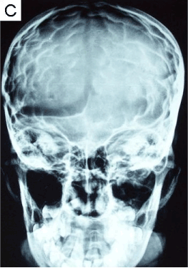
Figure 1. X-ray skull lateral view showing classical silver-beaten appearance of skull anteroposterior projections).
Digital X-ray skull showed heavy convolutional markings as a result of long-standing raised intracranial pressure giving a classical “Silver-beaten appearance of skull” (Figure 2,3). Patient was taken up for surgery. Strip craniectomy with barrel-stave osteotomy was done along with lax duraplasty. Patient had uneventful post-operative course and was doing well at follow-up visit six months after surgery.
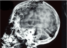
Figure 2. X-ray skull anteroposterior projections showing classical silver-beaten appearance of skull.
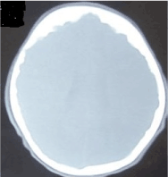
Figure 3. Non-contrast computerized tomography (NCCT) head, bone window view with reconstruction showing fusion of all calvarial sutures with irregular calvarial outlines in case of raised intracranial pressure.
Scaphocephaly is a type of craniostenosis, characterized by premature fusion of sagittal suture and causes deformed appearance of skull resembling keel of a boat as long and thin skull. It represents most common type of craniosynostosis, constituting more than half of all cases [2]. The Scaphocephaly is usually isolated and usually not associated with syndromes, but observed in premature infants.
As growth of the skull bones is prevented by e fused sagittal suture, however compensated by expansion at lambdoid the coronal sutures [3]. The skull appears as elongated and shortened in the biparietal diameter and palpable ridge of the sagittal suture as compensated frontal and occipital bones for the restricted lateral expansion of the parietal bones. It is associated with closed fontanelle [5].
X-ray skull shows presence of elongated skull. It may also show presence of “silver- beaten cranium” is also called as “beaten copper cranium“, which is postulated to be due to indentations in the bone from the underlying gyri due raised intracranial pressure and cerebrospinal fluid pulsations [1].The unyielding nature of calvarium in craniosynostosis produces a characteristic mottled appearance of the bone with lucencies of varying depth having round and poorly marginated edges.
The pathological mechanism of silver- beaten cranium is caused by sustained high intracranial pressure is causing increased inner table convolutional markings primary cause of copper beaten skull includes craniosynostosis, obstructive congenital hydrocephalus, hypophosphatasia and intracranial tumours.
The silver beaten appearance on imaging study needs different ion from convolution marking and lacunar skull [4,5]. The lacunar skull is also known as craniolacunae represents dysplasia of the membranous skull vault. Lacunar skull is associated with 70% cases Chiari malformation and typically inner table is more affected than the outer with intervening areas of apparent thinning. Lacunar skull appearance represents abnormal collagen development and ossification of membranous bone [2,4,5]. Typical radiological feature of lacunar skull is presence of a groups of oval or round pits on the inner surface of the vault, separated by ridges of bone and usually have predilection for thickest part of frontal, parietal and upper occipital bone [4,6].
The cranial computed tomography confirms the diagnosis of scaphocephaly. It can also show associated microcephaly and other associated pathology, like hydrocephalus, or chinked ventricle with effacement of arachnoid spaces [6,9].
Treatment of craniostenosis is surgery [2]. The surgical intervention should be carried out the earliest possible occasion as progressive raised intracranial pressure and cognitive impairment can be avoided at early surgery with good neurological outcome and leads to significant improvement in the head shape and allows permit re- growth and development of brain [3].
Early surgery can spare from effect of prolonged raised intracranial pressure and help in scholastic improvement. However neglected cases can develop typical scaphoid appearance of head and silver beaten appearance. Every case of craniostenosis should undergo surgery at infancy stage itself.
- Tuite GF, Evanson J, Chong WK, Thompson DN, Harkness WF, et al. (1996) The beaten copper cranium: a correlation between intracranial pressure, cranial radiographs, and computed tomographic scans in children with craniosynostosis. Neurosurgery 39: 691- 699.
- Ciurea AV, Toader C, Mihalache C (2011) Actual concepts in scaphocephaly : (an experience of 98 cases). J Med Life 4: 424-431. [Crossref]
- Farhad Afshar. The Craniosynostoses: Causes, Natural History and Management J R Soc Med. 1983 April; 76(4): 335
- Desai V, Priyadarshini SR, Sharma R (2014) Copper Beaten Skull! Can It be a Usual Appearance? Int J Clin Pediatr Dent. 7: 47-49. [Crossref]
- Rühli FJ, Nick2021 Copyright OAT. All rights reservase of beaten-copper cranium. J Neurosurg 106: 71-73. [Crossref]
- Shahdadpuri R, de Vries B, Pfundt R, de Leeuw N, Reardon W (2008) Pseudoarthrosis of the clavicle and copper beaten skull associated with chromosome 10p11.21p12.1 microdeletion. Am J Med Genet A 146A: 233-237. [Crossref]
- Agrawal D, Singh PK, Sinha S, Gupta DK, Satyarthee GD, et al. (2015) Remaining unconscious: The burden of traumatic brain injuries in India. J Neurosci Rural Pract. 6: 520-522.
- Satyarthee GD, Mahapatra AK (2015) Giant pediatric glioblastoma multiforme causing primary calvarial erosion and sutural diastasis presenting with enlarged head. J Pediatr Neurosci. 10: 290-293