Abstract
This study is carried on to investigate the effect of (UV) on the estimate of vitamin D production and the correlation ratio of vitamin D and its impact on the increase of blood glucose finally to prove the relationship via biochemical analysis of Vitamin D and glucose level. 90 adult rats were randomly divided into three groups. G1 (-ve control): This group contains 30 rats that were not exposed to the ultraviolet. Rats were eating daily/4 months. G2 (+ve control): This group contains 30 rats that were not exposed to the ultraviolet. Rats were fasting 2 days and eat the third day frequently (4 months). G3: Test group: To examine the exposed to the ultraviolet for 10 second to simulate sunrise and another 10 second to simulate sunset (using prototype cage). This group contains 30 rats, which were fasting 2 days and eat the third day frequently (4 months). Weights were recorder every week. The blood samples were collected through retro- orbital venous plexus in the zero time, every month, and at the end of four months and the obtained sera were subjected to the Glucose and D3 tests. The results showed that the body weights were reducing significantly in G1 compared with G3 at all measured periods (P < 0.000). Whereas no significant differences between G3 and G2 in all weeks. The Glucose levels in G1 and G3 were raised significantly in (1st, 2nd, 3rd and 4th month) (P< 0 .035), (P< 0 .036), (P< 0.030) and (P< 0.023). Also blood level of Glucose was raised significantly between G3 and G2 in all periods (P< 0 .008), (P< 0 .009), (P< 0.002) and (P< 0.001). Moreover the blood levels of Vitamin D between G1 and G3 were raised significantly in all tested periods (P< 0 .001), (P< 0 .000), (P< 0.000) and (P< 0.003). Also Vitamin D was raised significant (P< 0.000) in G3 compared with G2. This study was proved that vitamin D level was affected by the exposure to ultraviolet that intern affect the glucose level. Also ultraviolet (UV) extend to preservation of glucose homeostasis by raising glucose level. Accordingly ultraviolet (UV) raising vitamin D should be considered since this may help reduce the risk of glucose disorders.
Key words
vitamin D, ultraviolet, blood glucose
Introduction
Vitamin D is a group of fat-soluble seco-steroids, which is responsible for enhancing intestinal absorption of calcium, iron, magnesium, phosphate and zinc. Vitamin D occurs in many forms, but the most important compounds in this group are vitamin D3 (cholecalciferol) ("sunshine vitamin") and vitamin D2 (ergocalciferol) [1].
Glucose is a hexose sugar since it contains six carbon atoms, one of which is part of an aldehyde group, hence, is referred to as an aldohexose. It is also called blood sugar as it circulates in the blood at a concentration of 65-110 mg/dL (or 65-110 mg/100 ml) of blood [2].
Glucose also serves as an important metabolic intermediate of cellular respiration and glucose is stored as glycogen [3].
Vitamin D deficiency is an important risk factor for a variety of common diseases, including osteoporosis, severe asthma in children, cognitive impairment in older adults, cancer, autoimmune diseases and cardiovascular disease (CVD) [4].
Ultraviolet (UV) is electromagnetic radiation with a wavelength shorter than that of visible light but longer than X-rays. The UV wavelengths between 400 nm and 10 nm correspond to photon energies from 3.1 eV to 124 eV [5].
UV radiation is present in sunlight, and is produce by electric arcs and specialized lights such as mercury-vapor lamps, tanning lamps, and black lights. It can cause chemical reactions, and causes many substances to glow or fluoresce [6].
The mechanism of vitamin D is photosynthesized in the skin by the action of solar UVB radiation was 7-dehydrocholesterol convert to Vitamin D3 [7].
The humans need continuous glucose level in their blood, which allows the brain to work even with minimum function. So the aim of this work is to investigate the effect of (UV) on the estimate of vitamin D production. Study the correlation ratio of vitamin D and its impact on the increase in blood glucose and proved the relationship via biochemical analysis of Vitamin D and glucose level.
Materials and methods
Animal
Male albino wister rats were used in the present study. Animals were obtained from the animal house at the experimental animal unit at King Fahad research center, KAU, Jeddah, SA. Rats kept in the animal house for the whole period, where rats weighing between (270-350 g). Rats were acclimatized to the laboratory condition for 10 days prior to the initiation of experimental work. The animal were housed individually in special standard plastic cages under controlled laboratory conditions of humidity (65%), temperature (20 ± 1°C) and 12:12h light: dark cycle. Rats were feed normal rat chaw, and allow free access to water. All experimental procedures were care out in accordance with the guidelines of animal care and approved by the research ethics committee of King Abdul-Aziz University.
Experimental design
In the present study 90 adult rats were randomly divided into three groups. The groups were divided as follow:
G1: Negative control group (-ve): This group contains 30 rats which were not exposed to the ultraviolet. Rats were eating daily/4 months.
G2: Positive control group (+ve): This group contains 30 rats which were not exposed to the ultraviolet. Rats were fasting 2 days and eat the third day frequently (4 months).
G3: Test group: to examine the exposure to the ultraviolet for 10 second at sunrise and another 10 second at sunset (using prototype cage). This group contains 30 rats, which were fasting 2 days and eat the third day frequently (4 months).
Weights were recorder every 7 days (week) for all rats.
UVB irradiation
Four fluorescent lamps, TL 20W/12RS (Philips, Eindhoven, Holland) was used to irradiate the rats. These lamps emit a continuous spectrum from 275 to 390 nm, with a peak emission at 313 nm; f65% of that radiation is within the UVB wavelength range.
Blood sample collection
The blood samples were collected in the zero time before the start of the experiment, every months, and at the end of four months. Blood samples were withdrawn by capillary micro tubes (Micro Hematocrit Capillaries, Mucaps) through the retro orbital venous plexus sinuses in inner canthus of the eys; 5 ml of blood samples were collected in gel and clot activator tube at room temperature for half an hour and centrifuge at 3000 rpm for 10 min to obtain serum, which was subjected to the determination of Glucose and D3. The clear supernatant serum was separated and stored at -80ºC until analyzed for the biochemical analyses and hormone determinations.
Then animals were sacrificed under diethyl ether anesthesia.
Statistical analysis
Serum measurements for the 90 rats studied are presented as the mean ± SD and analyzed by One Way ANOVA test (LSD) which was used for comparison between different groups. Vitamin D and Glucose measured at 1st month, 2nd month, 3rd month and 4th month. The value of the detection level for each method was used for the statistical analysis. Using the IBM SPSS statistics version 20 (Armonk, NY, US). Graphs were made using Excel software version 7. For all tests, P- values of less than 0.05 (P˂ 0.05) were considered as significant.
Results
In the current study, we observed from our experiment that the rats which were exposed to the UVB showed healthy appearances, active movement and normal increase in weights.
Body weight
The results of the body weight which were measured in all groups at different periods, showed that the changes in the body weights were reducing significantly in negative control group compared with positive control group at periods 2nd week-16th week (P < 0.000). Moreover there are significant changes in the body weights that were reducing in negative control group compared with test group at periods 2nd week- 16th week (P < 0.000). While the body weight showed that there are no significant differences between test groups compared with positive control group in all weeks (Figure 1).
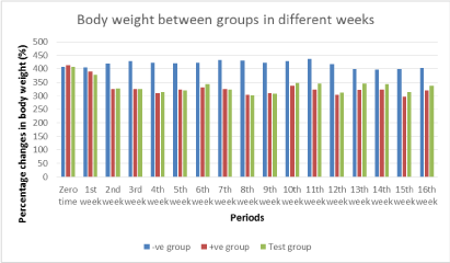
Figure 1. Comparing between body weights of all groups in different periods (negative control, positive control and test groups). Data are expressed as mean+/- SD
The life span was decreased in positive control group after 3rd week but in negative control group, test group no decreased in the number of animals to the end of the experiment just two or three in each group (Figure 2).
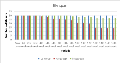
Figure 2. The life span in different periods decreased in positive control group after 3rd week comparing to negative control and test groups.
Serum glucose
The Glucose level showed insignificant changes between negative control group and positive control group in periods (zero time, 1st, 2nd and 3rd month) but in 4th month there was raised significantly between groups (P< 0.038). Moreover blood levels of Glucose between negative control group and test group was raised significantly in periods (1st, 2nd, 3rd and 4th month) (P< 0.035), (P< 0.036), (P< 0.030) and (P< 0.023) frequently. Also blood level of Glucose raised significantly between test group and positive control group in periods (1st, 2nd, 3rd and 4th month) (P< 0 .008), (P< 0 .009), (P< 0.002) and (P< 0.001) frequently (Figure 3).
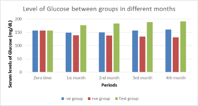
Figure 3. Blood levels of Glucose between Groups in different periods showing comparing between negative control, positive control and test groups. Data are expressed as mean+/- SD.
Serum's Vitamin D
Vitamin D level raised significant between negative control group and positive control group in periods (2nd, 3rd and 4th month) (P< .034), (P< 0.000) and (P< 0.000) frequently. Moreover blood levels of Vitamin D between negative control group and test group was raised significantly in periods (1st, 2nd, 3rd and 4th month) (P< 0 .001), (P< 0 .000), (P< 0.000) and (P< 0.003) frequently. Also Vitamin D raised significant (P< 0.000) in test group compared with positive control group in periods (1st, 2nd, 3rd and 4th month) (Figure 4).
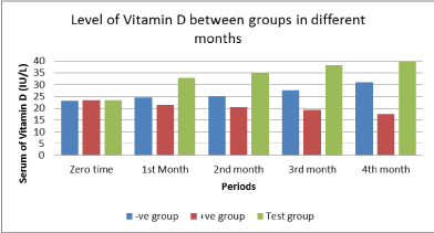
Figure 4. Blood levels of Vitamin D between Groups in different periods in comparing to each other (negative control, positive control and test groups). Data are expressed as mean+/- SD.
Discussion
This study is carried on to investigate the effect of (UV) on the estimate of vitamin D production and the correlation ratio of vitamin D and its impact on the increase of blood glucose finally we proved the relationship via biochemical analysis of Vitamin D and glucose level.
Many reports were recorded significant associations between sun exposure, vitamin D and health outcomes of life style. Vitamin D is likely to have a key role in these associations [8].
Moreover, Exposure to simulated sunlight that contains both UVB and UVA reduce cardiovascular risk factors and improve quality of life [9].
In our study during exposure of fasting animals to UVB we detected a significant increase in the serum level of vitamin D in all periods' times in comparing to non exposed animals.
These result are in agreement with other study which showed that vitamin D was increased with exposed to sun light (UVB) [10]. McCarroll et al., [11] recorded that spending time in the sun, useful strategies to improve 25(OH) D.
Our result showed that serum glucose level was significant increase in the group exposed to UVB compared with the groups none exposed in all periods' times.
2021 Copyright OAT. All rights reserv
Gluconeogenesis GNG is a metabolic pathway that results in the generation of glucose from non-carbohydrate carbon substrates such as lactate, glycerol, and glucogenic amino acids [12].
It is widely known that the liver is a central organ in lipogenesis, gluconeogenesis and cholesterol metabolism Ref. In the fasting state, the liver supplies the body with energy by breaking down glycogen, and following prolonged fasting by gluconeogenesis, which cleaves glucose from the glycogen polymer and produces glucose-1-phosphate, which is converted to glucose-6-phosphate by phosphoglucomutase.
Recent studies focuses on novel findings related to nuclear receptor signaling - including the vitamin D receptor and the liver receptor homolog 1 - in hepatic lipid and glucose uptake, storage and metabolism in the clinical context of liver regeneration [13].
On contrary, Bechmann et al., [14] reported that the vitamin D receptor in the liver interfere with hepatic lipid and glucose uptake, storage and metabolism.
Another approach that the animal's level of glucose in serum was increased in fasting rats from alternative substrata of glucose by glycogenesis process occurs in liver. The hypothesis that they survive from hypoglycemia but by inducing glycogenolysis, gluconeogenesis and lipolysis, it provided an energy source alternative to glucose.
This is in a good agreement with previous case of hypoglycemia and neuroglycopenia. They administered for treatment Ketogenic diet (KD), by reproducing body fuel mainly derives from beta-oxidation. The patients were free of epileptic crises, presented normalization of electroencephalogram (EEG), and showed a marked recover in psychological development and quality of life [14].
Conclusion
This study was proved that ultraviolet (UV) effect on increasing V-D level extend to preservation of glucose homeostasis by raising glucose level. Accordingly ultraviolet (UV) raising vitamin D should be considered since this may help reduce the risk of glucose disorders.
Acknowledgements
I would like to express my appreciation to my supervisor Prof. Faten Abdulrahman Khorshid, Professor of Cell Engineering and Cell Biology, Faculty of Science, Biology Department, King Abdulaziz University, whose help, advice and supervision was invaluable. My deep thanks to her constant help, guidance and countless hours of attention she devoted throughout the course of this work. Her priceless suggestions made this work interesting and learning for me.
References
- Seminario AL, Velan E (2016) Vitamin D and Dental Caries in Primary Dentition. J Dent Child 83: 114-119. [Crossref]
- Oh TJ, Lim S, Kim KM, Moon JH, Choi SH, et al. (2017) One-hour postload plasma glucose concentration in people with normal glucose homeostasis predicts future diabetes mellitus: a 12-year community-based cohort study. Clinical Endocrinology 86: 513-519.
- Kim GY, Lee YM, Kwon JH, Cho JH, Pan CJ, et al. (2017) Glycogen storage disease type Ia mice with< 2% of normal hepatic glucose-6-phosphatase-a activity restored are at risk of developing hepatic tumors. Mol Genet Metab. [Crossref]
- Holick MF (2007) Vitamin D deficiency. N Engl J Med 357: 266-281. [Crossref]
- Whitaker RJ (2003) Color and Light in Nature. School Science and Mathematics 103: 354-355.
- Dash K, Venkateswarlu G, Thangavel S, Rao S Chaurasia S (2011) Ultraviolet photolysis assisted mineralization and determination of trace levels of Cr, Cd, Cu, Sn, and Pb in isosulfan blue by ICP-MS. Microchemical Jour 98: 312-316.
- Christakos S, Dhawan P, Verstuyf A, Verlinden L, Carmeliet G (2016) Vitamin D: metabolism, molecular mechanism of action, and pleiotropic effects. Physiol rev 96: 365-408. [Crossref]
- Jelinek GA, Marck CH, Weiland TJ, Pereira N, van der meer DM, et al. (2015) Latitude, sun exposure and vitamin D supplementation: associations with quality of life and disease outcomes in a large international cohort of people with multiple sclerosis. BMC neurology 15: 132. [Crossref]
- Krause R, Stange R, Kaase H, Holick MF (2016) UV Irradiation and pleiotropic effects of vitamin D in chronic kidney disease–Benefits on cardiovascular comorbidities and quality of life. Anticancer res 36: 1403-1408. [Crossref]
- Osmancevic A, Gillstedt M, Landin-wilhelmsen K, Larkö AMW, Larkö O, et al. (2015) Size of the exposed body surface area, skin erythema and body mass index predict skin production of vitamin D. J Photochem Photobiol B 149: 224-229. [Crossref]
- Mccarroll K, Beirne A, Casey M, Mcnulty H, Ward M, et al. (2015) Determinants of 25-hydroxyvitamin D in older Irish adults. Age and Ageing 44: 847-853. [Crossref]
- Yoshida T, Okuno A, Takahashi K, Ogawa J, Hagisawa Y, et al. (2011) Contributions of hepatic gluconeogenesis suppression and compensative glycogenolysis on the glucose-lowering effect of CS-917, a fructose 1, 6-bisphosphatase inhibitor, in non-obese type 2 diabetes Goto-Kakizaki rats. Journal of Pharmacological Sciences 115: 329-335.
- Bechmann LP, Hannivoort RA, Gerken G, Hotamisligil GS, Trauner M, et al. (2012) The interaction of hepatic lipid and glucose metabolism in liver diseases. J Hepatol 56: 952-964. [Crossref]
- Maiorana A, Manganozzi L, Barbetti F, Bernabei S, Gallo G, et al. (2015) Ketogenic diet in a patient with congenital hyperinsulinism: a novel approach to prevent brain damage. Orphanet J Rare Dis 10: 120. [Crossref]