Abstract
Nasal bones are susceptible to trauma in view of their protruding nature. Motor vehicle accidents are the second most common cause for nasal fractures.
A previously healthy 30 year old gentleman was involved in a head- on motor vehicle accident, sustaining direct trauma to the nose causing a severe and unsightly nasal deformity. The patient described himself as “looking like a pug” since the nasal bones were pushed into the midface. This was the sole injury sustained by this belted patient. The patient was managaed conservatively with closed reduction under local anesthesia and was given antibiotics as this was an open nasal fracture.
This unsightly rare type of isolated nasal fracture was managed conservatively with good aesthetic and reasonably good functional results without the need of major rhinoplasty/ maxillofacial procedures. The patient is being followed up serially.
Key words
nasal fracture, nasal deformity, closed reduction, pug-like deformity.
Introduction
Nasal bones are the commonest facial bones to be fractured in assaults and motor vehicle accidents in view of their prominent position on the midface [1,2]. The degree of severity of fractures varies. There is no universal classification of nasal bone fractures.
This is a case report of a patient admitted to the Otorhinolaryngology, Head and Neck surgery unit in Mater Dei Hospital in Malta. Consent from the patient and the caring consultant was sought and granted. Serial examination of the patient as well as serial photographs were taken to have documentation of the patients’ progress.
Case report
A previously healthy 30 year old gentleman was involved in a head-on motor vehicle accident, sustaining direct trauma to the nose causing a severe and unsightly nasal deformity. The patient was a restrained driver. The patient described himself as “looking like a pug”. On inspection, the patient was thought to have a complex naso-orbital ethmoidal fracture, however on closer examination and work up; the patient was found to have an isolated nasal bone fracture. There was no traumatic telechantus and range of eye movements was normal. The patient was admitted for intravenous Co-amoxiclav (Amoxicillin/clavulanic acid) as this was an open nasal fracture. The fracture was reduced with close reduction. The skin was thoroughly cleaned with aqueous betadine solution and the lacero-contused wound sutured with polypropylene 4-0 suture under local anaesthesia on the same day of the injury. The patient was offerred anti-tetanus prophylaxis but was not given as he had taken it the previous year so he was still immunised.
Figures 1 and 2 shows the clinical findings a few hours after the accident while Figures 3 and 4 shows the computed tomography (CT) scan findings [3].
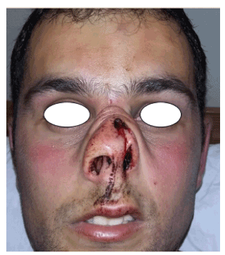
Figure 1. Frontal photograph showing left sided nasal skin invagination together with open fracture
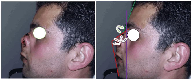
Figure 2. Left lateral photograph showing skin folds on the nasal dorsum due to skin invagination, more acute nasofrontal angle (green arrow), bigger nasomental angle (red arrow) and a bigger nasofacial angle (purple arrow).
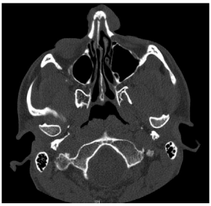
Figure 3. The CT scan axial views on admission showing a comminuted nasal fracture with buckling of cartilaginous nasal septum.
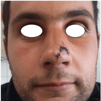
Figure 4. Frontal photograph showing restoration of normal contour of nasal dorsum.
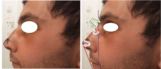
Figure 5. Lateral photograph showing restored nasofrontal angle (green arrow), nasomental angle (red arrow) and nasofacial angle (purple arrow).
The patient was seen 5 days after the injury for suture removal. The aesthetic appearance was very satisfying to the patient and thus far he is being managed conservatively. The patients’ only complaint was mild nasal obstruction on the left.
Discussion
This case represents a rare type of nasal fracture which is different to the routine nasal fractures we encounter on a day to day basis. The type of injury sustained by this patient looked like a naso-orbital ethmoidal fracture, however this was an isolated nasal bone fracture. No such case reports were identified on extensive online searches. In the beginning an open rhinoplasty supplimented with a possible bicoronal incision access was being contemplated in view of the complexity of the fracture; however upon doing a closed reduction the aesthetic and functional results were satisfactory. The importance of clinical examination cannot be overemphasised since missing associated complications, namely a septal haematoma would have had even more deleterious effects in such patient with severe cartilaginous and bony fractures. Antibiotic cover was prescribed in view of the fact that this was an open fracture.
Despite the unsighty look and rarity of such a nasal fracture, the suggestion is to attempt a closed reduction prior to considering a major rhinoplasty procedure as the outcomes from a closed reduction proved to be sufficient. The suggestion is, notably, based on the fact that a comprehensive imaging evaluation of facial bones is undertaken, and potential major complications are excluded.
Summary
- Motor vehicle accidents are the second most common cause for nasal fractures .
- No universal classification of nasal bone fractures exists.
2021 Copyright OAT. All rights reserv
- Despite the fact that the majority of nasal fractures are striaght forward, some fractures may pose a dilemma on the management of this rarer variety.
- Looks may be deceiving! This looked like a naso-orbital ethmoidal fracture however this was solely a nasal bone fracture.
- Clinical examination and exclusion of associated complications, namely septal haemtomas, cannot be overemphasised.
- The pug- like nasal deformity yielded good cosmetic and reasonably good functional results with simple closed reduction.
- The suggestion is, notably, based on the fact that a comprehensive imaging evaluation of facial bones is undertaken, and potential major complications are excluded.
References
- Haraldson JS (2016) Nasal fracture clinical presentation.
- Erdmann D, Follmar KE, Debruijn M, Bruno AD, Jung SH, et al. (2008) A retrospective analysis of facial fracture etiologies. Ann Plast Surg. 60: 398-403. [Crossref]
- Rawnsley. Your perfect nose.


