Key words
emergency department (ED), foreign body, abdominopelvic, laparotomy, sigmoid colon
Case presentation
Hospital presentation with foreign body retained in the rectum is occasionally observed in the emergency department (ED). Previous reports demonstrated that it is seen more frequently in male of 3rd or 4th decades. Various objects are presented with different size and shape. The diagnosis requires digital rectal exam as well as radiologic examination, such as abdominal X-ray and/or abdominopelvic CT scan to evaluate the retained material. Based on thorough evaluation, plan for management can be delineated.
Objectives
In this article, we present the cases with retained foreign body in the rectum.
Methodology
Between 2010 and 2016, 4 patients with foreign body in rectum.
Results
Case 1. A 50-year-old male presented in the ED, complaining inability to remove cylindrical plastic bottle in the rectum after 4 hours of initial insertion. On physical examination, anal bleeding as well as laceration was seen; however, abdominal symptom, such as pain, was not evident. Considering the size and shape, endoscopic removal was not possible (Figure 1). Under general anesthesia, transanal manual removal was attempted in lithotomy position. Postoperative colonoscopy showed no perforation site in the rectum, only two small lesions with mild erosion (Figure 2).
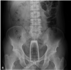
Figure 1. Preoperative KUB shows bottle in the rectum.
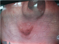
Figure 2. Colonoscopy shows mild erosion in the rectum.
Case 2. A 51-year-old male presented in the ED, complaining severe abdominal pain for 10 hours just after awakening from drunken state. On physical examination, no sign of peritoneal irritation was observed. On the radiologic examination, cylindrical object in the rectum sized 9.5cm by 6.5cm was detected. (Figure 3) Under the general anesthesia, the foreign object was removed transanally using rectal retractor in lithotomy position. (Figure 4)
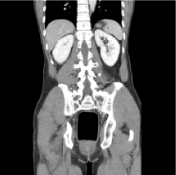
Figure 3. CT coronary view demonstrate cylindrical object in the rectum.
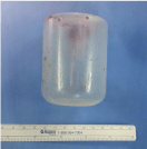
Figure 4.Case 3. A 54-year-old male came to the ED due to persistent anal pain after inserting a bottle of soy milk in 7 hours before. Grossly, any perianal wound was not observed. On the digital rectal examination, the foreign object was palpated at the tip of the index finger. Chest X-ray revealed intra-abdominal free air. Subsequent abdominal-pelvic CT scan demonstrated not only foreign body retained in the rectum but also perforation in the sigmoid colon (Figure 5). The patient underwent laparotomy for exploration under the general anesthesia. The foreign body was removed through the anus by squeezing the rectum intra-abdominally. For the perforated sigmoid colon, primary repair was performed.
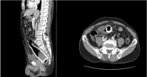
Figure 5. CT coronary and axial view shows bottle retained in the rectum and free air.
Case 4. A 59-year-old male came to the ED, complaining perianal pain with edema for a week. On digital rectal examination, sharp, needle-like foreign body was palpated in the rectum. On the abdominal-pelvic CT scan, 3cm sized linear object retained in the rectum was shown. On sigmoidoscopy, it was evident that the foreign body was fish bone, and it was successfully removed by using endoscopic forceps (Figure 6).
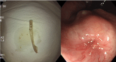
Figure 6. Removed foreign body and sigmoidoscopic finding after removal.
Conclusion
The management of retained foreign body requires sophisticated approach based on accurate information and thorough evaluation. The size, shape, and nature of the foreign object should be known before any attempt to remove. Appropriate method in various interventions should be chosen to least the injury to the rectum and anus.