Introduction: Osseous genioplasty is a versatile procedure which can predictably influence the profile, vertical length and esthetics of the chin. It can be performed as an isolated procedure or in conjunction with orthognathic surgery. With recent advances in imaging technology, three-dimensional computer assisted surgical simulation with images obtained from cone beam computed tomography are routinely used in treatment planning orthognathic surgery and genioplasty. To accurately duplicate the planned genioplasty movement on the operating table can be challenging due to strong muscle attachments on the genioplasty segment. We are proposing the use of a “foundation screw” which is a bone screw inserted perpendicular to the chin segment to provide better control of the genioplasty segment and improve surgical accuracy during fixation.
Method: In this retrospective study, thirty consecutive patients with bimaxillary orthognathic surgery and advancement genioplasty with the use of a “foundation screw” were included. The planned genioplasty advancement is compared with the actual advancement measured on lateral cephalometric radiographs.
Result: The mean value of the planned genioplasty advancement was 3.92 ± 1.86 mm. The actual advancement was 3.79 ± 1.81 mm. There is a good correlation between the planned and actual advancement (Pearson correlation r = 0.99, p < 0.001).
Conclusion: “Foundation screw” is a simple, yet effective tool in aiding fixation of the genioplasty segment. Surgeons can replicate the planned surgical movement on the operating table with ease using this technique.
genioplasty, fixation technique, surgical accuracy
With recent advances in imaging technology, there is a shift from 2D to 3D virtual surgical planning for orthognathic surgery [1,2]. Genioplasty can be planned along with orthognathic surgery to achieve the best esthetic outcomes [3-5]. To accurately duplicate the planned genioplasty movements on the operating table, however, can be challenging especially if the advancement is more than 2 mm or if an asymmetric advancement and/or vertical lengthening is planned. Very often, the surgeon positions the genioplasty segment to the desired position, and then the segment is held in place by the first assistant while fixation is applied. There is a large opportunity for error in positioning and fixation of the mobilized chin segment [4]. It is, therefore, important to establish a simple and effective method to aid in the fixation of the genioplasty segment.
This retrospective study was designed to investigate the accuracy of advancement genioplasty with the assistance of a “foundation screw” was approved by the Institutional Review Board at Chang Gung Memorial Hospital (201601216B0). Thirty consecutive dentofacial deformity patients who received bimaxillary orthognathic surgery and advancement genioplasty were included in this study. The results reported were based on a single surgeon’s experience between January of 2013 and December of 2015.
Bimaxillary Orthognathic Surgery and Genioplasty Planning
Pre-operative cone beam CT scan was performed with 1 mm cuts. Cone Beam CT Scan Images in DICOM (Digital Imaging and Communications in Medicine) was transferred to a computer workstation running 3D Surgery (Dolphin Imaging 11.0, Chatsworth, CA). The dental casts were scanned using a high resolution 3D optical scanner (Dental 3D scanner). Digital images of the scanned dental casts were then imported to the Dolphin software and superimposed on the CT images to produce better definition of the dental images. The virtual LeFort-I osteotomy surgical plan was generated first, followed by virtual surgical planning of the mandible with bilateral sagittal split osteotomy taking into consideration of the final occlusion. Finally, the genioplasty osteotomy was generated and the mobilized chin virtually repositioned according to the 3D surgical design.
The actual surgical movements of the osteotomy segments were executed according to the planned 3D surgical movements. The osseous genioplasty was approached with a mandibular labial sulcus incision about 1 cm from the mucogingival junction, followed by a subperiosteal dissection. Mental nerves were identified bilaterally. A vertical line representing the midline of the chin was marked on the mental protuberance. The genioplasty osteotomy line was then marked about 35mm from the incisal edge of the mandibular incisors and around 5-6 mm below the mental foramen. The relationship of the mental foramen to the mandibular canal was evaluated preoperatively on 3D CT and considered in the genioplasty design. Osseous genioplasty was then performed with a reciprocating saw. A caliper was used to mark the planned anterior-posterior advancement on the chin segment. A 2mm x10 mm bone screw, the “foundation screw,” was inserted at the point marked by the caliper, 7-8 mm into and perpendicular to the cut surface of the genioplasty segment. A horizontal osseous groove was created using a 1-mm round burr on the proximal segment to accommodate the screw head. The “foundation screw” was then used as an internal reference point to position the genioplasty segment; the amount of chin advancement, lateral shifting and yaw rotation can be more accurately determined. The permanent fixation was established with two lateral wires (Figure 1).
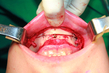
Figure 1. Asymmetric advancement of the genioplasty segment with the right side advanced 3mm and the left side only 1mm. The midline is shifted to the left by 1 mm.
Lateral cephalometric radiographs, obtained 1 week after surgery were analyzed for the horizontal distance of the advancement genioplasty and compared with the 3D pre-surgical treatment plan for each patient (Figure 2 and Figure 3). All measurements and data analysis were performed by two surgical colleagues who were not involved in the treatment planning or the surgical procedure. All measurements on the lateral cephalometric radiographs took into account the degree of magnification.
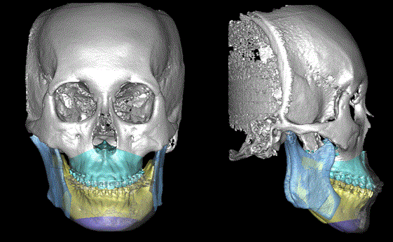
Figure 2. Presurgical treatment planning on 3D cone beam CT image of a patient included in this study. LeFort-I osteotomy, bilateral sagittal split osteotomies and genioplasty can be planned preoperatively. Noticed that the chin is planned to be advanced 3 mm.
a
b
c
d
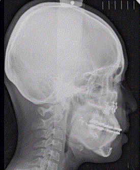
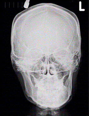
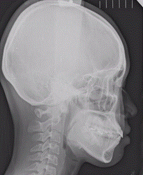
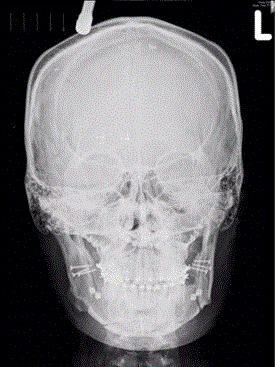
Figure 3. Cephalometric radiograph of the same patient in figures 2. (a) Preoperative anterior-posterior cephalometric radiograph; (b) Preoperative lateral cephalometric radiograph; (c) Postoperative anterior-posterior cephalometric radiograph taken one week after the operation; (d) Postoperative lateral cephalometric radiograph taken one week after the operation (The measurement of chin advancement was 2.86 mm).
Statistical Analysis
All data were presented as mean ± standard deviation. After the data points were collected, the planned advancement and actual advancement were compared with Pearson Correlation Test. The inter-examiner error was analyzed with interclass correlation coefficient.
Twelve males and eighteen females were included in this study. The interclass correlation coefficient of the two evaluators for chin advancement measurement reliability was 0.99 (p< 0.001). The mean for the planned virtual 3-D chin advancement was 3.92 ± 1.86 mm. The mean surgical chin advancement measured on lateral cephalogram was 3.79 ± 1.81 mm. Pearson Correlation test showed there was a high correlation between the 3-D planned chin advancement and lateral cephalometric measurement for genioplasty advancement (r=0.99, p< 0.001).
Genioplasty is the surgical correction of the bony contour of the chin which can be achieved through different surgical options including osseous genioplasty, chin contouring, alloplastic implant placement, or chin augmentation with fat or filler injections [6-8]. Osseous genioplasty through an intraoral approach was introduced by Richard Trauner and Hugo Obwegeser almost six decades ago [9,10]. It can be performed as an isolated procedure or in combination with orthognathic surgery to improve the profile, esthetics or symmetry of the lower third of the face. In many instances, after completion of bimaxillary osteotomy, the chin morphology remains asymmetrical or unsatisfactory in the anterior-posterior, vertical or horizontal dimensions. Osseous genioplasty is a versatile procedure that corrects various types of chin deformities with excellent postoperative stability [11-13]. It provides a predictable soft tissue response for advancement-extrusion movement and setback-impaction movement [14]. Osseous genioplasty, when combined with bimaxillary orthognathic surgery, is considered safe and can achieve the preferred pogonion position [15,16]. It is important to establish a simple and predictable genioplasty technique to avoid the scenario of a malpositioned chin. Lee et al reported 54 cases with secondary genioplasty after orthognathic surgery. They have emphasized that one should pay close attention to the chin contours to avoid secondary surgical revision [17].
An alternative to osseous genioplasty is the placement of an alloplastic implant. Alloplastic chin augmentation is used less frequently than osseous genioplasty in the setting of orthognathic surgery. This may be due to limited implant sizes precluding the precise control of chin advancement to satisfy each patient’s needs. The alloplastic implant provides chin advancement, but chin movements in other directions cannot be achieved by this method [18]. Moreover, with the wider soft tissue dissection associated with orthognathic surgery, there is also a concern for wound dehiscence and increased risk of infection associated with placement of the alloplastic implant. Bone resorption associated with alloplastic implant placement and secondary soft tissue deformities have also been reported in the literature [15,19]. Bertossi et al reported superior outcomes in terms of long term aesthetics and predictability with osseous genioplasty compared with alloplastic implant genioplasty [20].
Some have advocated the use of a computer-generated chin template to improve surgical accuracy in genioplasty [4]. Such templates are usually quite bulky and require more extensive soft tissue dissection and retraction during insertion. Others have recommended the use of pre-bent titanium plates using a 3D rapid-prototyping model and surgical cutting and drilling guides for genioplasty [21]. This technique can significantly increase the time spent on pre-surgical treatment planning and the total cost of the procedure. In this study, the “foundation screw” acted as a pivot point and internal reference for the surgeon to manipulate the chin segment and achieve the planned surgical movement. The genioplasty segments were then stabilized with lateral wires. A study by Precious et al. concluded that in cases where wire fixation was indicated, costs could be reduced by around $500 per case while providing adequate stability for osteosynthesis [22]. In cases where asymmetric genioplasty advancement was required, the use of the foundation screw and two lateral wires also allows better control and more precise surgical manipulation of the genioplasty segment during fixation.
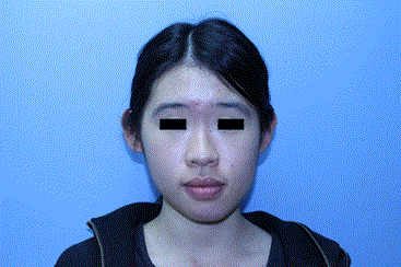
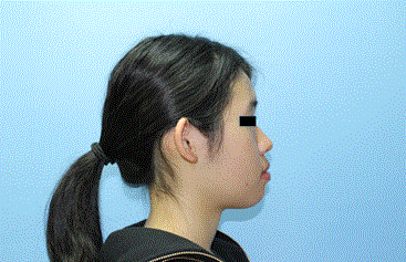
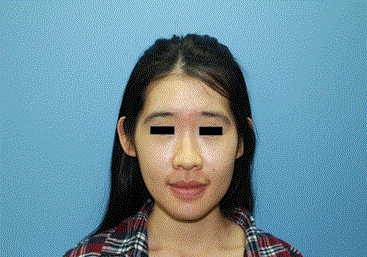
This method is technically simple and the results of this study have shown that the discrepancies between the actual and planned measurements are negligible with a mean difference of 0.14 mm. None of the patients included in this study developed post-operative complications such as infection, dental root injury or un-satisfactory esthetic outcomes that required revision. Preoperative and postoperative clinical photographs of a patient included in this study are shown in figure 4.
a
b
c
d
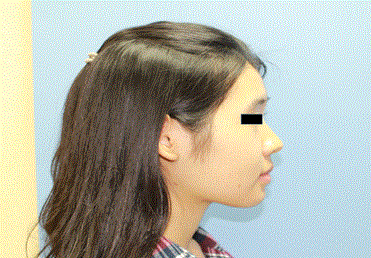
Figure 4. 20 year old female with facial asymmetry. (a) Pre-operative frontal view photo; (b) pre-operative lateral view photo; (c) six months post-operative frontal view photo; (d) six months post-operative lateral view photo.
A potential limitation of this technique is the occasional ability to palpate the “foundation screw” in patients with thin oral mucosa. This may be minimized by placing the screw head just 1-2 mm above the genioplasty segment and creating a horizontal groove on the outer cortex of the chin which acts as the resting point for the screw head. Adequate approximation of the mentalis muscle over the screw head prior to mucosa closure will often prevent this problem. In most cases, the “foundation screw” can be left in place to provide additional stability. If the screw head remains palpable due to thin vestibular soft tissue coverage, it can be removed after genioplasty fixation is completed.
2021 Copyright OAT. All rights reserv
In conclusion, genioplasty with the use of a “foundation screw” is a simple yet effective method to aid fixation in osseous genioplasty. The differences between the actual and planned advancement genioplasty measurements are negligible.
- Haas Jr OL, Becker OE, de Oliveira RB (2015) Computer-aided planning in orthognathic surgery-systematic review. Int J Oral Maxillofac Surg 44: 329-342. [Crossref]
- Zhang N, Liu S, Hu Z, Hu J, Zhu S, et al. (2016) Accuracy of virtual surgical planning in two-jaw orthognathic surgery: comparison of planned and actual results. Oral Surg Oral Med Oral Pathol Oral Radiol 122: 143-151. [Crossref]
- Edwards SP (2010) Computer-assisted craniomaxillofacial surgery. Oral Maxillofacial Surg Clinics North Amer 22: 117-134.
- Hsu SS, Gateno J, Bell RB, Hirsch DL, Markiewicz MR, et al. (2013) Accuracy of a computer-aided surgical simulation protocol for orthognathic surgery: a prospective multicenter study. J Oral Maxillofac Surg 71: 128-142. [Crossref]
- Scolozzi P (2015) Computer-aided design and computer-aided modeling (CAD/CAM) generated surgical splints, cutting guides and custom-made implants: Which indications in orthognathic surgery? Rev Stomatol Chir Maxillofac Chir Orale 116: 343-349.
- Park JY, Kim SG, Baik SM, Kim SY (2010) Comparison of genioplasty using Medpor and osteotomy. Oral Surg Oral Med Oral Pathol Oral Radiol Endod 109: e26-30. [Crossref]
- DeLorenzi C, Weinberg M, Solish N, Swift A (2009) The long-term efficacy and safety of a subcutaneously injected large-particle stabilized hyaluronic acid-based gel of nonanimal origin in esthetic facial contouring. Dermatol Surg 1: 313-321. [Crossref]
- Chang CS, Kang GC (2016) Achieving Ideal Lower Face Aesthetic Contours: Combination of Tridimensional Fat Grafting to the Chin with Masseter Botulinum Toxin Injection. Aesthet Surg J 36: 1093-1100. [Crossref]
- Trauner R, Obwegeser H (1957) The surgical correction of mandibular prognathism and retrognathia with consideration of genioplasty. I. Surgical procedures to correct mandibular prognathism and reshaping of the chin. Oral Surg Oral Med Oral Pathol 10: 677-689. [Crossref]
- Trauner R, Obwegeser H (1957) The surgical correction of mandibular prognathism and retrognathia with consideration of genioplasty. II. Operating methods for microgenia and distoclusion. Oral Surg Oral Med Oral Pathol 10: 787-792. [Crossref]
- Drissi Qeytoni H, Zribi A, Raphaël B, Lebeau J, Bettega G (2007) [Genioplasty: technique and applications]. Rev Stomatol Chir Maxillofac 108: 441-450. [Crossref]
- Gui L, Huang L, Zhang Z (2008) Genioplasty and chin augmentation with Medpore implants: a report of 650 cases. Aesthetic Plast Surg 32: 220-226. [Crossref]
- Talebzadeh N, Pogrel MA (2001) Long-term hard and soft tissue relapse rate after genioplasty. Oral Surg Oral Med Oral Pathol Oral Radiol Endod 91: 153-156. [crossref]
- San Miguel Moragas J, Oth O, Büttner M, Mommaerts MY (2015) A systematic review on soft-to-hard tissue ratios in orthognathic surgery part II: Chin procedures. J Craniomaxillofac Surg 43: 1530-1540. [Crossref]
- Posnick JC, Choi E, Chang RP (2016) Osseous genioplasty in conjunction with bimaxillary orthognathic surgery: a review of 262 consecutive cases. Int J Oral Maxillofac Surg 45: 904-913. [Crossref]
- Posnick JC, Choi E, Chavda A (2016) Operative Time, Airway Management, Need for Blood Transfusions, and In-Hospital Stay for Bimaxillary, Intranasal, and Osseous Genioplasty Surgery: Current Clinical Practices. J Oral Maxillofac Surg 74: 590-600. [Crossref]
- Lee SW, Ahn SH, Myung Y (2016) Secondary Genioplasties for the Treatment of Chin Deformities After Orthognathic Surgery in Asian Women: Defining the Aesthetic Importance of Managing the Chin Shape in Orthognathic Surgery. Annals of Plastic Surgery 76: 301-315.
- Lee EI (2013) Aesthetic alteration of the chin. Semin Plast Surg 27: 155-160. [Crossref]
- Stalder MW, St Hilaire H (2012) Immediate osseous genioplasty with Kirschner wire fixation for revision of infected alloplastic chin implant. J Craniofac Surg 23: e446-447. [Crossref]
- Bertossi D, Galzignato PF, Albanese M, Botti C, Botti G, et al. (2015) Chin Microgenia: A Clinical Comparative Study. Aesthetic Plast Surg 39: 651-658. [Crossref]
- Olszewski R, Tranduy K, Reychler H (2010) Innovative procedure for computer-assisted genioplasty: three-dimensional cephalometry, rapid-prototyping model and surgical splint. Int J Oral Maxillofac Surg 39: 721-724.
- Precious DS, Cardoso AB, Cardoso MC, Doucet JC (2014) Cost comparison of genioplasty: when indicated, wire osteosynthesis is more cost effective than plate and screw fixation. Oral Maxillofac Surg 18: 439-444. [Crossref]