Abstract
Introduction: Cervical radiculopathy is a condition caused by the compression of a nerve root in the cervical spine. Among the various pathologies which affect the nerve root, degenerative processes in the spine are the most common. The aim of this study was to compare anterior and posterior approaches in management of cervical radiculopathy.
Material and methods: Twenty-five patients performed anterior decompression and fusion of the affected level and thirteen patients underwent nerve root decompression through posterior open foraminotomy.
Results: There were 28 patients complained of neck and scapular pain and 10 patients with scapular pain before surgery. Thirty-three patients had no pain after surgery, 3 patients had persistent pain in the same sites. The mean (± standard deviation) score improved from 5 ± 3.6 to 16 ± 2.3 in the anterior decompression and fusion group and from 4 ± 4.1 to 15 ± 1.6 in the posterior forminotomy group. The increase of the score after surgery in each nerve root decompressed level was statistically insignificant (P>0.05).
Conclusion: Cervical radiculopathy can be a debilitating disease that can cause patients significant impairment. Rapid diagnosis and treatment of this condition is required to facilitate the return of the patient to their normal state of health. Anterior and posterior surgical approaches are equally effective in management of this condition.
Key words
cervical radiculopathy, surgical approach, scapular pain
Introduction
Cervical radiculopathy is a condition caused by the compression of a nerve root in the cervical spine. Among the various pathologies which affect the nerve root, degenerative processes in the spine are the most common. In 1926, Elliot described how radicular symptoms could arise through narrowing of the intervertebral foramen secondary to spur formation in either the Luschka or zygoapophyseal joint [1]. In 1928, Stookey described a clinical syndrome of cervical nerve root compression caused by herniated masses [2]. The clinical overlap between cervical radiculopathy and peripheral nerve entrapment syndromes and their nearly ubiquitous nature makes understanding of and identification of these entities mandatory for all practitioners [3,4]. Cervical radiculopathy can be a debilitating disease that can cause patients significant impairment. The effect of this can be significant both economically, from lost work and companseations, and psychologically, from prolonged pain and impaired social functioning [5,6]. Treatment of cervical radiculopathy ranges from conservative treatment which included collar, NSAIDS, bed rest, and cervical epidural injection to surgical treatment [7]. In the first half of the last century, only posterior surgery was performed for cervical spinal pathologies. In 1940s, posterior foraminotomy was introduced for managing cervical radiculopathy [8]. Although the intervention was small but insufficient visibility because of hemorrhage from extradural venous plexuses and traumatic handling of the nerve root when removing the disc herniation or spurs were problems [9]. In 1950s, anterior surgery was developed for the cervical spine. It gradually replaced the posterior foraminotomy for cervical radiculopathy. However, anterior fusion sometimes may lead to many complications [10,11]. In 1970, anterior surgery without fusion was introduced for cervical radiculopathy. It is less invasive, allow earlier rehabilitation but kyphotic deformity occurred frequently [12]. In 1980s, the development of microsurgical instruments and operating microscopes revived the posterior foraminotomy [13]. No difference in the results were reported previously between the anterior and posterior approaches [14,15]. In this series, we tried to compare both approaches in management of cervical radiculopathy.
Material and methods
Thirty-eight patients with cervical radiculopathy complained of neck pain beside symptoms in upper limb and fingers. The inclusion criteria cervical disc herniations manifesting as radiculopathy. Both computed tomography (CT) scanning and
magnetic resonance imaging (MRI) were performed for all patients. Patients with segmental instability, central disc herniation, ossification of the posterior longitudinal ligament (OPLL), or cervical myelopathy were excluded. Twenty-five patients performed anterior decompression and fusion of the affected level (Figure 1) and thirteen patients underwent nerve root decompression through posterior open foraminotomy (Figure 2). Surgery was performed in both groups after at least four months of conservative treatment. Informed consent was obtained from all patients before surgery. Age of the patients at surgery ranged from 32 to 67 years in anterior group
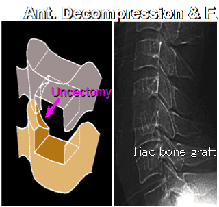
Figure 1. Anterior surgical approach for management of cervical radiculopathy
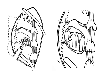
Figure 2. Diagram showing boundries of posterior foraminotomy
and from 34 to 71 years in the posterior group. The duration of symptoms before surgery ranged from 6 to 24 months in the anterior group (Average 9 months) and from 5 to 22 months in the posterior group (Average 8 months). Surgical complications such as hematoma, infection, dural damage, neurologic deficit, recurrence, and instability were documented during the follow-up period. Patients assessment was depending on cervical radiculopathy severity score of twenty points [16]. The system in which the normal score is 20, consists of four categories: (1) Subjective symptoms; (2) Ability to work; (3) Finger functions; and (4) Objective signs. This system scores the severity before and after treatment. Score 8 or less is an indication for surgery. All patients completed the follow up period of 3 years.
Statistical analysis: Comparison between the two groups was made using the independent two-sample t test for continuous variables. Fisher’s exact test was performed for categorical variables. A two-tailed p value of < 0.05 was considered significant.
Results
Age of the patients at surgery ranged from 32 to 67 years in anterior group (Mean 45 years) and from 34 to 71 years in the posterior group (Mean 46 years). There was no significant difference between groups for sex ratio, age, or symptom duration.
The involved nerve roots were C5 in 5 patients, C6 in 11 patients, C7 in 7 patients and C8 in 2 patients in the anterior group and were C5 in 2 patients, C6 in 7 patients, C7 in 2 patients, and C8 in 2 patients in the posterior group.
There were 28 patients complained of neck and scapular pain and 10 patients with scapular pain before surgery. Thirty three patients had no pain after surgery, 3 patients had persistent pain in the same sites, and two patients developed pain in a new sites in the nuchal and suprascapular regions (Figure 3).
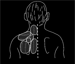
Figure 3. The 5 subgroups of neck and scapular pain
The mean (± standard deviation) score improved from 5 ± 3.6 to 16 ± 2.3 in the anterior decompression and fusion group and from 4 ± 4.1 to 15 ± 1.6 in the posterior forminotomy group. The score increased in all patients indicating that the decompression of the involved root was effective. The increase of the score after surgery in each nerve root decompressed level was statistically insignificant (P>0.05).
Discussion
In clinical studies on the neck and scapular pain, unavoidable difficulties are encountered in determining the site of the pain. Patients do not always describe the site because they seldom know its anatomical name. So, it is easier for the patient and to the doctor to choose the site of pain from 5 subregions in the neck and scapular regions. Through this approach, the painful sites were identified in all patients and also by comparing the preoperative and postoperative sites of pain, new pain that could develop after surgery was detected. This also help in detecting the source of pain and assessing the efficacy of the treatment. Yoss [14] divided the region into 3 subregions (nuchal, scapular and interscapular). Murphey [15] divided them into 2 (nuchal and scapular). The number of their subgroups are relatively low compared to the number of the nerve roots supplying the area.
Posterior foraminotomy decompresses the nerve root alone. Therefore, the root would prove to be the origin of the neck and scapular if the pain is relieved with the radicular symptoms in the arm and fingers.
Early postoperative assessment is essential for verification of the effect of surgical decompression of the nerve root. In the present study, Pain completely disappeared in 94% of the patients one month after surgery. Evaluation of pain in the other studies usually depends on the final follow up score. So, it is difficult to determine if the pain relieve was due to surgical decompression of the involved nerve root or a natural remission of pain originating from other sites [17].
No difference in results has been reported between anterior and posterior approaches [18]. Also, there are no difference in cases with bony spurs or those with disc herniation [7]. In our study, we reached also to the same conclusion.
Conclusion
Cervical radiculopathy can be a debilitating disease that can cause patients significant impairment. Rapid diagnosis and treatment of this condition is required to facilitate the return of the patient to their normal state of health. Anterior and posterior surgical approaches are equally effective in management of this condition.
References
2021 Copyright OAT. All rights reserv
- Elliot GR (1926) A contribution to spinal osteoarthritis involving the cervical region. J Bone Joint Surg 8: 42-52.
- Stookey B (1928) Compression of the spinal cord due to ventral extradural cervical chordomas. Arch Neurol Psych 20: 275-291.
- Carette S, Fehlings MG (2005) Clinical practice. Cervical radiculopathy. N Engl J Med 353: 392-399. [Crossref]
- Ellenberg MR, Honet JC, Treanor WJ (1994) Cervical radiculopathy. Arch Phys Med Rehabil 75: 342-352. [Crossref]
- Caridi JM, Pumberger M, Hughes AP (2011) Cervical radiculopathy: a review. HSS J 7: 265-272. [Crossref]
- Lees F, Turner Jw (1963) Natural history and prognosis of cervical spondylosis. Br Med J 2: 1607-1610. [Crossref]
- Lunsford LD, Bissonette DJ, Jannetta PJ, Sheptak PE, Zorub DS (1980) Anterior surgery for cervical disc disease. Part 1: Treatment of lateral cervical disc herniation in 253 cases. J Neurosurg 53: 1-11. [Crossref]
- Kokubun S, Sato T, Ishii Y, Tanaka Y (1996) Cervical myelopathy in the Japanese. Clin Orthop Relat Res 1996: 129-138. [Crossref]
- Smith GW, Robinson RA (1958) The treatment of certain cervical-spine disorders by anterior removal of the intervertebral disc and interbody fusion. J Bone Joint Surg Am 40-40A: 607-624. [Crossref]
- Cloward RB (1958) The anterior approach for removal of ruptured cervical disks. J Neurosurg 15: 602-617. [Crossref]
- Martins AN (1976) Anterior cervical discectomy with and without interbody bone graft. J Neurosurg 44: 290-295. [Crossref]
- Williams RW (1983) Microcervical foraminotomy. A surgical alternative for intractable radicular pain. Spine (Phila Pa 1976) 8: 708-716. [Crossref]
- Herkowitz HN, Kurz LT, Overholt DP (1990) Surgical management of cervical soft disc herniation. A comparison between the anterior and posterior approach. Spine (Phila Pa 1976) 15: 1026-1030. [Crossref]
- Yoss RE, Corbin KB, Maccarty CS, Love JG (1957) Significance of symptoms and signs in localization of involved root in cervical disk protrusion. Neurology 7: 673-683. [Crossref]
- Murphey F, Simmons JC, Brunson B (1973) Surgical treatment of laterally ruptured cervical disc. Review of 648 cases, 1939 to 1972. J Neurosurg 38: 679-683. [Crossref]
- Tanaka Y, Kokubun S, Sato T (1998) Cervical radiculopathy and its unsolved problems. Current orthopedics 12: 1-6.
- Aldrich F (1990) Posterolateral microdisectomy for cervical monoradiculopathy caused by posterolateral soft cervical disc sequestration. J Neurosurg 72: 370-377. [Crossref]
- Herkowitz HN, Kurz LT, Overholt DP (1990) Surgical management of cervical soft disc herniation. A comparison between the anterior and posterior approach. Spine (Phila Pa 1976) 15: 1026-1030. [Crossref]