Lipomatous Hypertrophy of the Interatrial Septum is (LHAS) an unusual and benign condition characterized by the excessive deposition of adipose tissue in the interatrial septum, which is most often detected as an incidental finding on echocardiography. The classic finding is a homogenous, bi-lobed configuration of the interatrial septum with sparing of the fossa ovalis. LHAS has been associated with various atrial arrhythmias, including multifocal atrial tachycardia, multiple premature atrial contractions, atrial fibrillation and rarely sudden death. Here the author describes a case report of a 57-year-old obese female who presents to the emergency department for sudden palpitation and dizziness due paroxysmal atrial fibrillation with rapid ventricular response. After successful cardioversion, a tranthoracic bidimensional echocardiography showed the typical finding of LHAS
lipomatous hypertrophy of the interatrial septum, obese woman, atrial fibrillation, echocardiography
Lipomatous Hypertrophy of the Interatrial Septum (LHAS) is an unusual condition, usually a benign condition and most often detected as an incidental finding on echocardiography. The classic finding is a homogenous, bi-lobed configuration of the interatrial septum with sparing of the fossa ovalis. This infiltration can also involve the septal tissue and has been associated with various atrial arrhythmias, including multifocal atrial tachycardia, multiple atrial premature contractions, atrial fibrillation and rarely even sudden death [1]. Interatrial septal thickness <2 cm is considered diagnostic of LHAS [2].
The prevalence of LHIS is estimated to be between 1-8%. It usually occurs in older, obese people and there may be a higher incidence in women [3].
Differential diagnoses should be thought of when there is sparing of the fossa ovalis.
Fat-containing neoplasms can arise in the atrial septum including cardiac lipoma, cardiac rhabdomyoma, cardiac myxoma, cardiac rhabdomyosarcoma and cardiac liposarcoma [4]. Microscopically, LHIS is characterized by fat infiltration between the myocardial fibers of the atrial septum. LHIS also can create a mass-like bulge. There is typical sparing of the fossa ovalis [5].
With this case, the author reports the clinical presentation and association between atrial arrhythmia and LHAS in an otherwise healthy obese woman.
A 57-year-old obese woman was referred to our emergency department with complaints of palpitations, dizziness, and fatigue of brief duration. Her family history was unremarkable. Physical examination disclosed obesity (weight 94 kg, height 162 cm BMI 35,8). Systemic blood pressure was 130/84 mm Hg; heart rate (apical) was 120 beats/min and sometimes irregular and respiratory rate was 22 breaths/min and slightly labored. The cardiac apex was palpable in the fourth intercostal space at the left anterior axillary line. A mild systolic ejection murmur was heard at the base of the heart; it was associated with normal carotid impulses bilaterally. No diastolic murmurs or sounds were heard. The 12 leads ECG showed atrial fibrillation with a rapid ventricular response and nonspecific ST-T-wave changes (Figure 1). The patient was treated with Flecainide intravenous and a normal sinus rhythm was restored quickly. A second ECG recorded in normal sinus rhythm disclosed a by fascicular block pattern (left anterior hemiblock (LAHB) and incomplete RBBB) with a secondary anomaly of ventricular repolarization (Figure 2). A chest x-ray was normal. Therefore, the patient was discharged in good health with an indication to further cardiologic examination. Subsequently Two-dimensional Trans-Thoracic echocardiography with subcostal four-chamber showed a hyperechogenic mass in the interatrial septum revealed by thickening and increased echogenicity of the inferior and superior portions of the atrial septum with sparing of the fossa ovalis. There was no decrease in flow velocities of the superior and inferior vena cava nor a flow disturbance in the pulmonary veins. These findings were typical for fatty infiltrate and results in a "dumbbell-shaped" appearance on Two dimensional trans-thoracic echocardiography (2D TT).
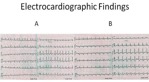
Figure 1. Showing Electrocardiographic tracings before and after pharmacological cardioversion. (A) Twelve-lead tracing shows atrial fibrillation with the fast ventricular response. (B) 12 lead tracing shows sinus rhythm with bi-fascicular block (i.e. LAHB and incomplete RBBB)
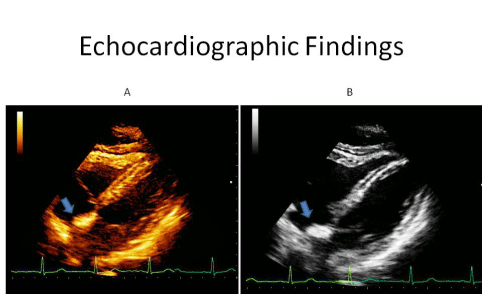
Figure 2. Panel A and B Trans-thoracic echocardiogram showing lipomatous hypertrophy of the intra-atrial septum (arrow). Subcostal 4 chamber apical long axis view shows classic lipomatous hypertrophy of the atrial septum (arrows).
Lipomatous hypertrophy of the heart is a benign and rare condition characterized by large fatty tissue deposits in the interatrial septum, usually extending to the atrial wall and rarely to the interventricular septum. The designation lipomatous is not properly correct because, unlike lipomas, the fatty lesions of LHIS are not encapsulated. Also, LHIS is characterized histologically by adipocyte hyperplasia, not hypertrophy. Furthermore, anatomic dissection studies have revealed that such ‘‘septal’’ fat is actually epicardial (extracardiac) [6]. This condition is more common than true cardiac lipomas, occurring almost exclusively in elderly, obese patients and is usually asymptomatic. Available data indicate that this condition is more likely to be seen in the seventh to the eighth decade of life, more commonly in women and with age-associated increases of epicardial fat. It is rarely seen in the younger age group which mandate more extensive investigation to rule out a true neoplasm. As above mentioned, it is characterized histologically by the presence of granular or vacuolated fetal adipocytes and non-encapsulated, non-neoplastic mature fat cells. The absence of encapsulated fat cells and the presence of fetal fat cells distinguish it from lipoma [7,8]. LHAS abnormality was first described by Prior in 1964 [9].
Besides being usually benign and asymptomatic, a significant number of patients have been reported to have unexplained arrhythmias such as atrial fibrillation, atrioventricular (AV) block and sudden death [10]. Therefore, in most patients, it represents an incidental echocardiographic finding requested for further examination after arrhythmias presentation [11]. Noninvasive modalities have led to the identification of these masses, which may be mistaken for a tumor or other neoplasm. Computed tomography (CT), magnetic resonance imaging (MRI), trans-thoracic echocardiograms (TTE) and trans-esophageal echocardiography (TEE) allow simple non-invasive diagnostic imaging of the interatrial septum [12].
TTE diagnosis of LHAS is based on the classic morphology of the thickened, echogenic bi-lobed septal mass. A variety of other cardiac lesions such as lipomas, liposarcomas, metastatic tumors, myxomas and amyloidosis can also present as a septal tumor mass. However, LHAS always spares the foramen ovale. Surgical excision of the LHAS is reserved for patients who have a superior vena cava (SVC) obstruction or an intractable rhythm disturbance [13,14].
In conclusion, this case confirms that LHAS is an uncommon anomaly, due to benign deposition of fatty tissue within the interatrial septum, often diagnosed incidentally and usually not requiring intervention. However, LHAS must be included in the differential diagnosis of any right atrial mass, or any fat-containing neoplasm, and suspected as the cause of paroxysmal atrial fibrillation in otherwise healthy, but obese persons, such as our patient. Surgical correction must be considered in cases of severe rhythm disorders or great vessel obstruction.
- Hutter AM Jr, Page DL (1971) Atrial arrhythmias and lipomatous hypertrophy of the cardiac interatrial septum. Am Heart J 82: 16-21. [Crossref]
- Shirani J, Roberts WC (1993) Clinical, electrocardiographic and morphologic features of massive fatty deposits ("lipomatous hypertrophy") in the atrial septum. J Am Coll Cardiol 22: 226-238. [Crossref]
- Xanthos T, Giannakopoulos N, Papadimitriou L (2007) Lipomatous hypertrophy of the interatrial septum: a pathological and clinical approach. Int J Cardiol 121: 4-8. [Crossref]
- Pugliatti P, Patanè S, De Gregorio C, Recupero A, Carerj S, et al. (2008) Lipomatous hypertrophy of the interatrial septum. Int J Cardiol 130: 294-295. [Crossref]
- Pérez Arroyuelos I, Berástegui Imaz M, Canteli Padilla B, et-al. (2007) Lipomatous hypertrophy of the interatrial septum associated with fatty replacement of the ventricular myocardium: a case report. J Magn Reson Imaging 26:152-4.
- Anderson RH, Webb S, Brown NA (1999) Clinical anatomy of the atrial septum with reference to its developmental components. Clin Anat 12: 362-374. [Crossref]
- Kozelj M, Angelski R, Pavcnik D (1995) Lipomatous hypertrophy of the interatrial septum: diagnosis by echocardiography and magnetic resonance imaging. A case report. Angiology 46: 863-866. [Crossref]
- Stone GW, O'Kell RT, Good TH, Hartzler GO (1990) Lipomatous hypertrophy of the interatrial septum: diagnosis by percutaneous transvenous biopsy. Am Heart J 119: 406-408. [Crossref]
- Prior JT (1964) Lipomatous hypertrophy of cardiac interatrial septum. A lesion resembling hibernoma, lipoblastomatosis and infiltrating lipoma. Arch Pathol 78: 11-15. [Crossref]
- Kusano KF, Ohe T (2002) Cardiac tumors that cause arrhythmias. Card Electrophysiol Rev 6: 174-177. [Crossref]
- Cohen IS, Raiker K (1993) Atrial lipomatous hypertrophy: lipomatous atrial hypertrophy with significant involvement of the right atrial wall. J Am Soc Echocardiogr 6:30-4.
- Kindman L A, Wright A, Tye T, Seale W, Appleton C (1988) Lipomatous Hypertrophy of the Interatrial Septum: Characterization by Transesophageal and Transthoracic Echocardiography, Magnetic Resonance Imaging and Computed Tomography. J Am Soc Echocardiography 1:450-4.
- Martin Breuer, Jens Wippermann, Ulrich Franke, Thorsten Wahlers (2002) Lipomatous hypertrophy of the interatrial septum and upper right atrial inflow obstruction. Eur J Cardiothoracic Surg 22:1023-1025.
- Zeebregts CJ, Hensens AG, Timmermans J, Pruszczynski MS, Lacquet LK (1997) Lipomatous hypertrophy of the interatrial septum: indication for surgery? Eur J Cardiothorac Surg 11: 785-787. [Crossref]
2021 Copyright OAT. All rights reserv