Abstract
Background: The commonly encountered unstable geriatric trochanteric hip fractures are frequently managed with a proximal femoral nail and spiral blade. Although well recognized risks and complications including cut-out/backing-out of the blade have been reported; current literature has put less emphasis into describing the intra-operative problems. Methods: We are reporting 2 cases to highlight the potential sources of inter-operative complications regarding the spiral blade. Results & conclusions: The authors advise that a good reduction prior to nail insertion is recommended; and proper implant assembly must be validated with specific emphasis placed on checking the locking mechanism in the blade. Multiple fluoroscopic views to assess the final implant position is also suggested and the use of adjunct BMPs in severely comminuted fractures may be considered.
Key words
hip fracture, PFNa, spiral blade, complication
Introduction
The proximal femoral nail antirotation (PFNa II) is commonly used for treating unstable geriatric trochanteric hip fractures [1]. Possible complications reported in literature include cut out/back out of the blade and non-union of fracture [2-4]. There have been no reported complications concerning intraoperative problems encountered during the spiral blade insertion and locking. 2 cases are reported here to highlightthe potential causes of intraoperative failure of the spiral blade insertion and locking mechanism. The authors emphasize that a good reduction prior to nail insertion is advised, proper implant assembly must be validated with emphasis on the locking mechanism in the blade, multiple fluoroscopy views are beneficial to confirm the final implant position and the use of BMPs in comminuted cases may be considered.
Geriatric hip fractureis one of the most common orthopaedic problems encountered nowadays due to aging population. Early reduction and internal fixation is recommended as it allows patients for early mobilization and rapid pain relief in order to prevent complications from prolonged immobilization like pressure sore, deep vein thrombosis and pulmonary embolism [5-6]. It also helps to improve the hospital length of stay in both acute and convalescence hospital [7]. Cephalomedullary device is commonly used for treating the unstable trochanteric fracture (AO fracture classification 31-A2 and 31-A3) nowadays and it could be performed in a minimally invasive and timely manner. Mal-functioning of implant during the operations can increase the operative time, cost, operative risks and be frustrated to surgeons. We reported 2 cases about the failure of spiral blade insertion and locking in this commonly used implant.
Case 1
The first case is a 79-year-old lady with hypertension, chronic obstructive airway disease admitted to hospital due to persistent left hip pain after fell 2 weeks ago. She had history of right femur osteomyelitis with multiple operations performed in China during childhood resulting in stiff right hip and knee joint. She could only walk with frame since then and with recurrent fall due to unsteady gait. She fell 2 weeks before admission and landed on her left buttock. She was unable to bear weight afterward and was admitted to emergency department with X-ray taken. No fracture was noted at that time and she was discharged with analgesics. She became chair-bound due to hip pain and was admitted to emergency department again 2 weeks later. Radiographs of her pelvis and left hip were repeated and displaced trochanteric fracture of her left femur with sub-trochanteric extension was noted (AO fracture classification 31-A2) (Figure 1 and 2). She was then admitted to our department for further management.
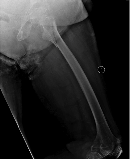
Figure 1. Displaced trochanteric fracture of left femur with sub-trochanteric extension was noted (lateral view).
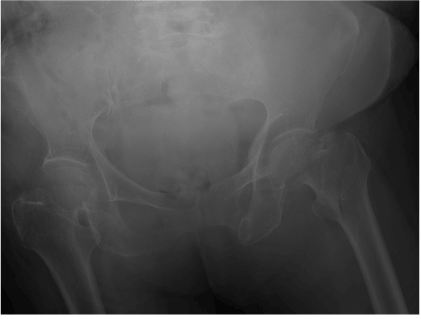
Figure 2. Displaced trochanteric fracture of left femur with sub-trochanteric extension was noted (AP view).
Closed reduction and fracture fixation with Proximal femoral nail antirotation (PFNa II) was arranged on the next day after admission. Patient was under spinal anaesthesia. She was put on traction table in supine position and closed reduction was performed under fluoroscopy. Routine skin preparation and draping of operative field was performed. Lateral skin incision proximal to greater trochanter was made and facialata was incised along the skin. Entry point was identified and prepared with curved cannulated awl. Guide rod was inserted. Proximal reaming was performed and a 130 degree, 10 mm x 240 mm PFNa II was inserted with the jig. All the instruments were assembled correctly and checked before insertion. The guide rod was removed. Correct aiming arm for PFNA blade was chosen and assembled. The buttress nut was screwed on the golden protection sleeve, and the golden drill sleeve and trocar were also inserted. The entry point of the blade was incised open and the soft tissue was dissected bluntly down to the bone. The entire sleeve assembly for PFNa II blade was advanced to the lateral femoral cortex. Guide pin was inserted. The required PFNa II blade length was measured to be 90 mm. The lateral femoral cortex was opened with the cannulated drill and reaming with the cannulated reamer was performed. The required PFNaII blade was checked for free rotation as recommended in the operative manual and the attached to the impactor. The blade-impactor assembly was inserted over the guide pin and hammered in place. The operation all along was smooth, but problem noticed when the surgeon tried to lock the blade. Normally, the gap between the rotating blade and the distal outer sleeve should be closed up with the locking mechanism in order to prevent rotation of the blade together with the femoral head. This can be checked under fluoroscopy. However, when the surgeon turned the impactor into the locked position (clockwise), the blade was unable to be tightened firmly. The impactor was loosen and could be removed easily before the gap was closed up. The blade was found to remaining unlocked (Figures 3 and 4) even the impactor was able to separate from it. The impactor was able to re-attach to the blade firmly under X-ray guidance, but the above situation reappeared when the surgeon tried to lock the blade by the impactor. The wound was enlarged and confirmed that no soft tissue was jammed. The whole assembly and jig was checked again and they were not loosened and there was no excessive angulation of the jig for femoral anteversion. The surgeon decided to extract out the blade and inserted another new PFNa II blade (85 mm). Before the new blade insertion, apart from checking the free rotation of the helical blade, it was checked for the successful locking unlocking to make sure that there was no mal-functioning of the locking mechanism. The bone purchase was good and the blade was able to lock smoothly (Figure 5 and 6) at this time. The total operative time was 2 hours and 46 minutes. Total blood loss was around 200ml.
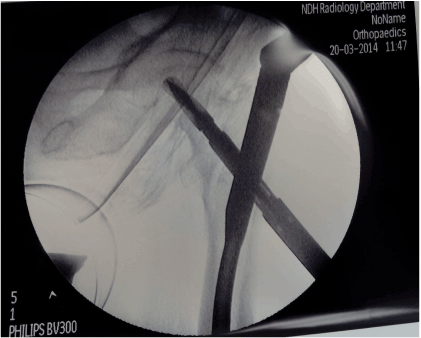
Figure 3. The blade was found to remaining unlocked in intraoperative X-ray screening
even the impactor was able to separate from the spiral blade (AP view).
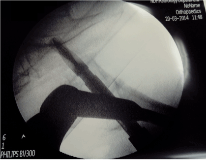
Figure 4. The blade was found to remaining unlocked in intraoperative X-ray screening even the impactor was able to separate from the spiral blade (lateral view).
Patient was stable all along post-operatively and tolerated full-weighted bearing walking exercise without any pain. Post-operative X-ray was performed showed the implant was in situ with good alignment (Figure 7 and 8). Examination of the spiral blade postoperatively noted there was an unusual metal mark on the body of the blade and it could not be fully locked. (Figure 9 and 10)
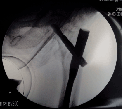
Figure 5. New spiral blade was able to lock smoothly, good implant position and alignment was confirmed with intraoperative screening (AP view).
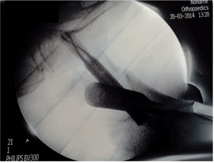
Figure 6. New spiral blade was able to lock smoothly, good implant position and alignment was confirmed with intraoperative screening (lateral view).
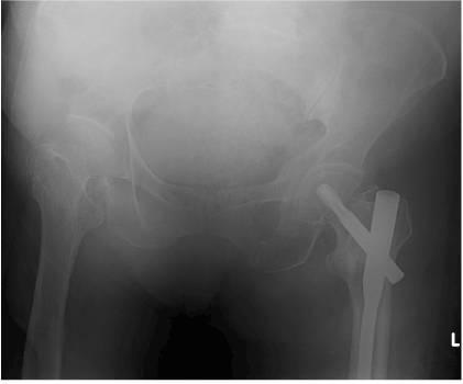
Figure 7. Post operative fracture alignment well and implant was in good position (AP view).
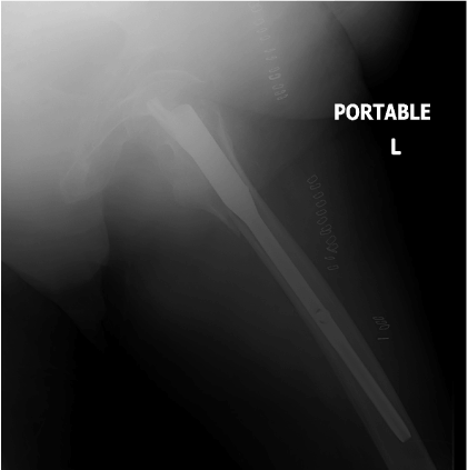
Figure 8. Post operative fracture alignment well and implant was in good position (lateral view).
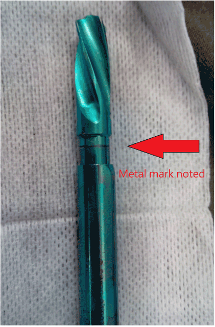
Figure 9. An unusual metal mark on the body of the blade .
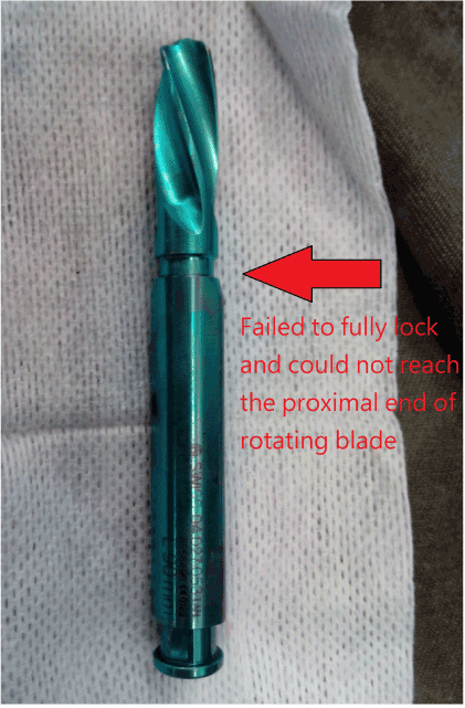
Figure 10. An unusual metal mark on the body of the blade, it could not be fully locked and reach the proximal end of the rotating blade part.
Case 2
A 62 year-old lady who premorbid was able to walk unaided, with history of hypertension, diabetes mellitus and end-stage renal failure required peritoneal dialysis, was presented for left hip injury and unable to bear weight after slipped and fell. Radiography was taken in Emergency department and showed subtrochanteric fracture of left femur (Figure 11). She was medically optimized after admission, and emergency operation was arranged 2 days after admission
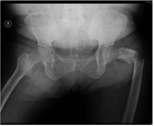
Figure 11. Subtrochanteric fracture of left femur showed in AP X-ray.
Closed reduction and fixation of left femur under X-ray guidance with proximal femoral nail antirotation (PFNaII) was performed. The incision, operative procedure and implant checking was same as the previous case. A 130 degree, 10 mm × 240 mm PFNa II was used with a 95mm spiral blade was inserted and locked under guidance of the standard provided jig and radiographic screening (Figure 12). Dynamic distal locking screw was inserted through jig also. The operation lasted total 3.5 hours as difficulty was encountered in maintaining satisfactory reduction. The part of implant insertion was quite smooth actually. She was discharged to general ward after operation. Clinically her vitals were stable after operation, haemoglobin level was static and drain output was not significant. Routine post-operative radiography was taken on 3 days after operation and showed the spiral blade of the PFNA was not in its expected position, also the femoral nail part was proximally migrated (Figure 13). Subsequent computerized tomography showed the spiral blade was actually passing anteriorly to the nail (Figure 14). The whole construct was lost and revision surgery was arranged after discussion with patient.
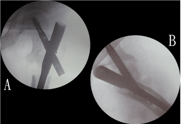
Figure 12. Spiral blade was inserted and locked under guidance of the standard provided jig and radiographic screening (AP + lateral view).
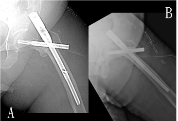
Figure 13. Routine post-operative radiography taken 3 days after operation and showed the spiral blade of the PFNA was not in its expected position; also the femoral nail part was proximally migrated (AP + lateral view).
Revision surgery was done 21 days after the initial operation after the patient was medically optimized and post-operative intensive care was arranged. Careful examination was performed intra-operatively, the spiral blade was found not passing through the femoral nail, with marked comminution over greater trochanter. No sign of infection was noted. The spiral blade and femoral nail were removed, with the blade track filled up with artificial bone substitute. This time open reduction was performed to achieve a better fracture alignment. A new 130 degrees, 10mm femoral nail with length 240mm was used, and a new 95mm spiral blade was inserted and locked through a new track with satisfactory bone purchase with the guidance of a jig. The defect over femoral lateral cortex was filled up with artificial bone substitute, and recombinant human bone morphogenetic proteins were applied to fracture site (Figure 15). Her post-operation progress was uneventful, with repeated radiography showed in-situ implant and static alignment.
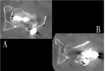
Figure 14. Computerized tomography showed the spiral blade was actually passing anteriorly to the nail, the whole construct was failed.
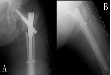
Figure 15. The defect over femoral lateral cortex was filled up with artificial bone substitute, and recombinant human bone morphogenetic proteins were applied to fracture site. Repeated radiography showed in-situ implant (AP + lateral view)
Patient was put on non-weight bearing walking for 6 weeks, then to partial-weight bearing walking for 6 weeks and then to full-weight bearing walking. The wound was healed and patient was fit for discharged home around 2 weeks after revision surgery. Patient was last seen around 3 months post revision operation, no significant hip pain was noted. Hip X-ray showed the fracture healing was in progress.
Discussion
Geriatric hip fracture is a very common nowadays and early fixation and mobilization is proven to decrease morbidity and mortality of the patient. It also helps to save money as it shortens the length of hospital stay significantly. The advantages of cephalomedullary devices over convention dynamic hip screws including minimally invasive surgical approach using stab wound incisions, easily inserted implant and superior fixation stability [8].
2021 Copyright OAT. All rights reserv
Common complications for using cephalomedullary devices fixation for proximal femur fracture included shortening, malrotation, malunion, non-union and implant failure or malposition [2,4,8]. Review by Paraschou in 2009, in 257 patients suffered from pertrochanteric fracture and treated with intramedullary nailing, 2 malunions, 1 nonunion, 1 cut out screw and 1 migration of the screw medially were reported. Also 2 out of 275 distal locking were misplaced using a commercially prepared jig9. In another literature by Aguado in 2013, 200 patients suffered from pertrochanteric fracture treated with PFNa were reviewed restropectively, 1 delay union, 2 nonunion, 2 cut out screw, 4 cases of helical blade sliding and 1 failure of distal locking procedure were reported [4]. Upon our literature review, So far no case of misplacement or failed locking of spiral blade was reported in the past.
For the first case, according to the official operative manual of PFNa II, after the insertion of the PFNa II blade with impactor, it should be locked tightly and the gap should be closed up in order to prevent further rotation of the blade and femoral head. Before insertion of the blade, it should be attached to the impactor correctly without being over tightened and the tip of the blade must be able to rotate freely in order to aim for advancement during insertion. We followed the operation steps strictly. However, the checking of successful blade locking was not mentioned in the operative manual and removal of the blade was needed in case of failure of locking after blade insertion. This problem not only increased the operative time and bleeding, but also increased implant cost and potentially jeopardized the bone purchase of the blade.
One possible reason for failure to lock the blade may be due to over-tighten of the blade. It may destroyed the thread within the blade and disturb the locking mechanism of impactor – blade assembly. On the other hand, jamming of soft tissue between the blade and impactor during advancement can prevent successful locking. If the blade was not attached to impactor correctly and the sleeve was not pushed to reach lateral cortex of femur, there might be possible that soft tissue jammed in the gap between the impactor and blade and disturbed the locking mechanism. The other possible mechanism was that the cancellous bone can be packed tightly around the blade during its advancement. The tightly packed bone around the blade might be tough enough to resist the proper rotation and locking of the blade. Finally it may due to that implant intrinsic problem, but we need to wait for the formal report of the manufacturer.
The blade needed to be removed and replaced by a new one. However, the tunnel created by the old blade will jeopardize the bone purchase of the new blade, particularly in those geriatric patients with poor bone quality. To avoid this, we can skip creating the drill hold for PFNa blade in those with poor bone quality or we can pack bone substitute or autograft/allograft into the tunnel before inserting the new blade.
Concerning the second case, the commercial prepared jig and nail were checked before operation and after removal in the revision surgery. No structural problem was noted and the jig worked perfectly well. One of the possible reasons for misplacement of the spiral blade might be due to loosening of the assembly. Intra-operatively, certain manipulations were done for reduction after nail insertion and before spiral blade insertion. Although the connection between nail and jig was checked and confirmed tight before insertion of the nail, the connection may be loosened during the manipulation. Therefore the jig might not be totally reliable for spiral blade insertion, especially for difficult case required lots of manipulation. In fact, reduction of fracture is recommended before inserting the nail, to prevent the drill and nail from taking an unwanted route, and subsequently locking the fragments in a displaced position. After inserting the nail, any attempt to further reduce the fracture would apply large torque towards the nail and aiming device, which could result in small but significant rotation of nail and aiming device. The minimal rotation could be enough to misplace the spiral blade. Besides, retightening of the lock again before inserting the spiral blade to ensure the connection between the nail and the jig is tight is also recommended by the operation guide provided, in order to avoid deviations during the insertion of the spiral blade through the aiming arm.
Furthermore, according to the operation guide, while advancing the sleeve assembly for spiral blade, one should ensure the sleeve assembly clicked into the aiming arm, and adjusted the position of the buttress nut if necessary; otherwise the position of the spiral blade would not be guaranteed. The sleeve assembly must be in contact with the bone during the entire blade implantation. The buttress nut should not be tightened too firmly as the precision of the insertion handle and sleeve assembly might be impaired.
To confirm the position of the spiral blade, different radiographic views should be obtained to ensure the blade is in position, especially a true lateral radiography of the hip. Reviewing our case retrospectively, the radiography screening already showed suspicious of misplacement of spiral blade under oblique view, but true lateral view was not obtained. High suspicious of complication should be remained, and any uncertainty of nail or blade position should be confirmed with different views of radiography, including true anteroposterior and true lateral views.
We also applied bone morphogenetic proteins (BMPs) to the fracture site in the second case. BMPs were belonged to a subfamily of the transforming growth factor-β (TGF-β). BMPs could be further divided into several groups, according to the amino acid sequences. BMPs were produced by different type of cells in bone. For example, osteoprogenitor cells, osteoblasts, chondrocytes and platelets. Different types of BMPs worked on different target cells and exerted it stimulatory or inhibitory effects. The effects of BMPs depended on the target cell type they acted on, its differentiation stage, the concentration of BMPs and the interactions with other proteins. BMPs induced a sequential cascade of events leading to chondrogenesis and osteogenesis, also they enhanced angiogenesis and synthesis of extracellular matrix in order to promote fracture healing. On literature review, some evidences suggested that BMP may be more effective than controls group for acute fracture healing, but the use of BMP for treating nonunion still remained unclear [10,11].
Take home messages
- Cephalomedullary device e.g. PFNa II is most commonly used implants for treating AO classification 31 - A2 hip fracture.
- Before insertion of the blade, apart from checking the free rotation of the blade, successful locking and un-locking should be checked as well.
- We need to make sure all the implants were assembled correctly and tightly before insertion.
- Different radiographic views are required to confirm good position of implant.
- BMPs may help to promote bone healing although still need further investigations.
Potential conflicts of interest
None
References
- Simmermacher RK, Ljungqvist J, Bail H, Hockertz T, Vochteloo AJ, Ochs U, et al. (2008) The new proximal femoral nail antirotation (PFNA) in daily practice: results of a multicentre clinical study. Injury 39: 932-939. [Crossref]
- Kish B, Regev A, Goren D, Shabat S, Nyska M (2005)Complication with the use of proximal femoral nail (P.F.N.) J Bone Joint Surg Br 87: 376.
- Zhou JQ, Chang SM (2012) Failure of PFNA: helical blade perforation and tip-apex distance. Injury 43:1227-1228. [Crossref]
- Aguado-Maestro I1, Escudero-Marcos R, García-García JM, Alonso-García N, Pérez-Bermejo DD, et al. (2013) Results and complications of pertrochanteric hip fractures using an intramedullary nail with a helical blade (proximal femoral nail antirotation) in 200 patients. Rev Esp Cir Ortop Traumatol 57: 201-207.
- Rogers FB, Shackford SR, Keller MS(1995) Early fixation reduces morbidity and mortality in elderly patients with hip fractures from low-impact falls. J Trauma 39: 261–265. [Crossref]
- Grimes JP1, Gregory PM, Noveck H, Butler MS, Carson JL (2002) The effects of time-to-surgery on mortality and morbidity in patients following hip fracture. Am J Med 112: 702–709. [Crossref]
- T.W. Lau, F. Leung, D. Siu et al. (2010) Geriatric hip fracture clinical pathway: the Hong Kong experience. Osteoporosis Int 21: 627-636. [Crossref]
- Sadic S, Custovic S, Jasarevic M, Fazlic M, Smajic N (2014) Proximal femoral nail antirotation in treatment of fractures of proximal femur. Med Arh 68: 173-177. [Crossref]
- Paraschou SAH., Rossas H, Papapanos A, Alexopoulos J, Karanikolas A, et al. (2009) Technical errors and complications of gamma nail and other cephalocondylic intramedullary nails in the treatment of pertrochanteric fractures: Prevention and treatment. Injury 40: S11-S12
- Garrison KR, Shemilt I, Donell S, Ryder JJ, Mugford M, et al. (2010) Bone morphogenetic protein (BMP) for fracture healing in adults. Cochrane Database Syst Rev: CD006950. [Crossref]
- Suzanne N, Lissenberg-Thunnissen, David J.J. de Gorter, et al. (2011) Use and efficacy of bone morphogenetic proteins in fracture healing. Int Orthop 35: 1271–1280. [Crossref]


