Emphysematous pyelonephritis (EPN) is a rare, severe, special form of pyonephritis with radiologic significance affecting female diabetes. It is characterized by severe, necrotizing, infection of the renal parenchyma, collecting ducts, and perinephric tissues by gas forming microorganisms (e.g. Escherichia coli, Klebsiella). Diverse pathologic features in EPN can cause diagnostic dilemma for pathologist.
We report a case of 75 year diabetic, hypertensive male with history of lacunar Infarcts and benign prostatic hyperplasia presented with generalized weakness. Laboratory examination revealed neutrophilic leukocytosis, hyperglycemia with Escherichia coli in urine and blood culture. Patient received culture directed antibiotics. Despite subsequent surveillance blood cultures were negative; his clinical condition deteriorated. CT scan of the abdomen showed air in the collecting system of the left kidney with no discrete abscess, consistent with EPN. Patient failed to improve and underwent radical nephrectomy.
Macroscopic examination of the kidney showed patchy areas of soft, tan/red areas of discoloration (0.7- 2.6 cm) with partial loss of corticomedullary junction and no obvious mass lesions. Histology showed neutrophilic abscesses, necrosis of renal parenchyma and emphysematous cystic spaces with no definite lining. Thrombosis, focal glomerulosclerosis and acute tubular necrosis was noted in adjacent kidney parenchyma. The differential consideration's mainly included analgesic nephropathy complicated by bacterial infection, xanthogranulomatous pyonephritis, neutrophilic infiltrate associated with myloma cast nephropathy, acute lobar nephronia, developmental hypoplasia (Ask Upmark kidney),and ischemia/infarction. The diagnosis of EPN was rendered considering the radiological and histopathological findings.
Age and sex
Emphysematous pyelonephritis (EPN) is preponderant in females (female-male ratio, 5.9:1), due to their high susptibility to the urinary tract infections. The mean age of occurrence was 60 years (age range 37 to 83 years) in a largest published retrospective study [2]. Another literature review of 135 patients with emphysematous cystitis showed similar findings female preponderance (64%) and mean age was 66 years [3]. Left kidney is affected more commonly than the right kidney due to preponderance of the right sided urinary tract infections [2].
Symptoms
EPN presents with symptoms of acute pyelonephritis which mainly includes fever with chills and rigors, dysuria, flank pain associated with nausea and vomiting. The clinical signs show flank tenderness, crepitus around the kidney area and rapid progression to septic shock may occur [2,4].
Laboratory findings
The hematologic evaluation shows leukocytosis in 70 -80 % of the cases with thrombocytopenia in 15-20% of the cases in a reported series of cases [5,6]. Urine examination revels hematuria, proteinuria and changes suggestive of acute renal failure. The biochemical findings show acid base disturbance and changes of acute kidney failure [2]. Patients having diabetes may show high levels of blood glucose.
The most common organisms cultured from urine and pus include lactate-fermenting organisms capable of producing gas include E. coli, Klebsiella pneumonia, Proteus, Candida, Clostridium, other organisms include anaerobic Streptococci and Pseudomonas.
Imaging features
Imaging shows gas accumulated within the renal collecting system and perinephric tissue. In some cases, gas may found extending into the scrotal sac and spermatic cord. The differential considerations of gas around the perinephric tissue include emphysematous pylitis, iatrogenic intervention of urinary bladder or pylonephric system and fistulas communication with bowel.
A plain radiograph can be used a screening tool for EPN and shows an abnormal gas shadow in the renal bed. Ultrasound of abdomen shows enlarged kidney with hypo echoic shadows within the renal calices and renal sinus and can give additional information about the urinary tract obstruction. CT scan is definitive modality for diagnosis of EPN preoperatively as it gives better information regarding extent of the renal and peri-renal involvement by the disease [9]. There are different classification schemes developed for EPN based on involvement of kidney parenchyma and perinephric tissue. The first classification was three tire system prosed by by Michaeli et al. [7] based on the plain abdominal film of kidney, ureter and bladder and intravenous pylogram and had bellow mentioned categories. I. Gas in renal parenchyma or perinephric tissue, II. Gas in kidney and its surroundings, III. Extension of gas through fascia or bilateral disease. Wan et al. [8] later categorized EPN in two tire system based on CT scan findings as I. Renal necrosis with presence of gas but no fluid, II. Parenchymal gas associated with fluid in renal parenchyma, perinephric space or collecting system. Most recent classification by Tseng H et al. [2] also graded this entity in four tire system based on CT scan findings as I. Gas in collecting system only. II. Parenchymal gas only, 3A. Extension of gas in perinephric space, 3B. Extension of gas in pararenal space, IV. EPN in solitary kidney or bilateral disease.
Etiology
Gas forming bacteria are main causative factor for EPN. In diabetic patients uncontrolled blood glucose levels.
Gross examination findings
Macroscopic examination shows kidney with cortical patchy, soft areas of tan red discoloration/infracts, papillary necrosis with partial or complete loss of corticomedullary junction. Multiple abscess can be seen and dilated cyst like spaces formed due to gas accumulation may or may not be identified grossly.
Microscopic examination findings
Histology shows neutrophilic abscesses, necrosis of renal parenchyma(Figure1 and 2) and emphysematous cystic spaces formed by gas bubbles with no definite lining. Patchy ares of cogaulative necrosis and bacterial colonies also can be noted [10]. These changes focally or diffusely involve kidney pernchyma and perirenal soft tisse involvement also an be seen depending on extent of the disease process. Thrombosis, glomerulosclerosis and acute tubular necrosis can be seen in adjacent kidney parenchyma. The differential consideration's mainly include analgesic nephropathy complicated by bacterial infection, xanthogranulomatous pyonephritis, neutrophilic infiltrate associated with myloma cast nephropathy, acute lobar nephronia, and ischemia/infarction [10].
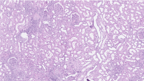
Figure 1. Low magnification view of emphysematous pyelonephritis: Image shows neutrophilic abscesses and glomerulosclerosis in adjacent kidney parenchyma.
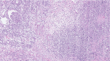
Figure 2. Low magnification view of emphysematous pyelonephritis: Image shows neutrophilic abscesses glomerulosclerosis in adjacent kidney parenchyma.
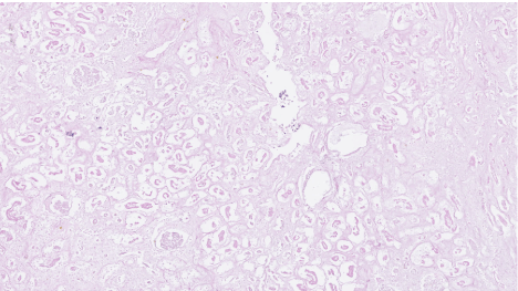
Figure 3. Low magnification view of emphysematous pyelonephritis: Image shows emphysematous cystic spaces with no definite lining, acute tubular necrosis in adjacent kidney parenchyma.
Treatment and prognosis
EPN is associated with significant mortality rate which may go up to 25%. The most appropriate management of EPN is still controversial and there are conflicting reports regarding conservative management versus nephrectomy.
EPN is a rare, potentially life threatening condition. Clinical information in correlation with imaging and pathologic findings is crucial to clinch the diagnosis.
2021 Copyright OAT. All rights reserv
- Pontin AR, Barnes RD (2009) Current management of emphysematous pyelonephritis. Nat Rev Urol 6: 272-9. [Crossref]
- Huang JJ, Tseng CC (2000) Emphysematous Pyelonephritis Clinicoradiological Classification, Management, Prognosis, and Pathogenesis. Arch Intern Med 160: 797-805. [Crossref]
- Thomas AA, Lane BR, Thomas AZ, Remer EM, Campbell SC (2007) Emphysematous cystitis: a review of 135 cases. BJU Int 100:17. [Crossref]
- Khaira A, Gupta A, Rana DS, Gupta A, Bhalla A, et al. (2009) Retrospective analysis of clinical profile, prognostic factors and outcomes of 19 patients of emphysematous pyelonephritis. Int Urol Nephrol 41: 959–66. [Crossref]
- Tang HJ, Li CM, Yen MY, Chen YS, Wann SR, et al. (2001) Clinical characteristics of emphysematous pyelonephritis. J Microbiol Immunol Infect 34: 125–30. [Crossref]
- Narlawar RS, Raut AA, Nagar A, Hira P, Hanchate V, et al. (2004) Imaging features and guided drainage in emphysematous pyelonephritis: a study of 11 cases. Clin Radiol 59: 192–7. [Crossref]
- Michaeli J, Mogle S, Perlberg S, Heiman S, Caine M (1984) Emphysematous pyelonephritis. J Urol 131: 203–7
- Wan YL, Lee TY, Bullard MJ, Tsai CC (1996) Acute gas-producing bacterial renal infection: correlation between imaging findings and clinical outcome. Radiology 198: 433–8
- Nayeemuddin M, Wiseman OJ, Turner AG (2005) Emphysematous pyelonephritis. Nature Clinical Practice Urology 2:108-112.
- David G. Bostwick, Liang Cheng, Urologic Surgical Pathology.