Abstract
Background: The inferior turbinate, as part of the nasal valve area, plays a key role in directing the airflow and moisturizing and heating the inspired air. Excessive resection of the inferior turbinate leads to a significant reduction in heating and humidification of inhaled air. Different types of turbinate surgery are described in the current literature. Pyriform Turbinoplasty is a new endoscopically performed procedure which includes a submucosal reduction of the bone of the frontal process of the maxilla and the lacrimal bone yet it preserves the mucosal surface. This new surgical technique is able to improve nasal airflow without hampering nasal climatization.
Objective: Effects of Nasal Wall Lateralization and Pyriform Turbinoplasty on nasal climatization were analyzed using Computational Fluid Dynamics.
Methods: A three dimensional model of the nasal cavity and paranasal sinuses was created basing on the CT-scans of a patient with nasal obstruction and hypertrophy of the inferior turbinates. The simulation was performed using transient boundary conditions and a breathing cycle with a length of 4.2 seconds.
Results: Nasal resistance was reduced after performing Nasal Wall Lateralization and Pyriform Turbinoplasty. The main area of airflow and humidification increased and was elevated from the inferior to the medial turbinate. A homogeneous distribution of airflow around all nasal turbinates was observed. In contrast to many other techniques that include partial resection of the inferior turbinate, heating and humidifying of respiratory air during inspiration takes place in the entire nasal cavity postoperatively.
Conclusion: Pyriform Turbinoplasty and Nasal Wall Lateralization are endoscopic procedures that widen the nasal valve area without mucosal resection. Computational Fluid Dynamics prove that these procedures produce a homogeneous distribution of airflow along the inferior and middle turbinate. The Pyriform Turbinoplasty and the nasal wall lateralization substantially improve nasal breathing without altering nasal climatization essentially.
Introduction
Many different types of turbinate surgery have been described including partial and total turbinectomy, turbinoplasty techniques, electrocautery, laser cautery, radiofrequency, laser therapy, microdebrider submucous reduction and ultrasound turbinate reduction. There is no general agreement or recommendation which surgical technique is most effective in improving nasal breathing and not harming nasal physiology [1,2].
In previous studies it has been demonstrated that the nasal valve area, which includes the head of inferior turbinate, is the most influential region to effect humidification and heating of inspired air [3–5].
The nasal valve area is formed by the septum medially, the caudal margin of the upper lateral cartilages laterally, the floor of the pyriform aperture and, finally, the head of the inferior turbinate. It represents a three-dimensional space [6].
The inferior turbinate, as part of the nasal valve area, plays a key role in directing the airflow and moisturizing and heating the inspired air. The inferior turbinate increases nasal resistance and simultaneously enables the mucosa to come into direct contact with large amounts of inspired air. For that reason, all surgical procedures regarding the inferior turbinate influence intranasal air conditioning, in a positive or negative sense. Additionally, the effect of the nasal cycle must be taken into consideration [7].
Excessive resection of the inferior turbinate leads to a significant reduction in heating and humidification of inhaled air [8–10]. Nasal air conditioning is more influenced by medial than inferior resection of the turbinate. Yet nasal resistance curves reveal no relevant changes between these in virtual surgery [11]. Partial reduction of hypertrophic turbinates results in improved nasal aerodynamics, which is most evident following resection of the lower third [12]. Partial reduction of the inferior turbinate can maintain its heating capacity whereas extensive or total turbinate resection can lead to significant impairment in the heating function of the nose [13].
Mucosa-preserving surgical techniques in turbinate surgery are able to improve both nasal airflow and air conditioning that has been demonstrated in recent in-vivo measurements [14,15].
Pyriform Turbinoplasty is a new endoscopically performed procedure which includes a submucosal reduction of the bone of the frontal process of the maxilla and the lacrimal bone (Figure 1a and 1b) yet it preserves the mucosal surface. Due to this technique, part of the lateral margin of the nasal valve area is opened by forming a mucosal flap. The resection of bone in this area can be extended by a “Nasal wall lateralization”. Here, the lacrimal bone that joins the uncinate process behind the lacrimal duct as well as the base of the inferior turbinate and the edge of the maxilla at the rim of the pyriform aperture is removed. This new surgical technique is able to improve nasal airflow [16].
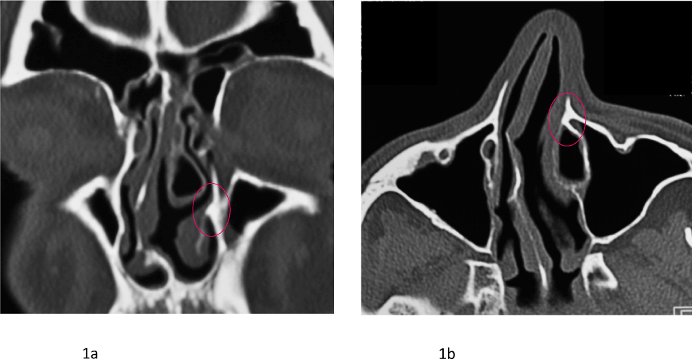
Figure 1. Figure 1a: A coronal CT-Scan showing the level of the “shoulder” of the inferior turbinate (red circle) and 1b the same area at the level of the pyriform aperture.
The tremendous advantage of this technique is minimal damage to the mucosa because the Pyriform Turbinoplasty is directed at the bone changing the architecture without removing any of the mucosa of the inferior turbinate. A significant improvement in ventilation after performing a Pyriform Turbinoplasty has been demonstrated [17]. The aim of this study was to analyze the effect of the Pyriform Turbinoplasty on intranasal heating and humidification of inspired air before and after surgery. This was realized by using Computational Fluid Dynamics (CFD) which are a valuable examination method regarding nasal physiology [5,7,18–20].
Materials and methods
Pyriform turbinoplasty
A 1 cm mucosal incision is made horizontally from the anterior lacrimal bulge towards the pyriform aperture. The head of the inferior turbinate is not easily lateralized as its bone is thick and often any lateralization is limited. The anterior aspect of the inferior turbinate is called the “shoulder” which comprises the frontal process of the maxilla and part of the lacrimal bone (Figures 1a and 1b). An inferior mucoperiosteal flap is folded downward by blunt dissection using a cottle elevator to expose the inferior part of the nasolacrimal canal and the maxillary conchal crest of the inferior turbinate (Figure 2a and 3b). This flap should ideally expose the piriform aperture.
A 3mm osteotome is used to remove the maxillary conchal crest of the inferior turbinate and the antero-medial bone of the inferior part of the nasolacrimal canal while the membranous of the nasolacrimal duct are preserved (Figures 2b and 3d). Only a gentle tap is needed to mobilize this fragment of bone before it is dissected free and removed. The lacrimal duct lies just behind this segment of bone and it is important not to damage this. The mucosa can then be replaced over this area (Figures 2c and 3c).
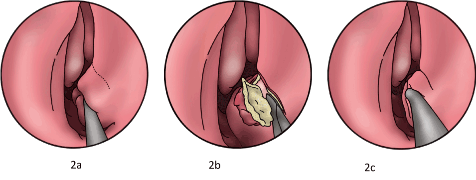
Figure 2. Diagrammatic representation of a pyriform turbinoplasty; left side. Figure 2a: To define where the incision should be made the inferior turbinate is lateralized as much as possible and this reveals the „shoulder“ which is too firm to outfracture with a freer‘s elevator. Figure 2b: A mucosal flap is elevated and an osteotome is used to mobilize the shoulder and a piece of thick bone is then removed. Figure 2c: The mucosal flap is replaced.
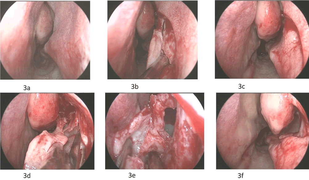
Figure 3. Preoperative view of the shoulder on the left side after a previous subtotal turbinectomy. The mucosal flap raised 3b, after bone removal the flap is replaced 3c. 3d Further elevation of the mucosal flap and exposure of the medial wall of the maxillary sinus. 3e Mobilizing bone with an osteotome. 3f Repositioning the mucosal flap after nasal wall lateralization in combination with a maxillary sinusotomy Type I.
Nasal wall lateralization
To gain space between the septum and the inferior turbinate one can continue the submucosal dissection more posteriorly to enable the entire inferior turbinate to be lateralized all the way back to its posterior attachment (Figure 4). Following a Pyriform Turbinoplasty the lacrimal duct is identified and pushed laterally to expose where the lacrimal fossa attaches onto the maxilla. The subperiosteal dissection can be extended more posteriorly by following the medial wall of the maxillary sinus until you reach the posterior attachment of the inferior turbinate to the pterygoid plate. The exposed bone posterior, lateral and inferior to the lacrimal duct is then freed using an osteotome. With gentle dissection this bone can be removed and then the mucosal flap can be repositioned (Figures 3d and 3f). This bone comprises the junction of the lacrimal fossa with the inferior insertion of the uncinate process and medial wall of the maxillary sinus. This results in a more substantial widening of the lateral nasal wall from the piriform aperture all the way back to the pterygoid bone.
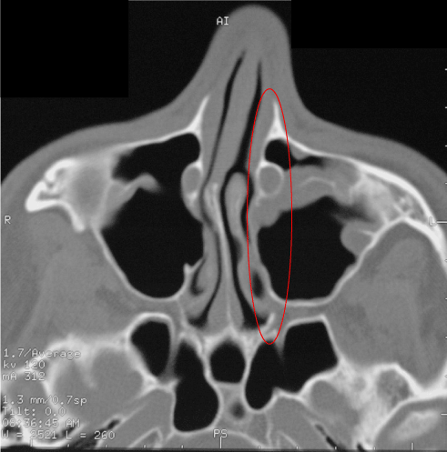
Figure 4. A medialized lateral nasal wall (red circle) on the left side shows assymmetry and narrowing.
If there is an indication for a maxillary sinusotomy this technique can be extended to expose the primary ostium of the maxillary sinus in what is called an anterior approach to the sinus ostium (Figure 3e). This allows a maxillary sinus ostioplasty to be done by the submucosal removal of bone from the medial wall of the sinus as well as creating mucosal flaps that can cover any exposed area inferiorly. It is an elegant way of lowering the maxillary sinusotomy and preserving the overlying mucosa. This provides an entrance through which it is possible to visualize and operate on the maxillary sinus with straight instruments. At the end of this procedure when mucosal flaps have been placed in position, check that the maxillary sinusotomy is lower than the middle turbinate. The advantages of this procedure are direct access and visualisation of the ostium and by removing the maxillary conchal crest the area is enlarged.
Computational fluid dynamics model
A three dimensional virtual model of the nasal cavity and paranasal sinuses was created basing on the CT-scans of a patient with nasal obstruction and hypertrophy of the inferior turbinates. Using these both a preoperative model and a postoperative one could be created. Humidity and temperature were defined in order to simulate physiological conditions. Using a physiological breathing cycle these properties varied over time. The boundary conditions were based on our own in vivo measurements [5,8,9,21,22]. A GE Medical Systems Discovery 690 Scanner with an axial slice thickness of 0.63 mm was used to perform the CT-Scan. Segmentation was performed using the software ITKSnap V. 3.0.0. In the end, a mesh with 3.1 million connected cells represented the finished virtual model (Figure 5). The Computational Fluid Dynamics-Calculation was performed using the ANSYS Fluent solver (ANSYS Workbench 14). To reproduce the physiological breathing cycle the following parameters were chosen. Inspired air had a temperature of 20°C and 30% relative humidity. Expired air was simulated at a temperature of 37°C and 100% relative humidity. The nasal wall temperature of the virtual model that represented the mucosal surface had a time-dependent temperature of 30-35°C and a moisture content of 100% assuming a continuous mucosal surface. The simulated sequence assumed that inspiration took 2 seconds and expiration 2.2 seconds. Two breathing cycles of 4.2 seconds were analyzed.
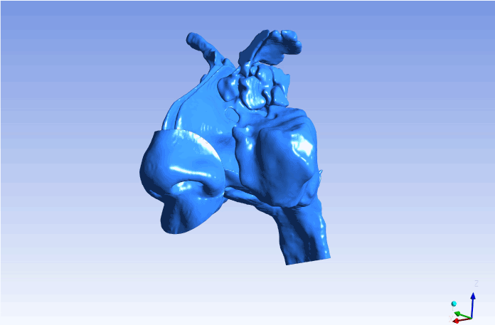
Figure 5. Three dimensional model based on the CT-Scan of the patient.
Results
As demonstrated in numerous previous studies, the highest velocity during inspiration was found in the nasal valve area or the transition area at the nasopharynx. Velocities of around 2 meters per second were demonstrated in the preoperative images. After performing the nasal wall lateralization and Pyriform Turbinoplasty velocities were decreased below 1.7 meters per second. The main flow area medial of the inferior turbinate and around the middle turbinate showed lower flow-speeds compared to the preoperative findings. The cross sectional area in which maximum flow speed was reached decreased significantly (Figure 6).
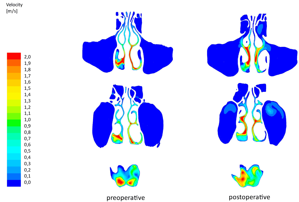
Figure 6. Coronal slices showing the nasal flow during maximum inspiration. The preoperative situs is visible on the left, the postoperative one on the right side.
Temperature within the maximum flow area in the middle of the nasal cavity was 26.5°C and reached nearly 33°C in the nasopharyngeal area and occurred during maximum inspiration (Figure 7). Compared to the preoperative model the temperature did not decrease significantly. Relative humidity reached 87% within the maximum flow area in the nasopharynx compared to 93% preoperatively (Figure 8). The main area of airflow and humidification increased from the medial to the inferior turbinate to those immediately below the middle turbinate. In summary a homogeneous distribution of airflow around all nasal turbinates was observed. In contrast to many other techniques which include partial resection of the inferior turbinate, heating and humidifying of respiratory air during inspiration takes place in the entire nasal cavity postoperatively.
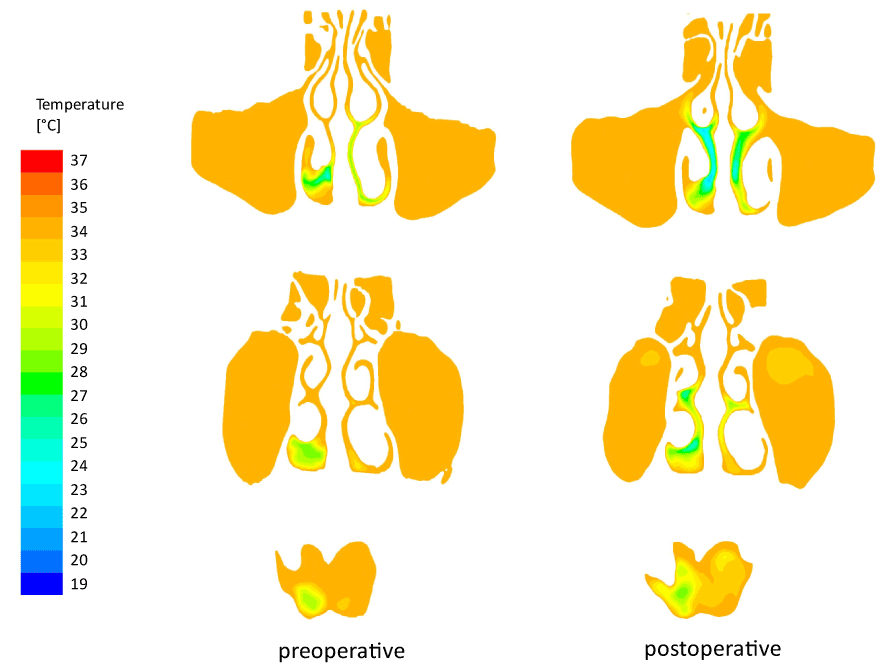
Figure 7. Coronal slices showing the temperature distribution during maximum inspiration.
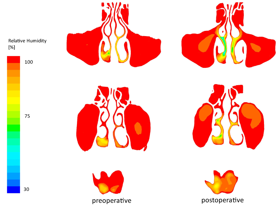
Figure 8. Coronal slices showing the humidity distribution during maximum inspiration.
The patient who underwent the Pyriform Turbinoplasty and nasal wall lateralization was free of complaints 4 weeks after surgery. Nasal blockage and dryness of the nose were not reported any more.
Discussion
The inferior turbinates are responsible for important functions including heating and humidification of the inspired air. Most of the applied techniques in turbinate surgery, especially partial or total resections, lead to a deterioration in nasal air conditioning [8–10].
A change in nasal air conditioning can be observed after any surgical intervention of the nasal turbinates. Excessive resection of these structures leads to nasal dryness with crusting and paradoxically a reduction in the sensation of airflow despite of an increased airflow.
Because the inferior turbinate is part of the nasal valve area, it is responsible for directing the incoming airflow. Extensive resection of the inferior turbinate results in an altered nasal valve and the detour of airflow along through the inferior meatus [8,9]. Patients often complain about the subjective feeling of a blocked nose that is caused by dryness of the nasal mucosa.
After resection of the2021 Copyright OAT. All rights reservuced contact of inspired air with mucosa is observed. This is a result of a more laminar airflow and a “channel” which transports most of the air. This strictly laminar airflow within the nasal cavity can be responsible for deterioration in the heat and water exchange between air and surrounding mucosa as demonstrated in previous numerical simulations [3-5]. These results confirm the close relationship between airflow and climatization regarding turbinate surgery.
To improve the bottleneck “nasal valve” all boundaries should be considered. This region is surrounded by the head of the inferior turbinate, the septum, the margins of the upper lateral cartilage, and the lateral aspect of the pyriform aperture. It is highly variable regarding the position and thickness of the frontal process of the maxilla, the lacrimal bone, the rim of the pyriform aperture and the anterior bony aspect of the inferior turbinate. The “Pyriform Turbinoplasty” is a new endoscopic technique that improves airflow by widening the nasal valve area without impairing physiological functions. This technique is based on the resection which makes up the bony structures of the lateral aspect of the pyriform aperture.
Theoretically the bony canal of the lacrimal duct can be damaged when resecting the shoulder of the inferior turbinate. In our experience this did not happen due to the thick and solid bone in this region. The mucosal flap which is raised to expose the bony shoulder should be wide enough to prevent wound healing disorders which might cause scarring close to the nasal valve area.
One indication for surgery to this area is narrowing of the nasal valve area where the pyriform aperture impinges on the nasal airway or mucosal hypertrophy restricts the nasal valve area in spite of medical treatment.
Of course, there are many surgical techniques to widen the nasal passage that do not affect nasal climatization severely when performed carefully. A septoplasty combined with submucosal anterior inferior turbinoplasty is a decent method to improve nasal flow. Most of modern procedures affect the mucosa of the lateral side of the turbinate or aim for a volume reduction by submucosal heating which again may lead to scarring and hypertrophy. The Pyriform Turbinoplasty does not affect the mucosa at all as it just reduces the bony shoulder of the inferior turbinate and therefore leads to a lateral widening of the nasal passage. Of course, this technique can be combined with a septoplasty when indicated.
Numerical simulation of airflow already demonstrated an improvement in airflow patterns and a reduction in nasal resistance within the entire nasal cavity after performing a Pyriform Turbinoplasty [17]. The airflow is distributed in an improved physiological manner as well as preserving the turbinate complex.
In contrast to total and even partial resections of the inferior turbinates nasal wall lateralization in combination with Pyriform Turbinoplasty did not adversely affect the nasal climatization process. The humidity and temperature of inhaled air was marginally lower when compared to the preoperative situation but distribution of airflow around the turbinates still was homogenously. Heating and humidifying during inspiration takes place within the whole nasal cavity.
In vivo studies support a negative correlation between mucosal temperature and nasal resistance measured by rhinomanometry. Changes in nasal patency influence nasal mucosal temperature. Within this context, nasal thermoreceptors might play an important role concerning the perception of nasal patency [23]. The high influence of mucosal cooling on nasal airway obstruction before and after surgery has been described by Sullivan et al. [24] by means of Computational Fluid Dynamics.
A higher nasal resistance usually leads to better humidification and warming as there is much more contact between air and mucosa. As we could demonstrate, the Pyriform Turbinoplasty lowered nasal resistance without influencing nasal climatization.
Conclusion
Pyriform Turbinoplasty and nasal wall lateralization are endoscopic submucosal procedures that widen the nasal valve area without any mucosal resection. Computational fluid dynamics prove that these procedures lead to a homogeneous distribution of airflow along the inferior and middle turbinate. Heating and humidifying of inspired air can take place within the entire nasal cavity. The Pyriform Turbinoplasty and the nasal wall lateralization substantially improve nasal breathing without altering nasal climatization essentially.
Acknowledgements
Fabian Sommer, Daniel Simmen are equally contributing authors.
References
- Hol MK, Huizing EH (2000) Treatment of inferior turbinate pathology: a review and critical evaluation of the different techniques. Rhinology 38: 157–166. [Crossref]
- Passàli D, Passàli FM, Damiani V, Passàli GC, Bellussi L (2003) Treatment of inferior turbinate hypertrophy: a randomized clinical trial. Ann Otol Rhinol Laryngol 112: 683-688. [Crossref]
- Lindemann J, Keck T, Wiesmiller K, Sander B, Brambs HJ, et al. (2006) Nasal air temperature and airflow during respiration in numerical simulation based on multislice computed tomography scan. Am J Rhinol 20: 219–223. [Crossref]
- Lindemann J, Keck T, Wiesmiller K, Sander B, Brambs HJ, et al. (2004) A numerical simulation of intranasal air temperature during inspiration. Laryngoscope 114: 1037-1041. [Crossref]
- Sommer F, Kroger R, Lindemann J (2012) Numerical simulation of humidification and heating during inspiration within an adult nose. Rhinology 50: 157-164. [Crossref]
- Kern EB (1978) Surgical approaches to abnormalities of the nasal valve. Rhinology 16: 165-189. [Crossref]
- Patel RG, Garcia GJM, Frank-Ito DO, Kimbell JS, Rhee JS (2015) Simulating the nasal cycle with computational fluid dynamics. Otolaryngol Head Neck Surg 152: 353–360. [Crossref]
- Lindemann J, Keck T, Wiesmiller KM, Rettinger G, Brambs HJ, et al. (2005) Numerical simulation of intranasal air flow and temperature after resection of the turbinates. Rhinology 43: 24–28. [Crossref]
- Lindemann J, Brambs HJ, Keck T, Wiesmiller KM, Rettinger G, et al. (2005) Numerical simulation of intranasal airflow after radical sinus surgery. Am J Otolaryngol 26: 175–180. [Crossref]
- Lindemann J, Leiacker R, Sikora T, Rettinger G, Keck T (2002) Impact of unilateral sinus surgery with resection of the turbinates by means of midfacial degloving on nasal air conditioning. Laryngoscope 112: 2062-2066. [Crossref]
- Hariri BM, Rhee JS, Garcia GJ (2015) Identifying patients who may benefit from inferior turbinate reduction using computer simulations. Laryngoscope 125: 2635–2641. [Crossref]
- Chen XB, Leong SC, Lee HP, Chong VF, Wang DY (2010) Aerodynamic effects of inferior turbinate surgery on nasal airflow--a computational fluid dynamics model. Rhinology 48: 394–400. [Crossref]
- Chen XB, Lee HP, Chong VF, Wang de Y (2010) Numerical simulation of the effects of inferior turbinate surgery on nasal airway heating capacity. Am J Rhinol Allergy 24: e118-122. [Crossref]
- Wiesmiller K, Keck T, Rettinger G, Leiacker R, Dzida R, et al. (2006) Nasal air conditioning in patients before and after septoplasty with bilateral turbinoplasty. Laryngoscope 116: 890-894. [Crossref]
- Rozsasi A, Leiacker R, Kühnemann S, Lindemann J, Kappe T, et al. (2007) The impact of septorhinoplasty and anterior turbinoplasty on nasal conditioning. Am J Rhinol 21: 302-306. [Crossref]
- Simmen D, Jones N (2013) Manual of Endoscopic and Skull Base Surgery. Thieme Verlag.
- Simmen D, Sommer F, Briner HR, Jones N, Kröger R, et al. (2015) The effect of "Pyriform Turbinoplasty" on nasal airflow using a virtual model. Rhinology 53: 242-248. [Crossref]
- Ishikawa S, Nakayama T, Watanabe M, Matsuzawa T (2006) Visualization of flow resistance in physiological nasal respiration: analysis of velocity and vorticities using numerical simulation. Arch Otolaryngol Head Neck Surg 132: 1203–1209. [Crossref]
- Tarabichi M, Fanous N (1993) Finite element analysis of airflow in the nasal valve. Arch Otolaryngol Head Neck Surg 119: 638-642. [Crossref]
- Keck T, Lindemann J (2010) Numerical simulation and nasal air-conditioning. GMS Curr Top Otorhinolaryngol Head Neck Surg 9: Doc08. [Crossref]
- Pless D, Keck T, Wiesmiller KM, Lamche R, Aschoff AJ, et al. (2004) Numerical simulation of airflow patterns and air temperature distribution during inspiration in a nose model with septal perforation. Am J Rhinol 18: 357–362. [Crossref]
- Pless D, Keck T, Wiesmiller K, Rettinger G, Aschoff AJ, et al. (2004) Numerical simulation of air temperature and airflow patterns in the human nose during expiration. Clin Otolaryngol Allied Sci 29: 642-647. [Crossref]
- Lindemann J, Keck T, Scheithauer MO, Leiacker R, Wiesmiller K (2007) Nasal mucosal temperature in relation to nasal airflow as measured by rhinomanometry. Am J Rhinol 21: 46-49. [Crossref]
- Sullivan CD, Garcia GJM, Frank-Ito DO, Kimbell JS, Rhee JS (2014) Perception of better nasal patency correlates with increased mucosal cooling after surgery for nasal obstruction. Otolaryngol Head Neck Surg 150: 139–147. [Crossref]