Abstract
Introduction: Cytodiagnosis of basal cell carcinoma (BCC) is easy to perform with different techniques. It is very useful in an out-patient setting as a rapid diagnosis method when a surgical excision is planned.
Material and methods: We carried out a prospective, observational and blinded to observer study. Cytologies of suspected BCCs were performed and compared with the classical gold-standard histological diagnosis. Three different cytological techniques were compared, scraping, scratching and imprinting. The results of Papanicolau and Diff-Quick® stains were also compared.
Results: A total of 83 tumors were studied, 58 were confirmed BCC histologically. Most of them were located in the head and neck area and were nodular. Diff-Quick exhibited better sensitivity than Papanicolau (84% vs. 69%, p >0.05). There were no significant differences in sensitivity between scraping and scratching (71% vs. 76%, p>0.005), but imprinting had lower results (31%, p>0.05). The best sensitivity combination was achieved with scratching technique and Diff-Quick stain (86%).
Discussion: Cytology appears as a simple method with good accuracy for rapid diagnosis. Nevertheless the sensitivity and specificity achieved in our study were lower than previous reports, may be due to inadequate sampling. This is the first study comparing two different stains and demonstrating the superiority of Diff-Quick to Papanicolau. The three different cytological techniques were not previously compared with better results obtained with scratching.
Conclusions: Cytology is a useful technique for the rapid diagnosis of BCC especially prior to surgical excision. Further studies are necessary to assess the accuracy of cytodiagnosis for BCC with adequate trained investigators.
Key words
basal cell carcinoma, cytodiagnosis, cytology, Diff-quick, Papapanicolau
Introduction
Cytology is a very simple and inexpensive technique although it is not used in current dermatological practice. Basal cell carcinoma (BCC) is the main indication for cytological diagnosis because of its high frequency and the high diagnosis reliability achieved by previous reports [1,2]. Cytology allows early diagnosis prior to conservative treatments as cryotherapy or topical treatments, in debilitated patients or when a punch biopsy is not indicated. Some investigators stated that the cytological findings of BCC are pathognomonic [3]”.
In a meta-analysis of the studies published of cytological diagnosis in dermatology the global sensitivity calculated for this technique was 97% (94-99%) with an 86% of specificity (80-91%) [4]. Cytodiagnosis of tumors can be performed by different techniques and different stains. The cells can be obtained by scrapping the tumor surface, by scratching a part of the tumor with a scalpel or by imprinting the tumor in a glass slide after swabbing the tumor using a curette. Also different stains have been used [5-10]. In this study we compared the results of these three different techniques for obtaining tumor cells (scraping, scratching and imprinting) and two stains (Papanicolau and Diff-Quick®) with the histopathological diagnosis in BCC.
Material and methods
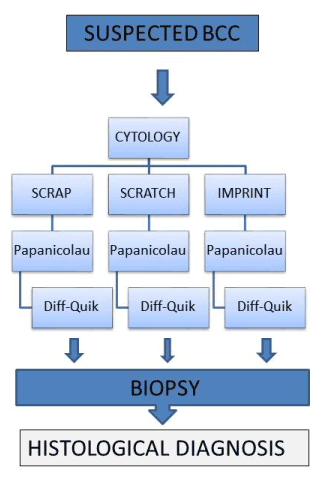
We carried out a prospective and observational study. The study was approved by the ethical committee of the hospital and all the patients included signed informed consent. Patients with clinically suspected BCC previous to surgical excision were selected in an out patients setting. The age, sex, site and size (cm) of the tumors were recorded. Once in the operating room, prior to extirpation, cytology of the tumor was performed after washing the surface with saline. The cytology was performed by three different techniques always in the same order: scrapping the tumor surface with a scalpel, scratching a part of the tumor with a scalpel or imprinting the tumor in a glass slide. The material obtained was extended in the slide with the scalpel blade trying to separate cell groups without smashing them avoiding damage. The purpose was to obtain a thin layer of visible cells without accumulation of erythrocytes. Imprinting was done supporting the glass slide on the tumor. Each of these procedures was applied in different sites of the tumor. Two glass slides were obtained in each site, one to be stained with Diff-Quick® and the other with Papanicolau (Figure 1). All the obtained material was fixed in the glass slide with a “citospray” (Labofix®) previous to staining. The assessment of the cytology was always done by the same pathologist trained in cytodiagnosis and blinded to the clinical suspected diagnosis. The cytological examination was classified in four different categories: positive for BCC, probably BCC, negative for BCC and insufficient for diagnosis. The results of cytological diagnosis were compared with the histological examination considered to be the gold standard. The sensitivity, the specificity and the accuracy of the different techniques and stains of the cytology were calculated with the Z-test.

Figure 1. Summary of the sampling of the study.
Results
A total of 83 lesions suspected for BCC were included in the study, 58 of them were confirmed histologically as BCC. Among the tumors not confirmed as BCC the diagnosis were Bowen´s disease, solar elastosis, squamous cell carcinoma, epidermoid cyst, actinic keratosis, seborrheic keratosis and pilomatrixoma. The characteristics of the patients and tumors are summarized in Table 1. The 58 BCC were found in 27 males and 31 females with a mean age of 71, 60 years (range 34-91) with a medium size of 1.36 cm (0.5-4 cm). Most of them were located in head and neck (46/58= 79%) and were nodular type BCC (52/58= 90%). The 25 not BCC tumors were found in 12 males and 13 females with a mean age of 79.08 (range 61-94), a medium size of 2.21 cm (0.5-5 cm), most of them located in head and neck (21/25=84%). The results of the accuracy of cytodiagnosis by stain and technique are summarized in table 2. The positive and probably BCC categories were classified as “positive” and the negative or insufficient categories were classified as “negative” for the analysis of the data. The results of the analysis for sensitivity and specificity are also summarized in Table 2. In our study, Diff- Quick® stain had better sensitivity than Papanicolau, 84% vs. 69% (p>0.005). Scrapping and scratching had similar sensitivity (71% and 76%, p<0.05) and both were better than imprinting. If Papanicolau stain and diff-quick would have been used together, the sensitivity obtained would not be higher than diff-quick alone (84% vs. 84%, p>0.05). The combination of scraping and scratching would not have been better than scratching alone (76% vs. 76%, p>0.05). So the best sensitivity was obtained by scratching the tumors and staining with Diff-Quick® (86%). The specificity was low in general with the exception of imprinting (96%) and scratching (72%).
Table 1. Characteristics of the patients and the tumors examined by cytological diagnosis
N=83 Suspected Basal Cell Carcinoma |
Histological Examination |
N=58 BCC |
N=25 NOT BCC |
Male/Female |
27/31 |
12/15 |
Mean age/ range |
71,60 (34-91) |
79,08 (61-94) |
Medium size /range |
1,36 cm (0,5-4 cm) |
2,21 (0,5-5 cm) |
Localization
|
46 (79%)
12 (21%) |
21 (84%)
4 (16%) |
Histological diagnosis |
-Nodular BCC 52 (90%)
-Superficial BCC 6 (10%) |
-Bowen disease [7]
-Elastosis [7]
-Squamous cell carcinoma [6]
- Epidermoid cyst [2]
-Actinic keratosis [1]
-Seborreic keratosis [1]
-Pilomatrixoma [1] |
Table 2. Results of the accuracy of cytodiagnosis by stain used and technique performed
Diagnosis
Technique |
Sensitivity |
Specificity |
P values |
PAPANICOLAU |
0.69 (0.57-0.81) |
0.64 (0.45-0.83) |
P >0.05 |
DIFF-QUIK |
0.84 (0.75-0.94) |
0.56 (0.37-0.75) |
SCRAP |
0.71 (0.59-0.82) |
0.64 ( 0.45-0.83) |
P<0.05 |
SCRATCH |
0.76 (0.65-0.87) |
0.72 (0.54-0.9) |
IMPRINT |
0.31 (0.17-0.45) |
0.96 (0.87-1.04) |
P >0.05 |
PAPANICOLAU+ DIFF QUIK |
0.84 (0.75-0.94) |
0.56 (0.37-0.75) |
P <0.05 |
SCRAP+SCRATCH |
0.76 (0.65-0.87) |
0.72 (0.52-0.9) |
P< 0.05 |
DIFF QUICK + SCRATCH |
0.86 (0.77-0.95) |
0.72 (0.54-0.9) |
|
Discussion
Cytological examination is easy to perform but it is not used widely by dermatologists. Cytodiagnosis has many advantages, it is simple, cost-effective, it could be performed in the initial visit, it allows pre-surgical diagnosis and it could be done even when a punch biopsy is inappropriate (cosmetic impact or elderly patients with comorbidities) [4].
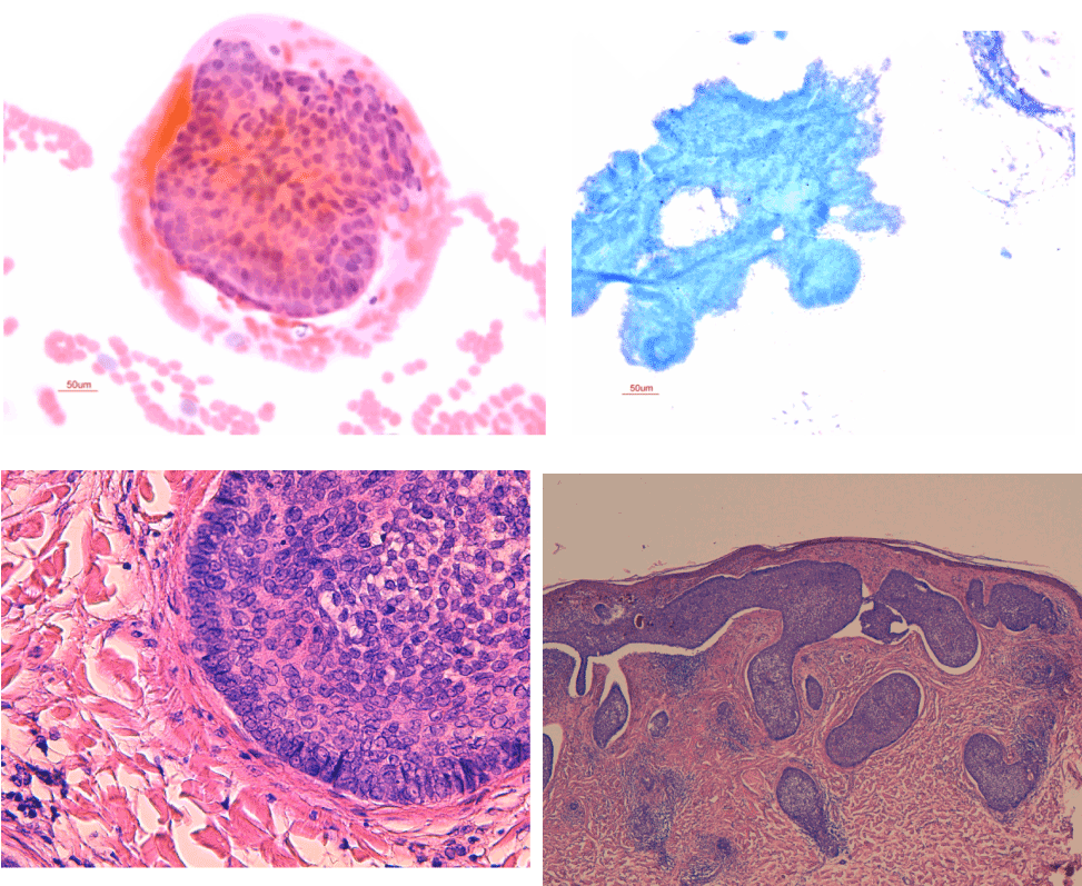
A possible use of cytodiagnosis in BCC is because of the high frequency of the tumor and the high degree of diagnosis reliability in previous studies with an estimated global sensitivity of 97% [4]. Furthermore, the cytological pattern of BCC has high sensitivity with numerous clusters of basaloid cells packed together. The palisade arrangement seen in histological slides of BCC is easier to recognize in the periphery of the groups of basaloid cells [3] (Figures 2 and 3). However cytodiagnosis of BCC is unable to differentiate BCC from other adnexal tumors like trichoepithelioma [8] and different subtypes of BCC [4]. Other limitation of cytology are that an adequate sampling is required with enough material, minimal contamination with blood, and optimal fixation, the dermatologist should carefully sample the material and the pathologist should be well trained in cytodiagnosis.
2021 Copyright OAT. All rights reserv

Figure 2. At the top in the left side: lusters of palisade basaloid cells (Diff-Quick®, 30x), and in the right side: tipical image of clusters of cells of BCC (Papanicolau, 30x). At the bottom histological images of the same BCC ( left 100X and right 30x
In our study the sensitivity and the specificity is lower than in the previous studies published [4-10]. Even with the best combination, scratch and Diff-Quick®, the sensitivity reached was 86% (77-95) and the specificity 72% (54-90). The data of a meta-analysis recently published based in eight studies and 1261 BCCs estimated the sensitivity and the specificity of cytodiagnosis in 87% (94-99) and 86% (80-91%) respectively. As the pathologist technicians perform cytodiagnosis staining routinely, we rather think that the dermatologists may not have been keen in obtaining the adequate sampling. A good sample with high cellularity and minimal blood and keratin scames (from cornified layer) contamination was essential for a correct diagnosis. In fact, when diagnosis could not be stablished in our study, the most common reason was “insufficient for diagnosis” or “hemorrhagic material that not allows diagnosis”.
This is the first study comparing different techniques to perform cytology in BCC and various stains. Imprinting has the advantage of less blood contamination and the disadvantage of less celullarity. Scraped and scratched samples were more cellular (and more hemorrhagic) and yielded minimal tissue fragments, very helpful for diagnosis. Among the three techniques studied, scraping and scratching proved the most favorable results with a slight superiority of scratching (p<0.05)
The inferiority of Papanicolau (Figure 3) in cytodiagnosis of BCC achieved in our study confirms the results of previous studies which obtained better results with Giemsa and its variants, like Diff-Quick® [1,3,10] ( Figure 2). Cytological details could be evaluated both in Diff-quick® and Papanicolau samples. Cohesive clusters with peripheral palisading were, in our experience, most easily identified in Diff-quick stain®, which takes the advantage of the possibility of immediate review while patient is in the consulting room [4]. Papanicolau stained seems to be more suitable for smears in which squamous differentiation is in doubt [10-14]. Parallel analysis in our study revealed that the best combination for cytodiagnosis of BCC in our group of patients was scratching and Diff-Quick®.
As basal cells rarely appear in smears from normal skin [11] diagnosis was established on identification of basal cells (small hyperchromatic cells with round or oval nuclei in cohesive sheets with indistinct cell borders and peripheral palisading) [11,12] assuming difficult cytological differentiation between basaloid benign and malignant lesions. Only one of our cases consisted on a benign basaloid lesion (pilomatrixoma) and smear was labeled as negative for malignancy.
Differentiate basal cell lesions form well differentiated squamous cell carcinoma is, in our experience, usually straightforward -in good smears- as desquamated well differentiated carcinoma cells are dyscohesive and shows evidence of squamous differentiation (abundant smooth and dense cytoplasm filled with keratin). Four of our cases were squamous carcinoma. One was correctly labeled as squamous cell carcinoma and three others were unsatisfactory for diagnosis because of consisting of blood and anucleated keratin squames only. None of our cases were moderately or poorly differentiated squamous cell carcinomas, in which case absence or minimal keratinization may pose more diagnostic problems. Nevertheless, experience with cytological smears from fine needle aspiration shows that, in these tumors, cells retain some specific characters that allow distinction from basaloid neoplasms such as large clusters of elongated cells with large nuclei, coarse chromatin texture and prominent nucleoli [13].
Concerning actinic keratosis, we think that, as occurs in other squamous intraepithelial lesions, if the whole epithelium is involved (KIN III), smears can look very similar to squamous cell carcinoma, although the background will be cleaner. On the other hand, when epithelial atypia is limited to lower epidermal stratum (KIN I) and superficial cells retain maturation, differential diagnosis with other benign entities with associated parakeratosis may be difficult. Only one of our cases was histologically actinic keratosis (KIN I) and smear was labeled as negative.
Actinic keratosis cells, on the other side, are not basaloid, so differential diagnosis from basal cell carcinoma is not difficult in our experience, although a case of actinic keratosis with basal cell hyperplasia misdiagnosed as basal cell carcinoma has been reported [11].
Some authors have found seborrheic keratosis smears quite different from basal cell carcinoma because of the presence of exfoliated superficial squamous cells and horn cysts [11]. We registered a false positive BCC diagnosis with an irritated seborrheic keratosis. In our opinion, as seborrheic keratosis consists basically in greatly increased epidermal basal cells as a result of maturation defect, cytological smears can be very similar to BCC ones (even more if horn cysts are not identificated) and confusion between both entities is possible.
Cytological diagnosis of basal cell carcinoma versus Bowen disease depends on the specific type of the last one. So, distinction from pagetoid variant (cells with clear cytoplasm) is easier while verrucous-hyperkeratotic or papillated variants are more difficult to differentiate from squamous cell carcinoma. Four of our cases corresponded histologically to Bowen disease. One of them was labeled as suspicious for malignancy (BCC), another two ones were labeled as squamous carcinoma and one was not adequate for diagnosis because of technical default.
Benign tumors originating in skin adnexal epithelium (tricoepithelioma, pilomatrixoma), the differential diagnosis is more troublesome because of the presence of similar basaloid aggregates [11,14]. Differential diagnosis can be extremely difficult in smears containing few cells (as well as in small biopsies). Particularly Pilomatrixoma has morphological features similar to BCC and must be ruled out when in doubt [14]. Only one of our cases consisted on a benign basaloid lesion (pilomatrixoma) and smear was labeled as negative for malignancy.
With respect to technical details, needless to say in any case, a good sample with high cellularity and minimal blood and keratin scames (from cornified layer) contamination was essential for a correct diagnosis. In fact, when diagnosis could not be stablished in our study, the most common reason was “insufficient for diagnosis” or “hemorrhagic material that not allows diagnosis”. Cytodiagnosis of BCC has significant potential for the dermatologist and allows easy, cheap and rapid diagnosis in out-patient setting. Cytodiagnosis of BCC is very useful when destructive therapies are planned but cannot differentiate tumor subtypes. Maybe the ideal use of this technique is by a trained dermatologist or pathologist in situ as to avoid the insufficient material for diagnosis sampling that unable further material obtention. However cytodiagnosis of BCC is no implemented as a routine technique for dermatologists and further studies are necessary to improve this promising technique.


