Background: Squamous cell carcinoma (SCC) arising from the nasal vestibule is a rare condition accounting for about 1% of head and neck malignancies. SCC develops as a result of exposure to sunlight and ultraviolet radiation, toxic agents, scars, compromised immune status, and genetic defects. It is characterized by several particularities concerning clinical presentation, staging and treatment. Through this case presentation, we report an unusual mode of revelation of a SCC of the collumella, eventually, we discuss through a brief review of the literature its various staging and management features.
Case presentation: We report a rare case of cutaneous squamous cell carcinoma of thecollumella in a 43-year-old Moroccan male who presented at first for an abscess of the columella of 10 days duration. After incision-drainage of the abscess, the histological examination of the biopsy specimen revealed cutaneous SCC. Complete surgical resection and bilateral lymph node dissection were performed. Reconstruction of columella defect was accomplished by a forehead flap after confirming disease-free surgical margins. Later, the patient underwent radiation therapy. There were no signs of recurrence or metastases at 3 years postoperatively.
Conclusions: despite being superficial and accessible, SCC’s diagnosis may be late due to its unspecific clinical presentation which causes an enormous problem of surgical management, because the removal of these tumors emerging in the face and the reconstruction of the extensive mutilations that follow, result in disastrous aesthetic outcomes and social consequences. Hence, the need to recognize symptoms in order to diagnose early for a better management and less damages.
Cutaneous squamous cell carcinoma, nasal vestibule, columella, surgical resection, forehead flap, radiotherapy, case report.
Cutaneous squamous cell carcinoma (SCC) is a typical non-melanoma skin cancer occurring in keratinocytes and the second most common skin cancer. It has a more invasive growth pattern, higher recurrence rate, and higher potential to metastasize than basal cell carcinoma (BCC) [1]. It mostly affects areas exposed to sunlight (face, neck). Through this case report, we report an unusual mode of revelation of a SCC of the face.
A 43-year-old male, originated from morocco, with no relevant medical history, presented to the ENT emergency department for an abscess of the columella evolving for 10 days. On physical examination, there was a fistulized abscess of the columella, and cervical examination found bilateral submandibular adenopathies (Figure 1). An incision-drainage was carried out, and the histological examination of the biopsy specimen revealed cutaneous squamous cell carcinoma. There was no evidence of perineural or vascular involvement. Nasoendoscopy resulted normal. A multidisciplinary (Otolaryngologist, Oncologist) decision concluded to perform a surgical excision with safety margin estimated to 5 mm, and bilateral lymph node dissection. Reconstruction of columellar defect was accomplished by a forehead flap after confirming disease-free surgical margins. CT scan of the chest and abdomen did not reveal any distant metastasis. General postoperative cosmetic result was good. The reconstructed columella had a proper width and satisfactory height and depth. Later, the patient has undergone radiation therapy sessions. An outpatient Follow-up was conducted 2, 4 and 6 months and 1, 2, and 3 years after discharge, with no evidence of the disease.
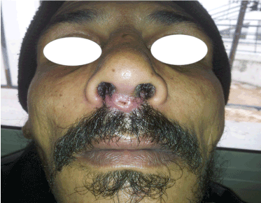
Figure 1. Squamous cell carcinoma of the columella revealed by an abscess.
Squamous cell carcinoma arising from the nasal vestibule is a rare condition accounting for about 1% of head and neck malignancies with several particularities concerning both staging and treatment [2]. It develops as a result of exposure to sunlight and ultraviolet radiation, toxic agents such as arsenic, scars caused by burns or injuries, chronic ulcer, extension of life expectancy, compromised immune status and genetic defects [3,4]. Certain viruses have been involved in the carcinogenesis of this tumor [5]. Men are twice affected as women.
In most cases, cutaneous SCC develops on precancerous lesions (actinic keratosis)or cancer in situ that appears as an erythematous nodule, with central ulceration, firm in palpation, often with irregular borders, covered with crusts bleeding easily on contact.
Local invasion concerns inevitably both surface as well as depth of the surrounding tissues, with a particular predilection for perineuralinvasion [6]. Lymph involvement is frequent which requires a systematic assessment of lymph node areas. Hematogenous spread of cancerous cellsresults in distant metastasis with the most frequent secondary locations: lung, liver, bones (especially the vertebra, the sternum, and the skull) and brain, in a decreasing order of frequency.
The biopsy or excision-biopsy allows the diagnosis of SCC through histological examination that informs on different features of the tumor: the degree of differentiation: keratinizing, non-keratinizing, well, moderately or poorly differentiated or anaplastic. The low- and high-risk of patients with SCC are classified according to tumor size and location, histological classification, tumor characteristics, and invasion of surrounding tissues [7].
The Wang T staging system showed its superiority on T&N staging used by the American Joint Committee on Cancer (AJCC) and seems at present to be the most adequate for SCC of the nasal vestibule. According to many authors, this staging system predicts prognosis of nasal vestibule tumors more accurately [8-11]. It is relatively simple and includes only three stages (T1: involving only vestibule skin; T2: invading subcutaneous tissue and cartilage; T3: invading bone).
Surgical resection is the treatment of choice for SCC. Taking into consideration the tumor size, it includes the tumor and a surrounding safety margin. However, controversy remains over what constitutes an adequate safety margin. Brodland and Zitelli recommend 4 mm for a low-risk tumor and 6 mm for a high-risk tumor [12]. Guidelines from the National Comprehensive Cancer Network recommend 4-6 mm for low-risk tumors and 10 mm for high-risk tumors [7]. Many plastic and reconstructive surgeons have focused only on the horizontal safety margin and surrounding tissues, whereas, Sam Yong Lee has established a formulae determining the vertical safety margin through an evaluation of the proportional relationship between the horizontal size and vertical depth of SCC [4]. Tumor depth is considered as the most important factor in predicting metastasis and relapses of SCC by many authors [4,13]. Thus, a complete surgical resection should be executed in order to lower the recurrence rate and the risk of distant metastasis, taking into account both horizontal and vertical extension of the tumor. Lymph node dissection depends on the size of the adenopathy, management of the cN0 neck is still controversial. Some authors do not recommend neck dissection in cT1, cT2, cN0.In our case, we performed a complete resection of the tumor with 5mm safety margins and bilateral neck dissection in order to diminish the risk of recurrence given the presence of bilateral cervical adenopathies at the first presentation.
Radiotherapy is the second most frequently employed therapeutic modality after surgery, with no settled standard so far. Radiotherapy and other nonsurgical treatments including cryotherapy, curettage and electrodesiccation are applied to patients who have difficulty in undergoing surgery due to a high failure rate in recurrent tumors or large tumors; these nonsurgical treatments have a high recurrence rate [14,15]. Radiotherapy was combined to surgery with the presence of neck metastases in our patient.
A combination of different treatment modalities (surgery, radiotherapy) should be considered in cases of locally advanced lesions and, most of all, clinical and/or pathological evidence of neck metastasis [2]. Chemotherapy has a limited place [16].
Columellar defects are the most difficult of all nasal esthetic subunits to reconstruct. Skin grafts and composite grafts are indicated only in cases of small losses. Forehead flaps and nasolabial flaps have been widely used for the reconstruction of large columellar defects [17]. A cheek-to-nose staged pedicle flap or a tunneled cheek-to-nose flap is a way to obtain an adequate full-thickness tissue of the area. A composite graft or cartilage graft with placement of a pedicle flap may be used when the extent of the loss of cartilage is significant and the stability of the remaining structure is threatened [18]. Reconstructions of the collumella are the most challenging because of the complexity of the area. In our context, reconstructions are made according to our possibilities.
Follow up modalities are not consensual, but a close monitoring should be applied within the first five years. The rate of tumor recurrence varies from 2.8 to 30.3%. Five-year recurrence of cutaneous SCC varies depending on tumor size. It reaches approximately 8% and 15% with small (diameter <2 cm) and large (diameter >2 cm) tumors, respectively. The overall survival rate range from 79 to 83.3% at 5 years. Three years follow-up, there was no evidence of recurrences or distant metastasis and our patient was satisfied with his clinical status.
Even if it arises in a superficial and explorable area, the diagnosis of squamous cell carcinoma of the columella can be late or missed which presents an enormous problem of surgical management, because the removal of these tumors and the reconstruction of the extensive mutilations that follow, result in a disastrous aesthetic results and social consequences. Hence, the need to diagnose early for a better management and less damages.
A written informed consent was obtained from the patient for publication of this case report and accompanying images.
2021 Copyright OAT. All rights reserv
The authors declare that all data supporting the findings of this study are available within the article.
The authors declare that they have no competing interests.
We are so grateful to Professor Roubal(primary surgeon) for his excellent work in the diagnosis and management of the patient.
Also, we thank Pr. Rouadi, Pr. Abada and Pr. Mahtar, Dr. Aboulfadl for their great influence in reviewing and publishing this article and for participating in the decision of the patient’s main therapeutic management.
- Johnson TM, Rowe DE, Nelson BR, Swanson NA (1992) Squamous cell carcinoma of the skin (excluding lip and oral mucosa). J Am Acad Dermatol 26: 467-484. [Crossref]
- Bussu F, Tagliaferri, Mattiucci G, Parrilla C, Dinapoli N, et al. (2016) Comparison of Interstitial Brachytherapy and Surgery as Primary Treatments for Nasal Vestibule Carcinomas. Laryngoscope 126: 367-371. [Crossref]
- Chung HG, Moon TK, Bang DS (1996) Clinical observation of cutaneous malignant tumors and premalignant lesions over 15 years (1982-1996). Korean J Dermatol 37: 1413-1422.
- Lee SY1, Hwang WJ1, Kim KP1, Kim HM1, Hwang J, et al. (2016) The Relationship between the Size and the Invasion Depth of Tumors in Head and Neck Cutaneous Squamous Cell Carcinoma. Arch PlastSurg 43: 538-543. [Crossref]
- Reychler H, Giroux JM, Cadotte M, et al. (1998) Pathologie tumorale cutanéo-muqueuse. In : Piette E, Reychler H éd. Traité de pathologies buccale et maxillo-faciale. De Boeck Université, Bruxelles 485-593.
- Carter RL, Foster CS, Dinsdale EA, Pittam MR (1983) Perineural spread by squamous carcinomas of the head and neck: a morphological study using antiaxonal and antimyelin monoclonal antibodies. J Clin Pathol 36: 269-275. [Crossref]
- Bichakjian CK, Olencki T, Aasi SZ, et al. (2015) NCCN clinical practice guidelines in oncology (NCCN Guidelines). Squamous cell skin cancer, version 1. Fort Washington: National Comprehensive Cancer Network, Inc.
- Jeannon JP, Riddle PJ, Irish J, O'sullivan B, Brown DH, et al. (2007) Prognostic indicators in carcinoma of the nasal vestibule. Clin Otolaryngol 32: 19-23. [Crossref]
- Wang CC (1976) Treatment of carcinoma of the nasal vestibule by irradiation. Cancer 38: 100-106. [Crossref]
- Agger A, von Buchwald C, Madsen AR, Yde J, Lesnikova I, et al. (2009) Squamous cell carcinoma of the nasal vestibule 1993-2002: a nationwide retrospective study from DAHANCA. Head Neck 31: 1593-1599. [Crossref]
- Levendag PC, Nijdam WM, van Moolenburgh SE, Tan L, Noever I, et al. (2006) Interstitial radiation therapy for early-stage nasal vestibule cancer: a continuing quest for optimal tumor control and cosmesis. Int J RadiatOncolBiolPhys 66: 160-169. [Crossref]
- Brodland DG, Zitelli JA (1992) Surgical margins for excision of primary cutaneous squamous cell carcinoma. J Am Acad Dermatol 27: 241-248. [Crossref]
- Bernstein SC, Lim KK, Brodland DG, Heidelberg KA (1996) The many faces of squamous cell carcinoma. Dermatol Surg 22: 243-254. [Crossref]
- Motley R, Kersey P, Lawrence C; British Association of Dermatologists; British Association of Plastic Surgeons; Royal College of Radiologists, Faculty of Clinical Oncology (2002) Multiprofessional guidelines for the management of the patient with primary cutaneous squamous cell carcinoma. Br J Dermatol 146: 18-25. [Crossref]
- Freeman RG, Knox JM, Heaton CL. The treatment of skin cancer: a statistical study of 1,341 skin tumors comparing results obtained with irradiation, surgery, and curettage followed by electrodesiccation. Cancer 17: 535-538. [Crossref]
- Beauvillain de Montreuil C., Dréno B, Tessier MHA (1998) Tumeurs bénignes et malignes des lèvres. Encyc Med Chir Otorhinolaryngologie 20: 625-A-10.
- Ayhan M, Sevin A, Aytug Z, Gorgu M, ErdoÄŸan B (2007) Reconstruction of congenital and acquired columellar defects: clinical review of 38 patients. J Craniofac Surg 18: 1500-1503. [Crossref]
- Goldman GD (2014) Reconstruction of the nasal infratip, columella, and soft triangle. Dermatol Surg 40 Suppl 9: S53-61. [Crossref]