Abstract
A diving mouthpiece has obvious relevance to oral tissues and conditions. A case has been reported where an injury by the mouthpiece was left undiagnosed for a few months. A 42-year-old male patient presented himself with complaint of recurrent swelling of the chin and mobility of tooth # 41 and # 31. After clinical and radiological examination it was shown to be an injury caused by an insufficiently disinfected and ill-fitting mouthpiece when diving at an exotic holiday resort. The injury allowed an entry for anaerobic bacterial colonies, causing damage to the buccal cortical wall and the pulp of tooth # 31. The abscess was incised and drained intra-orally and a root canal treatment was executed on tooth #31. Tooth # 41 remained its vitality. At the 6-month follow-up, there were no clinical complaints and an apical radiograph confirmed bony healing. Mouth injuries caused by mouthpieces are widely known. It is less known that an untreated injury could ultimately lead to tooth loss. Dentists have to be aware that during the clinical and radiological examination a detailed history should be performed to identify oral lesions due to maladaptive and poorly maintained diving equipment.
Key words
dental trauma, diving mouthpiece injury, diving dentistry
Introduction
A diving mouthpiece has obvious relevance to oral tissues and conditions. The scuba diver gets air from a compressed air tank, which is transmitted to the mouth via a regulator with a mouthpiece that is held between the teeth. There is argument theory that clenching on the mouthpiece, which may be increased due to emotional stress and the cold environment, may participate in the deterioration of dental restorations or mucosal injuries. Most of the reports regarding dental barotrauma may report that a tooth broke or shattered; a restoration fractured or lost retention [1]. The predisposing factors that appeared repeatedly in dental barotrauma reports were pre-existing leaked restorations and / or caries lesions in affected teeth [2]. Gunepin et al. [3] reported a unique case of fracture of a previously intact molar during flight. So far, and to our knowledge, there have been no reports of necrosis of intact incisors after mucosal damage during scuba diving.
Case report
A 42-year-old man was referred to the general practitioner dentist with recurrent swelling of the chin. His medical history revealed a healthy, athletic man who rarely takes any medication. The patient said he had been facing a swelling of the chin in combination with general fatigue, nausea, vomiting and fever when returning from a vacation on an exotic island three months earlier. He contacted his general practitioner who prescribed him antibiotics. The swelling disappeared after antibiotic treatment. Two months later, during another diving trip, the chin started to swell again. The patient took antibiotics on his own initiative and the symptoms disappeared. One month later the swelling recurred and the general practitioner was consulted for the second time. He referred the patient to the dentist.
Diagnosis and treatment
Clinical extra-oral examination revealed subcutaneous emphysema at the chin (Figure 1). Intra-oral examination revealed a healthy fully dentate mandible and maxilla. The mandible was intact and there were no pathological pockets present. On palpation, the swelling was diffuse, and there was no fistula present. The last cleaning dated 9 months ago and there was some tartar present. The patient is a non-smoker.
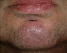
Figure 1.Facial view with swelling of the chin.
Radiological examination revealed a large radiolucent area at the level of the lower incisors #41 and #31 (Figure 2). The vitality test with carbon dioxide was positive for tooth # 42 and # 32 and negative for tooth # of 31 and # 41. The mandibular incisors were not sensitive to percussion and showed mobility degree 2 (2 mm horizontally). The patient is an avid scuba diver and has well developed jaws and facial muscles and a shallow vestibulum. There were no visible lesions.
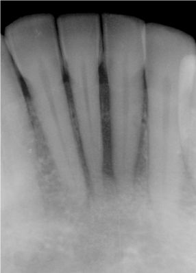
Figure 2.Apical radiograph of lower incisors revealing radiolucency at tooth # 41 and 31.
Under local anesthesia, the abscess was incised, pus was drained, the wound was irrigated, and a drain was inserted and sutured. Through this exploratory flap, a bony defect of almost 1 cm2 could be identified in loci #31 and #41. At the lower right incisor # 41, the entire apex was surrounded by inflammatory tissue while the apex of the lower left incisor # 31 was just surrounded by bone. There was no foreign body visualized but fibrous scar tissue at the level of the transition attached - nonattached gingiva was observed. After a few days the drain was removed. Test drilling on tooth # 31 proved the tooth to be vital and the cavity was filled with a composite filling. Tooth # 41 was tested to be nonvital. Subsequently, the necrotic tissue was removed and a root canal treatment was carried out in two stages. The patient was advised not to dive until the definitive treatment was completed in order to prevent the forcing of compressed air into the wound. A week later the swelling was gone (Figure 3). A control apical radiograph was taken after 6 months, which showed the healing of the bony lesion (Figure 4).
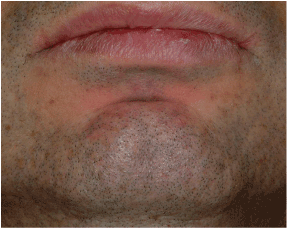
Figure 3.Facial view after healing.
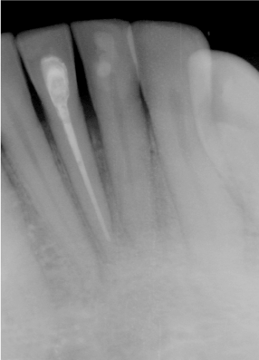
Figure 4.Apical X-ray after 6-months symptom free.
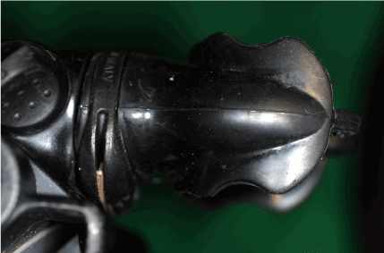
Figure 5.Pressurized air regulator with black mouthpiece.Uncustomized mouthpieces can damage the vestibulum and create an entry for anaerobic bacteria.
Discussion
Diving-mouthpiece-associated injuries are well defined (1). The diver gets air into the mouth from a tank via a regulator with a mouthpiece (Fig. 5) which he holds in place with his teeth (mostly canines and premolars). An airtight seal has to be created between the teeth and the lips. There are three types of mouthpiece designs: the standard or commercial mouthpieces, the semi-customized and the fully customized mouthpieces. They are manufactured from silicon or from soft acrylic resins. They are flexible but when biting hard on them or by strongly tightening the lips, damage to the teeth or surrounding tissue can arise [4]. When diving at exotic holiday centers, diving equipment with commercial mouthpieces can be rented. These are not always well-adjusted and can cause injuries. The hygiene is not always carefully monitored and exchange of mouthpieces between "diving buddies" is standard practice during training. The mouth piece has been identified as a possible vector for transmission of the herpes simplex virus [5].
This patient was probably injured and infected by the mouthpiece during the first diving vacation. The injury probably caused an entry for anaerobic bacteria that locally led to the destruction of the alveolar buccal wall at the level of the lower central incisors. In an experimental set-up, Bergenholtz & Lindhe [6] demonstrated that if the periodontal destruction is limited to half of the root, the nerve tissue did not show any alterations. However, if the periodontal destruction was severe, pathologic pulp alterations were observed and even pulp necrosis was observed. If the flare-up after two months, in this patient, was caused by the mouthpiece or by the necrotic pulp is unknown but it appeared again during a diving period. The leakage of the intracanal infected content to the periradicular tissues cannot be ruled out, although the patient reported no sign of barodontalgia , ie pain on descent or ascent while diving.
Given the immense popularity of snorkeling and diving it is likely that dentists are faced with these type of injuries. Therefore, they have to be able to inform the patient on mouthpiece design and the importance of disinfection and maintenance.
References
2021 Copyright OAT. All rights reserv
- Zadik Y, Drucker S (2011) Diving dentistry: a review of the dental implications of scuba diving. Austral dent J 56: 265-271. [Crossref]
- Zadik Y. Barodontalgia (2010) what have we learned in the past decade? Oral Surg Oral Pathol Oral Radiol Endod 109: e65-e69.
- 3.Gunepin M, Derachbe F, Audoual T (2010) Fracture of a sound tooth in a pilot under hyperbaric conditions. Aviat Space Environ Med 81: 691-693. [Crossref]
- Rogoff A (2010) Diving damage. J Am Dent Assoc 141: 15.
- Potasman I, Pick N (1997) Primary herpes labialis acquired during scuba diving course. J Travel Med 4: 144-145. [Crossref]
- Bergenholtz G, Lindhe J (1978) Effect of experimentally induced marginal periodontitis and periodontal scaling on the dental pulp. J Clin Periodontol 5: 59-73. [Crossref]