Abstract
Developmental process of late deceleration (LD) of fetal heart rate (FHR) was analyzed to clarify controversy LD record. The mechanical compression of pelvic large vessels by contracted uterine body develops LD, and fetal hypoxia is will be enhanced to severe level. Ppathologic LD should be searched. The loss of variability associated with LD was severe pathology. Actocardiogram, is useful to predict fetal outcome in LD cases.
Key words
fetal heart rate, late deceleration, large vessel compression, the loss of variability, fetal growth restriction, hypoxia index
Introduction
Transient FHR reduction (deceleration) was classified to U shaped variable one (VD) and V shaped periodic one, which was divided into early deceleration (ED) and late deceleration (LD), of which delay to uterine contraction is 20 or more S (Figure 1). LD was placental insufficiency. and its outcome was ominous [1]. Caldeyro- Barcia studied Type I dip, which was the same as ED, and Type 2 dip was LD [2]. Some cases, who were diagnosed as LD, received cesarean (C-) section according to FHR pattern theory expecting depressed neonate, while the neonate was vigorous, confusing doctor by the paradoxical results. Therefore, the author intended to study the mechanism to develop the LD in past reports [2-4]. The principle of some LDs was the compression of maternal pelvic lrge vessels with contracted uterus prior to the FHR deceleration with lag time, while it disappeared by the change of maternal
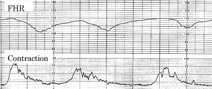
Figure 1. Typical LD and the loss of variability in the labor. Fetal hypoxia index was 25. One min Apgar score was 3. Three month infant died due to brain hemorrhage.
posture to lateral one [3].
Methods
The situations to develop delayed FHR deceleration after uterine contraction were searched in past literatures [1-4].
Results
Compression of large maternal pelvic blood vessels:
- Supine hypotension: Inferior vena cava was compressed by contracted pregnant uterus reducing the blood return to maternal heart and developed maternal hypotension followed by the reduction of placental arterial blood flow, developing hypoxia and delayed deceleration in FHR record. It is well known that the LD and supine hypotension disappear after changing maternal posture to lateral one from the supine.
- Pelvic large iliac arteries were compressed by contracted pregnant uterus in the labor at maternal supine posture, stopping placental arterial blood supply, developing fetal hypoxia and FHR deceleration after the contraction (Poseiro effect). The loss of iliac arterial flow due to the compression was confirmed by pelvic angiography. The LD, FHR deceleration after uterine contraction, disappeared after changing maternal posture to lateral from supine one [3]
Already existed fetal hypoxia was enhanced by repeated uterine contractions
Severe hypoxic FHR changes developed some weeks after early fetal hypoxia, detected by the loss of FHR
acceleration in fetal growth restriction (FGR). The severe hypoxia was composed of LD, fetal bradycardia and the
loss of variability, resulting heavy neonatal asphyxia and neonatal deaths [5], where the LD was pathologic in the
process, therefore, early delivery was recommended in the loss of acceleration found in FGR [5].
The loss of FHR variability
Fetal outcome was ominous even in very mild LD by Hon [1], however, mild LD was recognizable when FHR variability is lost [1]. As the loss of variability was severe fetal brain damage similar to anencephaly [6], fetal asphyxia is treated by the C-section before the loss of variability
Discussion
Causes of hypoxic FHR deceleration
- Fetal disorders Congenital or acquired fetal diseases develop fetal hypoxia and FHR deceleration, because the heart rate is parallel to PaO2 when il is lower than 50mmHg, which is common level of fetal arterial blood .
- Extrinsic causes Low maternal blood pressure, maternal hypoxia, abnormal placental oxygen transfer function, e.g. infarction in preeclampsia, intervillous space fibrin deposit, umbilical cord compression or uterine hypercontraction..
- Compression of maternal pelvic vessels
- Supine hypotension of mother
Maternal inferior vena cava is compressed by contracted pregnant uterus in maternal supine posture causing maternal hypotension by reduced blood return to the heart and fetal hypoxia causing late deceleration, which disappears after change of maternal posture to lateral posture.
Compression of iliac arteries
Contracted pregnant uterus in maternal supine posture stopps the arterial blood supply to the placenta causing fetal hypoxia. The mechanism was found by pelvic angiography (Poseiro effect) [ ]. Fetal deceleration occurs after the lag time, namely, late deceleration (LD) appears, where the LD disappears after maternal posture change to lateral one [3].
Management of late deceleration
As maternal lateral posture rejects the pelvic vessel compression caused by contracted pregnant uterus in maternal supine posture, i.e., the vessel compression is removed by maternal lateral posture increasing the blood supply to the placenta. As the effect of lateral posture is revealed by quick disappearance of LD [4], the mother has to take lateral posture immediately after recording LD in fetal monitoring to confirm whether the cause of LD is compressed pelvic large vessel or not, i.e. it should be known that the LD is caused by vessel compression with the contracted uterus, while staffs have to further search LD developing pathologic cause, if LD remains on FHR record after the change to lateral posture.
Tocolysis will be done in the uterine hypercontraction
Orcinoprenaline was effective to the LD in uterine hyper contraction in the past [4].. The tocolysis will be effective by the terbutaliine at present.
Early delivery
Severe LD case is cured by early delivery with C-section before the loss of variability preventing fetal brain damage, when intrauterine therapy was ineffective. Timely C-section will be performed before the loss of FHR variability, which is the sign of heavy fetal brain damage resulting neurological sequels, e.g. cerebralpalsy [6]. Timely C-section will be indicated by severe FHR changes, the loss of FHR acceleration, decreased variability [6], or when the hypoxia index was 20-24, as the index of cases of the loss of variability was 25-26. The index is determind by the sum of FHR bradycadia duration (min) x100, divided by the lowest bradycardia (bpm), because FHR lower than 110 bpm will be parallel to PaO2 lower than 50 mmHg..
Actocardiogram
2021 Copyright OAT. All rights reserv
The neonate was vigorous when fetal acceleration duration ratio to fetal movement burst duration (A/B ratio) was
higher than 1.0, despite repeated LDs were recorded, namely, fetal movement study was effective to estimate
favorable outcome even in the presence of LD [7].
Possibility of fetal/neonatal damage
Although FHR is parallel to the PaO2 under 50mmHg, where fetal bradycardia is recorded in monitoring record, it is not the same as fetal damage, namely, the bradycardia is caused by the neurologic excitation of parasympathicus center located in the medulla oblongata, and the experimental hypoxic bradycardia disappears after the narcosis of rabbit, and the apneic bradycrdia of anenephalic newborn who remains medulla oblongata disappeared by the infusion of oxygenated blood into the vessel. Fetal brain is damaged when FHR acceleration is lost an the variability lost by the hypoxia, namely fetal bradycardia shows only fetal environmental hypoxia, while hypoxic fetal brain damage is expressed by the loss of fetal brain response to fetal movement, i.e. FHR acceleration and the variability are fetal brain response to fetal movement burst and minor fetal movements, therefore the loss of acceleration to fetal movement is is early sign of fetal brain damage, while the loss of variability is advanced severe fetal brain damage, confirmed by actocardiographic studies [6,7]. Therefore, the early appearance of LD caused by the compression of large vessels which recovered after posture change cannot be fetal damage, therefore, the neonate is vigorous without brain damage, while sum of hypoxic effects in long repetition may be able to damage fetal brain if the LD lasted for several 10 min. The fetal damage could be estimated by the hypoxia index, which is the sum of duration of fetal bradycardia (min), multiplied by 100, and divided by the lowest FHR (bpm). As the Index after the loss of viability was 25-26, it must be around 20, if fetal brain damage would be prevented.. However, that is only an example of extreme case, and the author hope rapid cure of LD case in its earliest stage, e.g. the cure from LD caused by uterine body compression. Actocardiographic duration to fetalmovement duration
Conclusion
There would be several kinds in LD. Maternal posture change to lateral one should be tried after looking at LD on monitoring chart.. Although final decision will be cesarean section, as early as possible intrauterine therapy would be tried. The actocardiogram will be helpful to expect fetal outcome .by the FHR acceleration duration ratio to the duration of fetal movement (A/B ratio), i.e. the outcome is favorable, if the A/B ratiio is larger than 1. .
References
- Hon EH (1968) An atlas of fetal heart rate patterns. Harty Press.
- Caldeyro-Barcia R, Poseiro JJ, Mendez-Bauer C, Gulin LO (1967) Effects of Abnormal Uterine Contractions on Fetal Heart Rate in Labor. 9-27.
- Poseiro JJ, Mendez-Bauer C, Caldeyro-Barcia R et al. (1969) Effect of uterine contractions on maternal blood flow through the placenta. Perinatal factors affecting human development, Paho Advisary Committee. 161-171.
- Caldeyro-Barcia R, Magana JM, Poseiro, JJ et al. (1969) New Approach to the treatment of acute Intrapartum fetal distress. Perinatall factors affecting human development. Patho Advisary Committee, 248-253.
- Teshima N (1993) Non-reactive pattern diagnosed by ultrasonic Doppler fetal actocardiogram and outcome of the fetuses with non-reactive pattern. Acta Obstet Gynecol Jpn 45: 423-430. [Crossref]
- Maeda K (2014) Modalities of fetal evaluation to detect fetal compromise prior to the development of significant neurological damage. J Obstet Gynaecol Res 40: 2089-2094. [Crossref]
- Maeda K (2016) Fetal Actocardiogram. Analysis of Fetal Motion and Heart Rate, Jaypee Brothers, New Delhi, 2016.