We report a case of acute myelopathy in a patient with Burkitt lymphoma successfully treated by high-dose therapy (TBuCy) and autologous stem cell transplantation. Lymphoma involvement and methotrexate toxicity are discussed as potential causal mechanisms for neurological symptoms.
myelopathy, Burkitt lymphoma, methotrexate, autologous stem cell transplant
While neurological and meningeal tumoral involvement is frequent in aggressive B-cell lymphoma-and a hallmark of Burkitt lymphoma-elective spinal cord localization is uncommon. Here, we report the case of a 42-year-old female with stage IV Burkitt lymphoma who developed an early myelopathy on therapy with rapid progression.
She presented with acute weight-loss and asthenia along with B-symptoms including night sweats and fever. She also had diffuse bone pain and a typical numb chin syndrome but her neurological examination was normal. She had anemia (hemoglobin 86 g/l, normal range 120-170 g/l) and thrombocytopenia (platelets 59 × 109/l, normal range 150-450 × 109/l), and bone marrow biopsy revealed massive involvement by monomorphic medium-sized B-cells with morphological and immunophenotypic characteristics of Burkitt lymphoma. Routine blood tests showed a more than 20-fold increased lactate dehydrogenase (LDH) level (4878 UI/l, normal range 135-214 UI/l), moderate liver tests abnormalities (AST 146 UI/l, normal range 10-45 UI/l; ALT 38, normal range 10-35 UI/l; GGT 381 UI/l, normal range 7-35 UI/l; APL 232 UI/l, normal range 35-120 UI/l and total bilirubin 16.9 µM, normal range 3-17 µM) and a normal renal function (creatinine 38 µM, normal range 45-100 µM). Cerebrospinal fluid examination was normal (absence of visible cell; 0.33 g/l protein and 2 mM glycorachia with 3.6 mM glycemia). Brain and spine magnetic resonance imaging (MRI) were normal. No significant lymphatic or systemic involvement was observed on PET/CT scan. Finally, she was diagnosed with stage IV Burkitt lymphoma with bone marrow and possible meningeal involvement. She received emergency chemotherapy with a COP regimen including 460 mg cyclophosphamide day 1; 1.5 mg vincristine day 1 and 60 mg prednisone day 1 through 5, along with three therapeutic lumbar punctures (15 mg methotrexate, 40 mg cytarabine and 20 mg hydrocortisone) during the first six days of treatment. An improvement of chin dysesthesia was observed at this stage. She had an excellent response to COP as day 7 bone marrow examination showed less than 5% of lymphoma infiltration. Then she received vincristine 2 mg day 1, methotrexate 4300 mg day 1, doxorubicine 86 mg day 2, cyclophosphamide 720 mg days 2 through 4, prednisolone 90 mg day 1 through 6 (the COPADM regimen), rituximab 600 mg at days 6 and 14 and 3 additional intrathecal injections. At day 7 from the onset of COPADM, she experienced sudden lower limb deficiency, bladder dysfunction and T10-T11 sensitive level with a complete loss of proprioception, pain and temperature sensation, along with the recurrence of numb chin syndrome. Emergency spine MRI excluded a spinal compression or myelitis. A cerebrospinal fluid examination showed an increased protein level (1.6 g/l) without lymphoma cells. In the absence of other cause of paraplegia and sensory loss, and in a context of progressive numb chin syndrome, she was considered as having a neurological widespread of her disease and received additional high-dose methotrexate (11 500 mg). A few days later, improvements of her legs and chin sensitivity was observed. Then she received CYVE (cytarabine 70 mg over 12 h day 1 through 5; cytarabine 4300 mg over 3 h day 2 through 5; etoposide 290 mg day 2 through 5) followed by a stem cell collection upon G-CSF mobilization. Unfortunately, her neurological condition worsened during this treatment, with the occurrence of lower limbs hyperalgesia and a progression of her sensory loss to a T6 level. At this time, spine MRI showed features of conus medullaris hemorrhagic infarction and a C6-C7 intramedullary FLAIR hyperintensity suggesting a spinal cord ischemia (Figure 1A). She received a salvage therapy by CYM (methotrexate 4300 mg day 1 and cytarabine 145 mg over 24 h, day 2 through 6). Ten days after this therapy, she was referred to intensive care unit for mechanical ventilation due to the extension of her neurological defect to a T4 level with acute respiratory failure. After discussion with the patient, her family and the medical teams, conditioning regimen for autologous stem cell transplantation was given using thiotepa 360 mg days 1-3; busulfan 38 mg for a total of 10 infusions during 3 days and cyclophosphamide 2820 mg days 7-8 (TBuCy regimen), and autologous stem cells were injected 48 h after chemotherapy completion. She had a partial hematopoietic recovery 8 days after stem cell transplant. She recovered a spontaneous ventilation allowing a withdrawn of mechanical ventilation 11 days after transplant. Follow-up spine MRI showed extensive T2 hyperintensities from C7 to the conus medullaris (Figure 1B), with spontaneous T1 hyperintensities suggesting hemorragic transformation of ischemic lesions (Figures 1C and 1D). Then, she underwent a 6 month-long in-hospital neurological, psychological and social rehabilitation. She is now two years from transplant in persistent complete remission of her lymphoma, and lives at home with environment accommodations and daily nurse care.
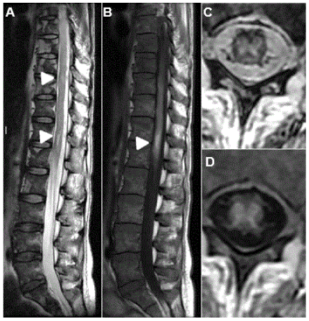
Figure 1. Follow-up MRI spine imaging. A) Extensive T2 hyperintense signals appeared on follow-up (head arrows). Of note, the heterogeneity of the vertebrae, suggesting a lymphomatous bone infiltration. B) Extensive abnormalities of the spinal cord (from C7 to the conus medullary) with spontaneous T1 hyperintense signals centered on the conus medullary, without enhancement, suggesting hemorrhagic lesions. C) Two-dimension Axial T2 showed a global T2 hyperintense signal of the spinal cord’s grey matter. D) Spontaneous T1 hyperintense signals suggesting hemmorhagic transformation of the ischemic lesion.
Sudden onset of neurological sign associated with normal initial MRI suggested spinal cord ischemia. Characteristics of one-month MRI signal were consistent with this mechanism, but the large extension of the spinal T2 hyperintensity - beyond the segmentation of spinal arterial vascularization - observed during follow-up is uncommon. Hence, we discussed a potential neurological toxicity of methotrexate, which hypothesis was reinforced by the detection of brain MRI FLAIR white matter hyperintensities that may have been cause by high-dose methotrexate therapy. However, most cases of methotrexate-related myelopathies were reported with intrathecal administration [1-4], suggesting a local rather of systemic mechanism for these toxicities. The concomitance of progressive numb chin syndrome and the transient improvement of neurological symptoms upon high-dose methotrexate rather suggested that neurological symptoms were lymphoma-related. The absence of extrinsic or intrinsic tumor compression suggested intravascular lymphoma diffusion, while this topography is only reported in intravascular lymphoma (IVL), a rare diffuse large B-cell lymphoma variant [5]. We first used high-dose methotrexate and cytarabine, either combined or sequentially administrated for their diffusion into central nervous system [6]. However, the hypothesis of methotrexate CNS toxicity, even as a concomitant factor prompted us to change our therapeutic strategy. We thus performed a salvage autologous stem cell transplantation using a blood-brain barrier disrupting conditioning chemotherapy. The TBuCy regimen is a widely approved regimen for CNS lymphoma, allowing long-term disease control even in diseases refractory to chemotherapy [7]. Moreover, this regimen is associated with acute toxicities (particularly neutropenic fever and infection) responsible of a 5-7% toxicity-related mortality, but is not associated with acute or late neurological toxicities [8,9]. In our case, we observed a rapid neurological response after autologous transplant, as attested by mechanical ventilation withdrawn and gradual improvement of motor functions, suggesting that her neurological symptoms were mostly lymphoma-related.
With a two-year post-transplantation follow-up, our patient is still in complete remission, and had a slow but progressive improvement of her autonomy. She and her family now consider they have a good quality of life despite her persistent disability. Our current report supports the use of repeated central nervous system imaging in case of neurological symptoms in lymphoma patients. Because distinguishing spine toxicity of methotrexate from disease-specific involvement is challenging, we suggest considering autologous stem cell transplantation using non-neurotoxic blood-brain barrier disrupting conditioning chemotherapy as frontline treatment in lymphoma patients with progressive neurological symptoms.
- Cachia D, Kamiya-Matsuoka C, Pinnix CC, Chi L, Kantarjian HM, et al. (2015) Myelopathy following intrathecal chemotherapy in adults: a single institution experience. J Neurooncol 122: 391-398. [Crossref]
- Counsel P, Khangure M (2007) Myelopathy due to intrathecal chemotherapy: magnetic resonance imaging findings. Clin Radiol 62: 172-176. [Crossref]
- Joseph PJ, Reyes MR (2014) Dorsal column myelopathy following intrathecal chemotherapy for acute lymphoblastic leukemia. J Spinal Cord Med 37: 107-113. [Crossref]
- Murata KY, Maeba A, Yamanegi M, Nakanishi I, Ito H (2015) Methotrexate myelopathy after intrathecal chemotherapy: a case report. J Med Case Rep 9: 135. [Crossref]
- Tahsili-Fahadan P, Rashidi A, Cimino PJ, Bucelli RC, Keyrouz SG (2016) Neurologic manifestations of intravascular large B-cell lymphoma. Neurol Clin Pract 6: 55-60. [Crossref]
- Ferreri AJ (2011) How I treat primary CNS lymphoma. Blood 118: 510-522. [Crossref]
- Soussain C, Hoang-Xuan K, Taillandier L, Fourme E, Choquet S, et al. (2008) Intensive chemotherapy followed by hematopoietic stem-cell rescue for refractory and recurrent primary CNS and intraocular lymphoma: Societe Francaise de Greffe de Moelle Osseuse-Therapie Cellulaire. J Clin Oncol 26: 2512-2518. [Crossref]
- Scordo M, Bhatt V, Hsu M, Omuro AM, Matasar MJ, et al. (2017) A Comprehensive Assessment of Toxicities in Patients with Central Nervous System Lymphoma Undergoing Autologous Stem Cell Transplantation Using Thiotepa, Busulfan, and Cyclophosphamide Conditioning. Biol Blood Marrow Transplant 23: 38-43.
- Soussain C, Choquet S, Fourme E, Delgadillo D, Bouabdallah K, et al. (2012) Intensive chemotherapy with thiotepa, busulfan and cyclophosphamide and hematopoietic stem cell rescue in relapsed or refractory primary central nervous system lymphoma and intraocular lymphoma: a retrospective study of 79 cases. Haematologica 97: 1751-1756. [Crossref]