Abstract
Background: Castleman Disease (CD) describes a rare group of heterogenous diseases characterized by lymph node enlargement. It has been associated with various neoplastic, autoimmune, and infectious processes. CD has two main histological forms, hyaline vascular and plasma cell type. Clinically, there are two forms of CD - unicentric and multicentric. CD involvement of the central nervous system (CNS) is rare. We present a rare case of unicentric CD that presented as leptomeningeal enhancement and a review on the previous literature of similar cases.
Case Report: A 41-year-old man with no past medical history presented with a first time seizure with persistent numbness of his left face, left arm, and left leg. MRI brain showed leptomeningeal involvement. Extensive workup done for malignant, autoimmune and infectious etiologies during admission was unremarkable. The patient was started on antiepileptics with clinical improvement and discharged. As an outpatient, he underwent meningeal biopsy showing CD of hyaline-vascular subtype. He was then seen in hematology clinic and the plan for the patient was to monitor disease progression with MRI. Radiation oncology and neurosurgery were then consulted, who stated that no intervention should be done at this time as the patient was asymptomatic. The patient remains clinically stable.
Conclusion: Leptomeningeal enhancement is a rare presentation of CD and further evidence-based data is warranted to better guide management. In cases like ours in which the patient had unresectable disease, we opted for conservative management with close clinical monitoring.
Keywords
castleman disease, leptomeningeal enhancement, hyaline-vascular, unicentric
Introduction
Castleman Disease (CD) is a rare, heterogenous, group of diseases characterized by enlargement of lymph nodes and surrounding tissues. It often presents in the mediastinum [1]. Histopathologic features including atrophic or hyperplastic germinal centers, prominent follicular dendritic cells, hypervascularization, polyclonal lymphoproliferation, and/or polytypic plasmacytosis [2]. There are around 6500 to 7700 cases of CD diagnosed each year in the United States [3]. The two main histological forms of Castleman disease are hyaline-vascular CD and plasma cell CD. Hyaline-vascular CD is characterized as interfollicular microvascular proliferation and represents about 90 percent of cases of CD [4]. Plasma cell variant CD consists of an abundance of plasma cells and often is associated with systemic manifestations [4]. Current data suggest that hyaline variant Castleman Disease is more likely a clonal neoplastic process [5]. It has been postulated that the most likely cell of origin is stromal, specifically the follicular dendritic cell [6-9]. Recently a study using next-generation sequencing of nodal tissue from Castleman Disease revealed somatic platelet-derived growth factor receptor β mutations in nearly 20% of cases; these mutations were localized to CD45negative cells, likely representing stromal cell [10].
CD can also be categorized by the areas involved on presentation. Unicentric CD affects a localized set of lymph nodes and its surrounding tissues. Unicentric Castleman's disease most commonly involves the mediastinal lymph nodes in the chest [4]. Multicentric CD affects lymph nodes and surrounding tissues throughout the body.
CD has been associated with neoplastic, autoimmune, and infectious etiologies. In particular, it is associated with human herpes virus type 8 (HHV8), which is almost exclusively found in patients with human immunodeficiency virus (HIV) infection. Presentations include fever, cough, abdominal pain, enlarged spleen, ascites, pleural effusion, xerostomia, autoimmune hemolytic anemia, rash, nasal obstruction, elevated C-reactive protein level (CRP), and elevated erythrocyte sedimentation rate (ESR) [4]. Presentations with localized CNS involvement are rare; case reports have documented symptoms such as aphasia, seizures, and pseudotumor cerebri. Diagnosis is usually made by biopsy.
Treatment options include surgical resection, radiation, or systemic therapy targeting CD20 or IL-6.4 This report is of a 41-year-old man who presented with seizures and numbness of the left side, with imaging finding of a solitary right-sided leptomeningeal mass, diagnosed on histology as unilateral hyaline-vascular Castleman's disease.
Case Presentation
A 41-year-old male without prior medical history presented to the emergency department with complaints of new onset time seizure, accompanied by persistent numbness in his left lower face, left arm, and left leg. He noted hyperesthesia of his left lower extremity and a posterior headache. He denied vision changes, changes in speech, changes in gait, or history of head trauma. Lab work was remarkable for white blood cell count 8.8 K/cumm, hemoglobin 14 g/dL, and platelet count of 484 K/cumm. Levetiracetam was initiated as an antiepileptic. Magnetic resonance imaging (MRI) with and without contrast showed abnormal signal intensity of the pachymeninges and leptomeninges, particularly on the right side of the vertex (Figures 13 and 14). The differential diagnosis at this time included infectious (such as meningitis), inflammatory conditions (such as sarcoidosis or systemic lupus erythematosus), or malignancy. Cerebral spinal fluid from the lumbar puncture was unremarkable and his serum erythrocyte sedimentation rate and C-reactive protein levels were elevated, suggesting an inflammatory syndrome. Rheumatology consultation recommended against steroids in the absence of systemic symptoms. Ophthalmology evaluation did not identify ocular vasculitis involvement. Studies from a second lumbar puncture were unremarkable. Given the patient’s stable clinical status, he was discharged home with levetiracetam after a weeklong hospitalization with outpatient neurosurgery and neurology follow up.
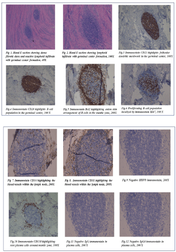
Figures 1-12:
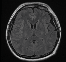
Figure 13. Axial T1-weighted MRI of the brain showing leptomeningeal enhancement, right worse than left
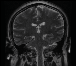
Figure 14. Coronal T1-weighted MRI for the brain with findings consistent with pachymeningeal enhancement bilaterally, worse on the right, and leptomeningeal enhancement over the right cerebral convexity
A meningeal biopsy was subsequently performed; histopathology from the dura was consistent with hyaline variant of CD (see full pathology report below). Serologies for HIV and HHV8 were negative. The patient was evaluated in hematology clinic where it was determined that he did not meet criteria for multicentric disease and recommended repeat MRI and labs. There was no systemic therapy offered at this time as patient was asymptomatic. Patient was then referred to radiation oncology and neurosurgery for possible interventions. A subsequent MRI demonstrated the leptomeningeal enhancement seen on prior MRI studies. Radiation oncology determined that radiation was not indicated given the localized nature of the CD and the patient’s clinical stability on levetiracetam. Given the lack of an obvious resectable mass on imaging, clinical improvement, and stable MRI findings, neurosurgery recommended against surgical intervention.
Pathology
Pathological and immunophenotypic findings:The biopsy specimen from the dura revealed the characteristic findings of hyaline vascular Castleman Disease. Microscopic examination of permanent sections showed a dense fibrotic dura and reactive lymphoid infiltrate with germinal center formation (Figure 1 and 2). A plethora of fine capillaries in the center of the lymphatic follicles and in surrounding mantle zone was identified. A characteristic onion-skin arrangement of the expanded mantle zone lymphocytes was demonstrated. The differential diagnosis included Castleman’s disease, low grade B cell neoplasm, vasculitis, an infectious etiology.
A comprehensive panel of immunostains were performed. No aberrant lymphoid population was present based on CD3, CD5, CD20, PAX5, CD21, CD10, Bcl6, Bcl-2, and Ki-67 immunostaining. The germinal centers contain polyclonal B cells, a peripheral zone of T cells with very few admixed plasma cells and an irregular follicular dendritic meshwork.
The meshwork of follicular dendritic cells in the germinal centers was highlighted by CD21 staining (Figure 3). Cells constituting the expanded mantle zones expressed CD20 and PAX5 (Figure 4). Bcl-2 staining also indicated small and mature lymphocytes in the mantle zone. The onion-skin arrangement was appreciated via Bcl-2 immunohistochemical staining (Figure 5). Ki-67 staining indicated proliferating cells, which were mainly observed in the B cell aggregate (Figure 6). CD31 and CD34 showed vessels within the lymphoid aggregate (Figure 7 and 8). Immunostaining with HHV-8 was negative (Figure 9). Furthermore, CD138 and MUM1 localized very few plasma cells (Figure10); with rare positivity with IgG and IgG4 (Figure11 and 12). Therefore, a diagnosis of a hyaline variant of Castleman Disease was rendered.
Discussion
We present a case of a 41-year-old man who had a localized form of CD manifesting as leptomeningeal enhancement on MRI. It is important to note that CD can present without a discrete mass. Given the rare presentation, we conducted a comprehensive literature search using the PubMed database and the search terms “unicentric, Castleman Disease, neurological, intracranial, and leptomeningeal” from 1980 to 2021. An initial thirteen titles and abstracts were found (Table 1). An additional five references were found in the titles with similar presentations to our case (unicentric CD) and were included in this review. Multicentric cases as well as pediatric cases were excluded.
On our literature search of cases of unicentric CD with neurological involvement (Table 1), CD was also diagnosed by biopsy and all the cases involved surgery. The mean age of the cases was 54 years old. The most common symptom was seizures (both focal and generalized tonic-clinic), which occurred in 7/17 or 41.1 percent of patients. Other symptomology included headache, dizziness, and vision changes. Of the cases that identified the histological type of CD, five were the hyaline-vascular variant and four were the plasma cell variant. HHV8 was only documented in two of the cases and was negative. In many of these cases, the abnormal neurological or seizure activity subsided once surgical resection was done. One patient received radiation. In one case documented in Severson,et al. the patient presented with localized motor seizures in his left arm, became more frequent over a two-month period, and he subsequently developed grand mal seizures despite being on anti-epileptics. He was found to have a 6.5-centimeter mass that was then surgically resected. This is different compared to our case, where the patient had numbness in his left lower face, left arm, and left leg and hyperesthesia of his left lower extremity which improved on levetiracetam [11-24].
Table 1.Literature Review of CD case reports
M: male; F: female
What makes our case unique is the initial MRI finding of leptomeningeal enhancement rather than a discrete, resectable mass. This finding poses the question on how to manage meningeal involvement of CD. Preferred treatment of unicentric CD is surgical resection regardless of intracranial presentation [3,25]. Unresectable disease can undergo radiation therapy, rituximab/cyclophosphamide/prednisone, or embolization therapy. Surgery for unicentric CD is usually curative [25]. Radiation therapy has noted to be a reasonable alternative in unicentric CD patients, with complete response in 35% of patients and partial response in 41% of patients [26]. Systemic chemotherapy has been used in some cases of unresectable unicentric CD with 55% response rate [27]. Embolization has only been described in a handful of cases and is an emerging option for large vascular lymph nodes in unicentric CD to reduce the size to render surgery safer [28]. IL-6 targeted therapy such as siltuximab or tocilizumab is an option in symptomatic unicentric CD with inflammatory symptoms (fevers, weight loss, anorexia) as IL-6 has been documented as the main driver of symptomatology in multicentric CD patients. Tocilizumab was used in four patients with CD with inflammatory symptoms and one patient had complete response and three had partial response but was not effective when used for a patient with refractory unresectable unicentric CD [27,29]. Our patient represents a unique case of intracranial CD that was deemed unresectable by neurosurgery, and with no recommendation for radiation by radiation oncology or systemic therapy by hematology.
Treatment will be guided by the patient’s symptomatology and close follow up. If the patient exhibits worsening symptoms, we can consider intervention with IL-6-targeted therapy such as siltuximab. Given limited literature on focal neurological involvement of CD, we emphasize further study and investigation regarding this rare condition and to help gain more knowledge of developing treatment strategies for CD.
Conclusion
This case describes a rare case of CD that presented as a first-time seizure with only leptomeningeal enhancement seen on imaging that was unresectable. This case is unique as the previous cases described in our literature review had a discrete mass. The purpose of our literature search was to summarize treatment summaries for similar presentations given the paucity of information for unicentric CD in the leptomeninges and whether these could be applied to our patient. Given that the patient did not have a resectable mass, we decided on conservative management given that the patient remains clinically stable. We emphasize considering CD in the differential for patients who have new onset of neurological symptoms and the need for further study into this rare condition to gain more knowledge to develop treatment strategies.
Financial support and disclosures
There was no financial support secured for this project. All authors have no disclosures to declare.
Authorship and Contributorship
The authors SL and BH wrote and edited this manuscript. The authors TD, JY, and ST edited and revised the manuscript.
References
- Castleman B, Iverson L, Menendez VP (1956) Localized mediastinal lymph node hyperplasia resembling thymoma. Cancer9:822-830. [Crossref]
- Menke DM, Tiemann M, Camoriano JK, Chang SF, Madan A,et al. (1996) Diagnosis of Castleman's disease by identification of an immunophenotypically aberrant population of mantle zone B lymphocytes in paraffin-embedded lymph node biopsies. Am J Clin Pathol105: 268-276. [Crossref]
- Munshi N, Mehra M, van de Velde H, Desai A, Potluri R, et al. (2015) Use of a claims database to characterize and estimate the incidence rate for Castleman disease. Leuk Lymphoma56:1252-60. [Crossref]
- Abramson JS (2019)Diagnosis and Management of Castleman Disease, Journal of the National Comprehensive Cancer Network.J Natl ComprCancNetw 17: 1417-1419. [Crossref]
- Chang KC, Wang YC, Hung Y, Wan-Ting H, Jen-Hui T,et al. Monoclonality and cytogenetic abnormalities in hyaline vascular Castleman disease. Mod Pathol27:823-831.[Crossref]
- Cokelaere KK (2002) Hyaline vascular Castleman's disease with HMGIC rearrangement in follicular dendritic cells: molecular evidence of mesenchymal tumorigenesis. Am J Surg Pathol26: 662-669. [Crossref
- Pauwels P, Dal Cin P, Vlasveld LT, Aleva RM, van Erp WF,et al. (2000) A chromosomal abnormality in hyaline vascular Castleman's disease: evidence for clonal proliferation of dysplastic stromal cells. Am J Surg Pathol24: 882-888. [Crossref]
- Reichard KK, Robinett S,Foucar MK (2011) Clonal cytogenetic abnormalities in the plasma cell variant of Castleman disease. Cancer Genet 204: 323-327. [Crossref]
- Chang KC, Wang YC, Hung LY,Huang W, Tsou J,et al. Monoclonality and cytogenetic abnormalities in hyaline vascular Castleman disease. Mod Pathol27: 823-831. [Crossref]
- Li Z, Lan X, Li C, Zhang Y, Wang Y, et al. (2019) Recurrent PDGFRB mutations in unicentric Castleman disease. Leukemia 33:1035-1038.[Crossref]
- Lacombe MJ, Poirier J, Caron JP (1983) Intracranial lesion resembling giant lymph node hyperplasia. Am J Clin Pathol80:721-723.[Crossref]
- Severson GS, Harrington DS, Weisenburger DD, McComb RD, Casey JH, et al. (1988)Castlemans disease of the leptomeninges. Report of three cases. J Neurosurg69:283-286. [Crossref]
- Gianaris PG, Leestma JE, Cerullo LJ, Butler A (1898) Castleman's disease manifesting in the central nervous system: case report with immunological studies. Neurosurgery24:608-13.[Crossref]
- Gulati P, Sun NC, Herman BK, Said JW, Cornford ME (1998) Isolated leptomeningeal Castleman's disease with viral particles in the follicular dendritic cells. Arch Pathol Lab Med122:1026-9.[Crossref]
- Hashimoto H, Iida J, Hironaka Y, Sakaki T (1999) Intracranial Castleman's disease of solitary form. Case report. J Neurosurg90:563-6.[Crossref]
- Ropponen KM, Kankkunen JP, Ronkainen A, Vihavainen M, Alafuzoff I (2002) Castleman's disease of the leptomeninges--immunohistochemical findings in 2 cases. Clin Neuropathol21:278-83.[Crossref]
- Cummings TJ, Gong JZ, Friedman AH, McLendon RE (2000) Castleman's disease confined to the leptomeninges. Ann Clin Lab Sci30:278-82.[Crossref]
- Delmont E, Lonjon M, Michiels JF, Chanalet S, Bourg V, et al. (2003) Castleman localisée au systèmenerveux central [Castleman's disease located in the central nervous system]. Rev Neurol (Paris)159:581-5. [Crossref]
- Matsumura K, Nakasu S, Tanaka T, Nioka H, Matsuda M (2005) Intracranial localized Castleman's disease. Case report. Neurol Medchir45:59-65.[Crossref]
- Ozono K, Fujimoto T, Hirose M, Kawahara I, Uchihashi K (2017) A Case of Intracranial Localized Castleman's Disease Mimicking Convexity Meningioma. No ShinkeiGeka45:39-45.[Crossref]
- Mallik AA, Katchy KC, Clotan N (2007) Solitary intracranial Castleman's disease, plasma cell variant: a case report. Med PrincPract16:226-9. [Crossref]
- Turek G, Reszeć J, Kochanowicz J, Hermanowicz A, Mariak Z (2013) A case of a solitary form of Castleman disease: ten-year follow-up. Neurol Neurochir Pol47:179-83.[Crossref]
- Escribano Paredes JB, Carrasco Moro R, López Gutiérrez M, Arias HP, García-Cosío M, et al. (2019) Radiologic and Histopathologic Features in an Intracranial Localized Castleman Disease: A Case Report and Review of Literature. Neurologist24:33-36.[Crossref]
- Wang Y, Zhou L, Wang W, Cheng H (2020) Intracranial Castleman Disease Presenting as a Meningioma. World Neurosurg142:420-422. [Crossref]
- Talat N, Belgaumkar AP, Schulte KM (2012) Surgery in Castleman’s disease: a systematic review of 404 published cases. Ann Surg255:677–684.[Crossref]
- Chan KL, Lade S, Prince HM, Harrison SJ (2016) Update and new approaches in the treatment of Castleman disease. J Blood Med7:145-158. [Crossref]
- Boutboul D, Fadlallah J, Chawki S, Fieschi C, Malphettes M, et al. (2019) Treatment and outcome of Unicentric Castleman Disease: a retrospective analysis of 71 cases. Br J Haematol186:269-273. [Crossref]
- van Rhee F, Oksenhendler E, Srkalovic G, et al. International evidence-based consensus diagnostic and treatment guidelines for unicentric Castleman disease. Blood Adv4:6039-6050.[Crossref]
- Abid MB, Peck R, Abid MA, Al-Sakkaf W, Zhang Y, et al. (2018) Is tocilizumab a potential therapeutic option for refractory unicentric Castleman disease? Hematol Oncol36:320-323.[Crossref]