Excessive fluoride intake through teeth development stages can lead to dental fluorosis. Fluoridated water used for drinking is a main potential source for this developmental tooth disorder. White opaque spots with secondary brown stain caused by the surface hypermineralization and subsurface hypomineralization as fluoride alterations is the primary cause. Prosthodontic intervention and esthetic rehabilitation can be used to restore the natural appearance of the teeth and its function. In this presented case, a treatment option of porcelain veneers was shown to be a satisfactory approach for the aesthetic treatment of moderate fluorosis.
25 years old male patient presented to the clinic with chief complaint “I want to restore my front teeth and have a better smile”. Intraoral and preoperative panoramic radiograph examination revealed generalized staining in upper and lower teeth (Figures 1 and 2), multiple carious teeth, badly broken tooth and partial edentulism. The patient used to live in Hail city which known with high fluoride percentage in ground water [1]. According to Dean’s fluorosis index, patient was diagnosed as severe fluorosis [2]. Treatment plan was discussed with the patient and we agreed to go with ceramic veneers in upper teeth after smile analysis was done. Upper and Lower alginate impression were taken to make upper wax up (Figure 3). After that mockup was done using putty index and placed in the patient mouth using “Systemp®.c&b II Ivoclar vivadent” (Figure 4). Patient was satisfied with the temporary, so we proceeded with the treatment. Teeth preparation was done using the depth-cutting bur over the mockup. Teeth were evaluated after preparation and there was no more fluorosis (Figure 5). Final impression were taken for the upper teeth using 3M ESPE Vinyl Polysiloxane (VPS) Regular Set Impression Materials and 3M Express Light Body Regular Set (Figure 6). The selected shade B1 IPS e.max Ivoclar vivadent (Figure 7). Finally, ceramic veneers were cemented with Variolink Esthetic LC System kit e.max and the cement shade was Neutral (Figure 8). The patient was given the pre and post-operative photographs for comparison which he showed high level of satisfaction and appreciation (Figure 9).
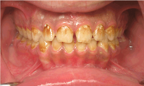
Figure 1. Preoperative intraoral view.
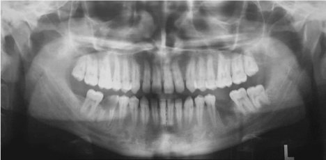
Figure 2. Panoramic radiograph.
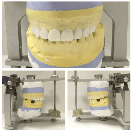
Figure 3. Diagnostic wax-up
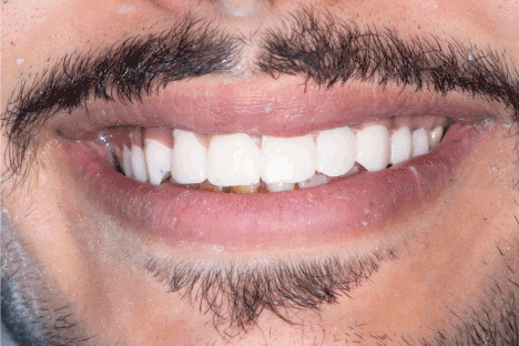
Figure 4. Provisional restorations in place
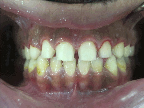
Figure 5. Veneer preparation showing stains removed
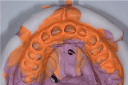
Figure 6. polyvinyl siloxane impression material
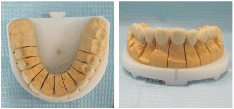
Figure 7. Porcelain veneers
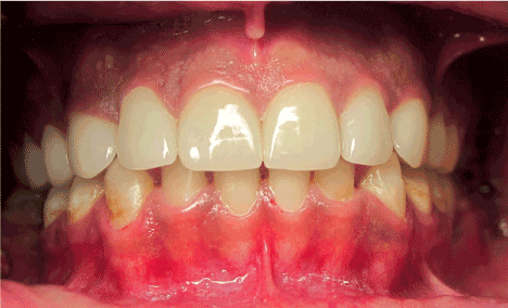
Figure 8. Veneers cemented
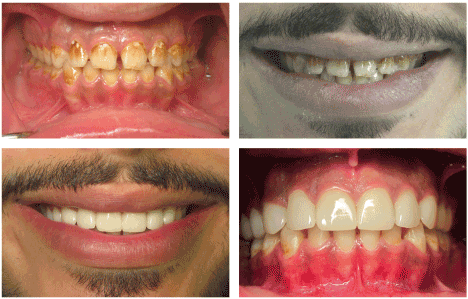
Figure 9. Pre and postoperative smile photograph
Treatment options to enhance a smile with esthetic dentistry include bleaching, enamel microabrasion, composite veneers, ceramic veneers and full crown coverage. Enamel microabrasion is a conservative treatment option that can improve the esthetic in selective cases [3]. In 1980, Enamel microabrasion was used first to eliminate teeth discoloration [4]. Treatment option varies and the decision depends on the fluorosis severity [5]. In case of minimal malalignments or spacing, prosthetic or restorative correction of the teeth shape and contour is required, in these cases microabrasion can’t achieve the desired outcome and the results of esthetic treatment will be compromised. Using indirect porcelain restorations can be used as the fracture resistance, wear resistance and color stability of indirect porcelain restorations is Higher than direct composite veneers porcelain restorations [6]. Bonding to the enamel affected by fluorosis require some modifications in the teeth preparation by preparing the fluorotic enamel surface to remove the hyperminerlized layer, acid etching time [7,8]. The best etching time effect were obtained at thirty seconds for teeth with moderate enamel fluorosis. However, increasing the etching time for teeth with severe fluorosis result in less retentive surface [9]. The bond strength of etch-and-rinse systems is known to provide the maximum bond strength to fluorotic enamel [10]. Although the use of the two-step self-etch adhesive system was reported and showed that a reliable bonding can be obtained [11,12].
Improvement in the techniques, ceramic materials, and bonding materials made porcelain veneers the most accepted, more conservative treatment option for esthetic rehabilitation of the anterior teeth over full coverage crowns.
- Alabdulaaly AI, Al-Zarah AI, Khan MA (2013) Occurrence of fluoride in ground waters of Saudi Arabia. Applied Water Science 3: 589-595.
- Dean HT (1942) The investigation of physi- ological effects by the epidemiological method. In: Moulton FR, ed. FIuorine and dental health. Washington, DC: American Association for the Advance- ment of Science, pub no 19:23-31.
- Lynch CD, McConnell RJ (2003) The use of microabrasion to remove discolored enamel: a clinical report. J Prosthet Dent 90: 417-419. [Crossref]
- Croll TP (1995) Enamel microabrasion: 10 years’ experience. Asian J Aesthet Dent 3: 9-15. [Crossref]
- Denbesten P, Li W (2011) Chronic fluoride toxicity: dental fluorosis. Monogr Oral Sci 22: 81-96. [Crossref]
- Peumans M, Van Meerbeek B, Lambrechts P, Vanherle G (2000) Porcelain veneers: a review of the literature. J Dent 28: 163-177. [Crossref]
- Ermis R, Demunck J, Cardoso M, Coutinho E, Vanlanduyt K, et al. (2007) Bonding to ground versus unground enamel in fluorosed teeth. Dental Mater 23: 1250-1255. [Crossref]
- Weerasinghe DS, Nikaido T, Wettasinghe KA, Abayakoon JB, Tagami J (2005) Micro-shear bond strength and morphological analysis of a self-etching primer adhesive system to fluorosed enamel. J Dent 33: 419-426. [Crossref]
- Torres-Gallegos I, Zavala-Alonso V, Patiño-Marín N, Martinez-Castañon GA, et al. (2012) Enamel roughness and depth profile after phosphoric acid etching of healthy and fluorotic enamel. Aust Dent J 57: 151-156. [Crossref]
- Ertuğrul F, Türkün M, Türkün LS, Toman M, Cal E (2009) Bond strength of different dentin bonding systems to fluorotic enamel. J Adhes Dent 11: 299-303. [Crossref]
- Waidyasekera PG, Nikaido T, Weerasinghe DD, Tagami J (2007) Bonding of acid-etch and self-etch adhesives to human fluorosed dentine. J Dent 35: 915-922. [Crossref]
- ErmiÅŸ RB, Gokay N (2003) Effect of fluorosis on dentine shear bond strength of a self-etching bonding system. J Oral Rehabil 30: 1090-1094. [Crossref]