Abstract
Aims: Hypoxic fetal compromise is cured by early cesarean delivery at present, while the surgery causes maternal morbidity by uterine scar. Vaginal delivery of normal fetus after hypoxia treatment in utero will cure the fetus reducing maternal morbidity.
Methods: Although various methods were proposed, maternal lateral posture should not be overlooked.
Results and Conclusion: Although there will be many topics , most will be tried in the future, while fibrin deposit was diagnosed by high gray-level histogram width (GLHW) in fetal growth restriction, which is treated by fibrin solution with maternal heparin treatment.
Key words
fetal hypoxia, brain damage, cerebral palsy, C-section, FHR, placenta, fibrin deposit, high-intensity ultrasound, stem cell, gene therapy
Introduction
As fetal arterial blood oxygen pressure (PaO2) was half of adult, and umbilical arterial PO2 was 50 or lower mmHg [1], fetus tends to be hypoxic, due to the oxygen supply from maternal arterial blood through placental villi. Fetal hypoxia damaged central neuronal cells [2]. Placenta villi prepares active transfer function of nutritive materials for fetal growth, e.g. glucose concentration of fetal blood is higher than maternal one, while oxygen and carbon dioxide gasses are transferred by passive transfer function. Therefore, the active transfer function is damaged forming fetal growth restriction (FGR), preserving simple passive gas transfer [3], therefore fetal hypoxia appears in FGR after the reduction of estimated fetal weight, and fetal heart rate (FHR) acceleration disappears against fetal movement in early stage of hypoxia, where FHR variability is preserved [4]. As the variabilty developed by the reaction of fetal brain to minor fetal movements, severe hypoxia appears some weeks after the loss of acceleration, when the fetal heart rate (FHR) shows the loss of variability [8] and severe FHR changes appear after the loss of acceleration [4]. Although cases of severe fetal asphyxia received cesarean delivery, the outcome was ominous, developing severe neonatal asphyxia or death, while FGR cases of normal acceleration was born with normal vaginal delivery [4]. The hypoxic fetal brain damage should be prevented by early caesarean delivery (CD) performed before the loss of FHR variability [8]. However, maternal morbidity developed forming the scar in the uterus.
Methods and results
Maternal oxygen inhalation
Treatment of fibrin deposit (Figure 1) in placental intervillous space
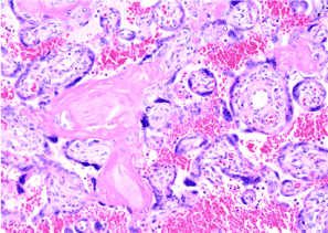
Figure 1. Microscopic fibrin deposit in intervillous space of a FGR placenta. H-E, 200X (Utsu M).
Figure 2. Placental infarction in a preeclampsis complicated by fetal death. (Maeda 1965)
Utsu & others [6] found high echogenesity and high gray level histogram width (GLHW) tissue characterization in a FGR case, who had positive cardiolypin antigen, in the second trimester, detecting fibrin deposit in intervillous space by high ultrasound echogenesity and high GLHW, treated with 5,000 U heparin every day in 17 to 31 weeks of pregnancy, where estimated fetal weight increased to normal level, no hypoxic FHR sign appeared, and normal neonate was achieved, instead the fetus died in previous pregnancy of the same mother [6]. Therefore, a FGR case would be treated with fibrin solving therapy, if fibrin deposit is diagnosed by fetal growth restriction (FGR), high ultrasound echogenesity and high GLHW tissue characterization. The fibrin deposit would be treated even in the loss of FHR acceleration in FGR, to prevent severe hypoxia, where the recovery of acceleration should be confirmed. If not, C-delivery will be indicated. The FHR in the loss of FHR variability is such severe fetal brain damage as the anencephalic fetus, so that C-delivery should be performed before the loss of variability in fetal monitoring [9].
Disappearance of late decelerations after the change to lateral posture from supine one
Since late deceralation (LD) appear by the compression of iliac arteries with contracted pregnant uterus, stopping placental circulation, the LD disappears after changing to lateral posture from supine one [12, 13]. Thus, maternal posture should be changed to lateral one, when there is LD in the FHR. Another cause of LD must be studied, if the posture change is ineffective. Supine hypotension develops pathologic deceleration, also lateral posture should be tried.
Improved umbilical cord circulation in variable deceleration by maternal posture change
It is a common clinical procedure in fetal monitoring in labor to change maternal posture to lateral, in order to remove the compression of umbilical cord, when the FHR showed variable deceleration. The procedure was often effective to keep normal FHR. New vaginal labor anesthesia reduced FHR variable deceleration by the reduction of pelvic floor resistance in vaginal delivery [10].
Tocolysis in uterine hypercontraction
As uterine contraction in the labor reduces the inflow of maternal arterial blood into placenta, uterine hypercontraction decreases placental maternal blood circulation, then produces hypoxic fetal environment. Therefore, the tocolysis with terbutaline in drip infusion is recommended, if intrauterine pressure is high or contraction interval is shorter than 1 min, accompanying hypoxic FHR changes, e.g. late deceleration or bradycardia. Oxytocin infusion in the labor induction was stopped to reduce uterine contraction.
Intensive focused ultrasound ablation
The ablation of nutritive vessels of acardiac twin (TRAP sequence) was achieved by the non-invasive high-intensity focused ultrasound [5]. Various fetal abnormalities will be noninvasively treated by the technique, where associated hypoxia will be treated.
Future fetal distress treatments
- Pharmaceutical treatment
The application of antiglutamate [7], free radical scavenger and others have been reported to cure fetal distress, while their definite effect has not been reported to remove fetal hypoxic changes.
- Treatment of hypoxia due to uterine artery constriction in preeclampsia
The constriction of uterine artery, radial and spiral arteries reduce maternal arterial blood supply to the placenta resulting fetal hypoxia, then finally placental infarction, and fetal death due to the sympathicotonic preelampsia [11]. Therefore, anti-sympathetic treatment will increase placental maternal blood supply, to prevent fetal hypoxia in the future.
- Congenital diseases treated by stem cells and genetic procedures
Congenital diseases develop fetal hypoxia where stem cell therapy will treat tissue defect and neurological damage.
- Treatment of genetic diseases
Genetic diseases will be treated by chromosomal engineering [14] and gene editing [15] in the future, where associated hypoxia will be removed.
- Preterm labor treatment
Possible developing of preterm labor contraction is as follows; A positive feedback loop of labor composed of uterine contraction - the nerve from the uterus to brain – hypothalamus – hypophysis - oxytocin secretion and uterine contraction develops the oscillation of labor contractions [18]. Thus, preterm labor is suppressed, if a feed-back component is rejected. However, no fetal hypoxia was discussed in preterm labor.
Results
Fetal body weight increased to normal level, no hypoxic FHR sign was recorded, and normal newborn was achieved after fibrin deposit solution therapy with heparin administration to the mother in a case of FGR, which associated placental fibrin deposit, while the fetus, who received no fibrin deposit solution, died in previous pregnancy of the same mother [4].
Maternal posture change was effective to improve variable deceleration reducing umbilical cord compression, treating maternal supine hypotension and preventing FHR deceleration caused by the compression of umbilical cord. Late decelerations, developed by the compression of illiac arteies by contracted pregnant uterus at supine posture (Poseiro effect), promptly disappeared after maternal taking lateral posture [12]. FHR deceleration developed by supine hypotension caused by the pressure of inferior vena cava dissapeared by maternal lateral posture too, along with the recovery of blood pressure. The donor twin damage in the TRAP sequence disappeared after the coagulation of nourishing acardiac anomaly with high intensity focused ultrasound [5]
Discussion
Caesarean delivery rate performed by obstetric indication was about 8 % and by fetal hypoxia was 3% of all births in 1981 Japan, namely, actual C-delivery number in one million births will be about 30,000 cases in a year Japan. The effort to reduce C-delivery under the diagnosis of NRFS (fetal distress) will be useful to lower the maternal morbidity.
Since fetal hypoxia is primarily caused by decreased placental villi oxygen transfer from maternal blood, maternal arterial blood flow reducing inter-villous fibrin deposit should be treated. Also, the increase of maternal blood flow in the placenta removing sympathicotonic constriction of uterine artery will improve fetal oxygenation in preeclampsia. Maternal hypoxia or hypotension caused by maternal disorders, massive hemorrhage and supine posture will be improved by maternal treatment or posture change.
2021 Copyright OAT. All rights reserv
Secondly, various fetal disorders, congenital diseases and fetal cardiac failure will develop fetal hypotension reducing fetal placental circulation and oxygen intake. Therefore, the treatment of fetal disorders will improve fetal hypoxia. Noninvasive high-intensity focused ultrasound ablated nourishing vessel of a TRAP sequence would improve the donor twin [5]. Various fetal diseases will be treated improving fetal disorders and hypoxia. Such genetic disorders as trisomies would be treated by chromosomal technology [12] and gene editing [13] improving fetal hypoxia in fetal disorders in the future.
In preterm labor, periventricular echo density (PVE), which is hyperechogenesity around the ventricle, preceded neonatal PVL and CP, when the PVE lasted until preterm birth, but no brain damage was noted in the full term delivery [19]. Therefore, normal infant is expected without hypoxia if preterm labor is suppressed by tocolysis until full term delivery.
Conclusion
Although the treatment of fetal hypoxia usually depends on C-delivery at present, obstetricians would endeavor to find such non-surgical therapy as the solution of fibrin deposit in placental intervillous space, or antisympathetic therapy in preeclampsia. Various maternal, fetal or genetic diseases will be treated in the future to achieve normal vaginal delivery. Preterm labor will also be reduced preventing CP.
References
- Maeda K, Kimura S, Nakano H, Fukui Y, Ozawa S, et al. (1969) Pathophysiology of Fetus. Fukuoka Printing, Proc, 21st JSOG Conv, Shukudai lecture.
- Windle WF (1966) An experimental approach to prevention or reduction of the brain poseirodamage of birth asphyxia. Develop Med Child Neurol 8: 129-140.
- Maeda K (1977) Fetal development and placental function during pregnany. Automedica 2: 13-25.
- Teshima N (1993) Non-reactive pattern diagnosed by ultrasonic Doppler fetal actocardiogram and outcome of the fetuses with non-reactive pattern. Acta Obstet Gynecol Jpn 45: 423-430.
- Okai T, Ichizuka K, Hasegawa J, Matsuoka R, Nakamura M, et al. (2013) First successful case of non-invasive in-utero treatment of twin reversed arterial perfusion sequence by high-intensity focused ultrasound. Ultrasound Obstet Gynecol 42: 112-114. [Crossref]
- Maeda K, Utsu M, Kihaile PE (1998) Quantification of sonographic echogenicity with grey-level histogram width: A clinical tissue characterization. Ultrasound Med Biol 24: 225-234. [Crossref]
- Kochhar A, Justin A, Patrick D, Mazzarella V (1988) Glutamate antagonist therapy reduces neurologic deficits produced by focal central nervous system ischemia. Arch Neurol 45: 148-153. [Crossref]
- Maeda K (2014) Origin of the long-term variability and acceleration of FHR studied for the prevention of cerebral palsy in fetal hypoxia and general insults. J Perinat Med 42: 401-403. [Crossref]
- Maeda K (2014) Modalities of fetal evaluation to detect fetal compromise prior to the development of significant neurological damage. J Obstet Gynaecol Res 40: 2089-2094. [Crossref]
- Utsu M, Kato Y, Takehara K, Maeda K (2016) Safe labor analgesia with vaginal submucosal injection and pudendal nerve block. J Global Anesthesiology 2: 11-13.
- Maeda K (2014) Preeclampsia is caused by continuous sympathetic center excitation due to an enlarged pregnant uterus. J Perinat Med 42: 233-237. [Crossref]
- Poseiro JJ, Mendez-Bauer SV, Caldeyro-Barcia R (1969) Effect of uterine contractions on maternal blood flow through the placenta. 8 PAHO Advisary Committee on Medical Research 161-171.
- Caldeyro-Barcia R, Poseiro JJ, Mendez-Bauer, Gulin LO (1967) Effects of abnormal uterine contrations on fetal heart rate during labor. Proc 5th World Cong Gynaecol Obstet, Sydney, 1967, 9-29.
- Oshimura M, Uno N, Kazuki Y, Katoh M, Inoue T (2015) A pathway from chromosome transfer to engineering resulting in human and mouse artificial chromosomes for a variety of applications to bio-medical challenges. Chromosome Res 23: 111-133. [Crossref]
- Kamminski R, Chen Y, Fischer T, Tedaldi E, Napoli A, et al. (2016) Negative feedback regulation of HIV-1 by gene editing strategy. Sci Rep 6: 31527. [Crossref]
- Yamamoto N, Utsu M, Serizawa M, Ohki S, Murakoshi T, et al. (2000) Neonatal periventricular leukomalacia preceded by fetal periventricular echodensity. Fetal Diagn Ther 15: 198-208. [Crossref]
- Utsu M, Kato Y, Takehara K, Maeda K (2016) Safe paracervical block and pudendal nerve block anesthesia in the labor. J Global Anesthesiology 2016; 2: 11-13.
- Maeda K (2013) Uterine contractions in normal labor developed by a positive feed-back and oscillation. J Health Med Informat 4: 130.
- Yamamoto N, Utsu M, Serizawa M, Ohki S, Murakoshi T, et al. (2000) Neonatal periventricular leukomalacia preceded by fetal periventricular echodensity. Fetal Diagn Ther 15: 198-208. [Crossref]