Abstract
A ventriculoperitoneal shunt is not an absolute contraindication to peritoneal dialysis. However, peritoneal dialysis in patients with a ventriculoperitoneal shunt is rare because the ventriculoperitoneal shunt may need to be removed if peritonitis occurs. Peritonitis may occur after the insertion of a catheter during peritoneal dialysis; this can be due to percutaneous contamination, transluminal bacterial shifting, or occult intra-abdominal abscesses. Treatment depends on the etiology of the peritonitis, as determined by a dialysate culture. The percutaneous method of catheter insertion is commonly used to establish the peritoneal dialysis pathway and is commonly associated with lower infection rates compared with the surgical method of catheter insertion. We present a rare case of peritonitis secondary to abscess in the psoas muscle after percutaneous peritoneal dialysis catheter insertion in a patient with hydrocephalus and a ventriculoperitoneal shunt. We began initial treatment with an intraperitoneal antibiotic for 4 weeks; the patient's peritonitis was cured, the size of the psoas muscle abscess decreased, and the ventriculoperitoneal shunt was preserved.
Key words
peritoneal dialysis, ventriculoperitoneal shunt, peritonitis, psoas muscle abscess
Introduction
Peritonitis is a complication that must be avoided when a catheter is inserted during peritoneal dialysis. Microsurgical techniques tend to yield lower rates of peritonitis compared with traditional open surgical techniques; however, peritoneal dialysis can induce peritonitis through a number of mechanisms, including transluminal bacterial shifting due to abnormal bowel movements or occult intra-abdominal abscesses. The treatment of peritonitis is determined by its etiology. A patient's condition may influence the decision on whether to proceed with peritoneal dialysis. For instance, peritoneal dialysis is rarely administered to patients with a ventriculoperitoneal shunt. We present a case of peritonitis secondary to occult abscess in the psoas muscle after percutaneous peritoneal dialysis catheter insertion in a patient with hydrocephalus and a ventriculoperitoneal shunt; the peritonitis was successfully treated with a course of intraperitoneal antibiotics.
Case Presentation
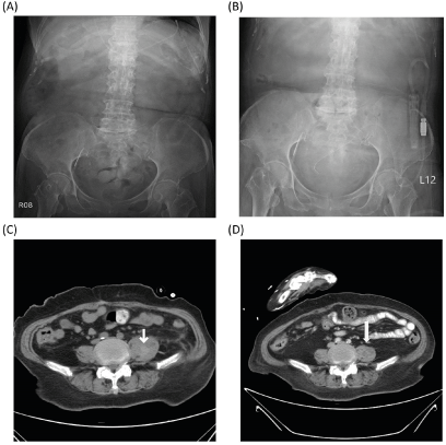
A 68-year-old woman with a history of stroke with hydrocephalus s/p and a ventriculoperitoneal shunt was admitted with end-stage renal disease. A biochemical examination revealed azotemia, normocytic anemia, and metabolic acidosis. A kidney, ureter, and bladder X-ray revealed a ventriculoperitoneal shunt in the upper abdomen area (Figure 1A). A Tenckhoff catheter was successfully inserted using an echo-guided percutaneous technique for peritoneal dialysis. The catheter tip was positioned in the pelvis, and the dialysate performed the in-and-out drainage function effectively (Figure 1B). However, turbid dialysate was noted on the next day of insertion. Biochemical analysis of the dialysate indicated peritonitis with neutrophil dominance, and the dialysate culture showed infection with Enterobacter cloacae. Abdominal computed tomography was performed to identify the etiology of the peritonitis and incidentally identified a left psoas muscle abscess with hematoma (Figure 1C). The ventriculoperitoneal shunt may cause the Enterobacter cloacae to spread, which may result in meningitis, thus the cerebrospinal fluid was checked, and no inflammatory reaction was found. Because it was difficult to approach the abscess to do a tissue aspiration biopsy, we began initial treatment with an intraperitoneal antibiotic. After a four-week intraperitoneal Ciprofloxacin treatment, the peritonitis subsided, and the left psoas muscle abscess with hematoma decreased in size (Figure 1D). The patient was asymptomatic and in good health and continued with peritoneal dialysis.

Figure 1. (A) Kidney, Ureter, and Bladder X-ray Indicating a Ventriculoperitoneal Shunt (white arrow); (B) Kidney, Ureter, and Bladder X-ray Indicating a Peritoneal Dialysis Catheter (white arrow); (C) Abdominal Computed Tomography Indicating a Left Psoas Muscle Abscess with Hematoma (white arrow); (D) Abdominal Computed Tomography Indicating the Decrease in Size of Left Psoas Muscle Abscess with Hematoma (white arrow)
Discussion
Peritoneal dialysis is a common renal replacement therapy for patients with end-stage renal disease. Three methods for tube insertion in peritoneal dialysis are available: open surgery, laparoscopic surgery, and percutaneous insertion (Seldinger). Studies on peritoneal dialysis in patients with a ventriculoperitoneal shunt are rare, although peritoneal dialysis is not an absolute contraindication in patients with a ventriculoperitoneal shunt. Only a few case reports have shared similar experiences [1]. In this case, a percutaneous peritoneal tube was successful inserted without bowel injury or cerebrospinal fluid leak from the exit site of the peritoneal dialysis. The worst-case scenario that we envisioned was having to remove the ventriculoperitoneal shunt once peritonitis occurred. Although peritonitis occurred within a day of the insertion of the peritoneal dialysis catheter, we chose to preserve the catheter and treat the peritonitis.
Contamination by percutaneous procedure is the most common etiology of peritonitis that occurs within a day of insertion. Gram-positive cocci are often isolated in dialysate, and Staphylococcus aureus is the most common bacteria. In this patient, Gram-positive cocci infection by procedure was excluded according to the dialysate culture, and the occult psoas muscle abscess was determined to be cause of the peritonitis. Risk factors for primary psoas muscle abscesses include diabetes, chronic kidney disease, and a compromised immune system. Trauma and hematoma formation may be predisposing factors for secondary psoas muscle abscesses. Klebsiella pneumoniae is a common cause of psoas abscesses in Taiwan, especially in patients with diabetes [2]. Streptococcus pneumoniae, Streptobacillus moniliformis, Staphylococcus lugdunensis, Actinomyces israeli, Salmonella, and Candida albicans have also been cited as causes of psoas abscesses [3-8]. Enterobacteriaceae peritonitis is a serious complication in peritoneal dialysis, and the most common species is Escherichia coli. Enterobacter cloacae is uncommon in Enterobacteriaceae peritonitis, and the percentage of Enterobacter cloacae in peritonitis was only 2.4% in a previous study [9]. Enterobacter cloacae in a psoas muscle abscess with hematoma is rarely seen in the general population and seldom results in peritonitis.
Enterobacteriaceae are a large, heterogeneous family of Gram-negative bacteria. Two of its well-known species, Enterobacter aerogenes and Enterobacter cloacae have taken on clinical significance as opportunistic bacteria and have emerged as nosocomial pathogens from intensive care patients [5]. Enterobacter cloacae is naturally resistant to ampicillin, amoxicillin–clavulanic acid, cephalothin, and cefoxitin because the production of constitutive AmpC β-lactamase has led to β-lactam resistance [10]. Enterobacter cloacae is usually sensitive to fluoroquinolones and Carbapenems. Thus, we chose Ciprofloxacin for intraperitoneal therapy following the results from the dialysate culture sensitivity test (Table 1).
Table 1. Fixed Effects of amputation rates across various regions
Enterobacter cloacae |
+ |
MIC |
Amikacin (AN) |
S |
≤ 8 |
Gentamicin (GM) |
S |
≤ 2 |
Imipenem (IPM) |
S |
0.5 |
Trinethoprim-sulf amethoxazole (SXT) |
S |
≤ 0.5/9.5 |
Ampicillin (AM) |
R |
>16 |
Ciprofloxacin (CIP) |
S |
≤ 0.5 |
Cefotaxime (CTX) |
R |
>32 |
Cefmetazole (CMZ) |
R |
>32 |
Ceftazidime (CAZ) |
R |
>16 |
Ceftriaxone (CRO) |
R |
>32 |
Piperacillin-tazobactam (TZP) |
R |
>64/4 |
Cefepime (FEP) |
I |
4 |
Levofloxacin (LVX) |
S |
≤ 1 |
Meropenem (MEM) |
S |
≤ 0.25 |
Sulbactam/Ampicillin (SAM) |
R |
>16/8 |
Cefazolin (CZ) |
R |
>16 |
Tigecycline (TGC) |
S |
2 |
In conclusion, peritoneal dialysis peritonitis secondary to psoas muscle abscess is a rare and challenging condition when it occurs in a patient with a ventriculoperitoneal shunt. Although the peritoneal and psoas muscle abscess should be treated immediately using intraperitoneal antibiotics, the ventriculoperitoneal shunt should be preserved if possible.
References
- Zapata AZ, Montilla LAL, Restrepo JMR, Acevedo RAG (2015) Ventriculoperitoneal shunt and peritoneal dialysis:“A paradigm for the health team”. Report of 4 cases. Rev Colombiana de Nefrología 2: 152-156.
- Ricci MA, Rose FB, Meyer KK (1986) Pyogenic psoas abscess: worldwide variations in etiology. World J Surg 10: 834-843. [Crossref]
- Aoyama M, Nemoto D, Matsumura T, Hitomi S (2015) A fatal case of iliopsoas abscess caused by Salmonella enterica serovar Choleraesuis that heterogeneously formed mucoid colonies. J Infect Chemother 21: 395-397. [Crossref]
- Tamargo Delpón M, Demelo-Rodríguez P, Cano Ballesteros JC, Vela de la Cruz L (2016) Absceso de psoas por Staphylococcus lugdunensis [Psoas abscess caused by Staphylococcus lugdunensis]. Rev Argent Microbiol 48: 119-121. [Crossref]
- Dubois D, Robin F, Bouvier D, Delmas J, Bonnet R, et al. (2008) Streptobacillus moniliformis as the causative agent in spondylodiscitis and psoas abscess after rooster scratches. J Clin Microbiol 46: 2820-2821. [Crossref]
- Giladi M, Sada MJ, Spotkov J, Bayer AS (1996) Pneumococcal psoas abscess: report of a case and review of the world literature. Isr J Med Sci 32: 771-774. [Crossref]
- Lin MF, Lau YJ, Hu BS, Shi ZY, Lin YH (1999) Pyogenic psoas abscess: analysis of 27 cases. J Microbiol Immunol Infect 32: 261-268. [Crossref]
- Yamada Y, Kinoshita C, Nakagawa H (2019) Lumbar vertebral osteomyelitis and psoas abscess caused by Actinomyces israelii after an operation under general anesthesia in a patient with end-stage renal disease: a case report. J Med Case Rep 13: 351. [Crossref]
- Szeto CC, Chow VC, Chow KM, Lai RW, Chung KY, et al. (2006) Enterobacteriaceae peritonitis complicating peritoneal dialysis: a review of 210 consecutive cases. Kidney Int 69: 1245-1252. [Crossref]
- Davin-Regli A, Pagès JM (2015) Enterobacter aerogenes and Enterobacter cloacae; versatile bacterial pathogens confronting antibiotic treatment. Front Microbiol 6: 392. [Crossref]

