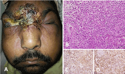
A 36-year-old man presented with crusted lesions on his forehead, dorsum of nose and right eye that had developed over a period of 2 months. He had occasional bloody and purulent discharge, without pain, and did not have constitutional or other systemic symptoms. Ocular examination revealed bilateral peri-orbital edema with erythema, conjunctival chemosis with right upper eyelid completely replaced by crusts, exposing the cornea (Figure 1A) resulting in absent vision on the right side. A clinical diagnosis of lethal midline granuloma was considered. Hematological investigations revealed leucocytosis with relative lymphocytosis and raised ESR. Urine microscopy (for casts or hematuria) and sputum analysis were normal. Biochemical tests for c-ANCA, p-ANCA and HIV were negative. Chest X-ray was normal. Computed tomography revealed soft tissue thickening and bony involvement with no lymphadenopathy or metastases. Multiple skin biopsies eventually confirmed NK/T cell lymphoma (Figures 1B-1D). The patient was admitted and SMILE regimen chemotherapy was started. However, the patient developed immunosuppression and meningitis with disease progression leading to death during hospital stay.

Figure 1A. Clinical image showing crusted lesion over forehead, root of nose and right eye with necrotic slough and blood crusts and exposed cornea on the right side, Figure 1B-1D. Photomicrograph showing an infiltrate comprising of intermediate sized atypical lymphoid cells (B, H&E x400) expressing NK- T cell markers CD56 (C) and Granzyme (D).
Informed written consent was obtained from the patient’s father.
Nil
Article Type
Image Article
Publication history
2021 Copyright OAT. All rights reserv
Received date: July 10, 2017
Accepted date: July 13, 2017
Published date: July 18, 2017
Copyright
© 2017 Sakthivel P. This is an open-access article distributed under the terms of the Creative Commons Attribution License, which permits unrestricted use, distribution, and reproduction in any medium, provided the original author and source are credited.
Citation
Sakthivel P, Rajeshwari M (2017) Lethal midline granuloma. Glob Imaging Insights 2: DOI: 10.15761/GII.1000128
Corresponding author
Pirabu Sakthivel, M.S., DNB. ENT
Department of Otorhinolaryngology and Head & Neck surgery, All India Institute of Medical Sciences, New Delhi – 110029, India
E-mail : bhuvaneswari.bibleraaj@uhsm.nhs.uk

