Purpose: To evaluate and compare the immediate effects of rapid maxillary expansion (RME) on the nasal cavity dimensions in subjects treated with Haas-type and Hyrax-type expanders using cone-beam computed tomography (CBCT).
Methods: Thirty-one growing patients (mean age=10.8 years old) with transverse maxillary deficiency were randomly divided into two groups: Haas group (n=16) and Hyrax group (n=15). All patients had RME with initial screw activation of 4 quarter turns followed by activations of ½ turns per day (0.4mm) until the expansion reached 8mm opening at the screw. CBCT scans were taken before RME (T1) and at the end of the active phase of expansion (T2). Vertical and transverse measurements were performed at different regions of nasal cavity for comparison between groups and treatment times. Intraclass correlation coefficient (ICC), paired Student's t-test and repeated measures ANOVA (α-level p<0.05) were calculated.
Results: RME procedure increased every vertical and lateral nasal cavity dimensions (p<0.001). The most significant gains were observed in the lower portions of the nasal cavity located at the anterior and medial regions.
Conclusions: Both appliances were able to create significant changes in the dimensions of the nasal cavity immediately after RME. However, the Hyrax-type expander presented greater transverse effects on the airway.
cone-beam computed tomography, palatal expansion technique, nasal cavity
Respiratory function can influence the development of facial growth and dental occlusion. In individuals with nasal obstructions and reduced airflow, a lower position of the tongue and mandible can facilitate the passage of air through the mouth [1]. During growth, mouth breathing can create an imbalance between the forces leading to muscular and postural alteration which may result in dentoskeletal changes [2,3]. Patients with transverse maxillary deficiency usually associated to constricted nasal dimensions, trends to present greater resistance to nasal airflow and mouth breathing habit when compared to patients with normal maxillary arch [4]. The application of orthodontic or orthopedic forces for maxillary expansion in growth period could restore the normal development of the face, as well as maxillomandibular and occlusal relationships. When RME is used, tension effects on bone formation occur directly in the maxillary sutures and, by indirect transmission. In this case, the effect of the force applied during the expansion procedure can reach other facial bone sutures in adjacent locations [5]. For example, maxillary expansion can move the walls of nasal cavity laterally and increase the nasal dimensions [4-17]. As a result, nasal breathing can be improved [4,5,14-17].
Different types of maxillary expansion appliances have been reported in the literature. Among these, the most popular are Haas-type and Hyrax-type expanders, which differ basically in the method used for anchorage and delivery of intraoral forces [9,10]. Previous reports using conventional radiographs did not observe differences on how these appliances act on the nasal cavity [4,9]. Three-dimensional imaging methods allow the assessment of the upper airways and nasal structures with no distortion and magnification of images, or superimposition of other structures [18]. The advent of cone-beam computed tomography (CBCT) has made the assessment of the nasomaxillary region possible with less radiation risk to the patient in comparison to multislice CT [19-22].
The purpose of this retrospective study using CBCT was to evaluate and compare the immediate effects on the nasal cavity (NC) produced by Haas and Hyrax expanders in patients with transverse maxillary deficiency.
Ethical approval was obtained from Pontifical Catholic University of Rio Grande do Sul (PUCRS), Research Committee (08/04147). The sample was composed of patients who sought orthodontic treatment at PUCRS School of Dentistry. The inclusion criteria were transverse maxillary deficiency, mixed or permanent dentition, patients younger than 15 years old, with no previous orthodontic or orthopedic treatment, no adenoidectomy or tonsillectomy, as well as no syndromes or craniofacial anomalies. Patients should have pre and post expansion CBCTs. Thirty-one patients were selected (20 girls and 11 boys) and belonged to two groups according to the type of expander appliances: Haas group (n=16), and Hyrax group (n=15) (Figure 1). Skeletal and chronological mean ages where 10.9 and 10.8 years old, respectively (Table 1).
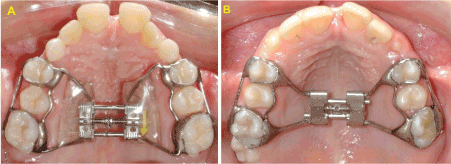
Figure 1. In occlusal view, the expander appliances used in this clinical trial: a Haas-type and b Hyrax-type.
Table 1. Sample characteristics.
|
n |
Chronological age mean |
Skeletal age mean |
Male |
Female |
Haas Group |
16 |
10.6y |
11y |
5 |
11 |
Hyrax Group |
15 |
11y |
10.9y |
6 |
9 |
Total |
31 |
10.8y |
10.9y |
11 |
20 |
The RME activation protocol was the same for both groups, with daily activation of 2 turns, until reaching 8mm of expansion in the screw, usually achieved in 19 days.
CBCT scans were taken in upright position before RME (T1) and immediately after the active phase of expansion (T2) using i-CAT scanner (Imaging Sciences Int, Hatfield, PA, USA) set at 120 kV, 8 mA, 40 seconds of scanning time and 0.3mm voxel size. The patient data were reconstructed and exported as DICOM (Digital Imaging and Communication in Medicine) files. All measurements were performed by a blinded operator (A.M.M.), using InVivoDental Software (Anatomage, San Jose, CA, USA).
Images of each patient's head were first reoriented in axial, coronal and sagittal planes according to anatomical references as shown in Figure 2.

Figure 2. Orientation of the patient's head: a axial plane, midpoint of the anterior border of the foramen magnum and the nasion point (N); b coronal plane, right and left orbital points; c sagittal plane, palatal plane.
In axial view, the nasal cavity (NC) was divided in 3 different regions, using the following anteroposterior references (Figure 3).
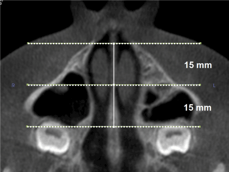
Figure 3. Axial image of the maxilla showing the location of the sections used as anteroposterior reference planes for nasal cavity evaluation.
- Anterior: Corresponding to the pyriform aperture (region 1)
- Medial: 15mm posterior to the region 1 (region 2)
- Posterior: 15mm posterior to the region 2 (region 3)
Transverse measurements at the NC were performed in coronal sections. Every NC regions located at anterior, medial and posterior sites were subdivided horizontally into three thirds from upper to lower limits of the nasal contour and lower, middle and upper transverse dimensions were measured (Figures 4A-4C). Also, vertical dimension was evaluated at the anterior region of NC (Figure 4D), an area that shows well-defined bone boundaries at the coronal view. Horizontal and vertical assessments of the NC were performed using linear measurements as described in Table 2.
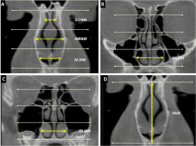
Figure 4. Division of the nasal cavity into thirds; a anterior, b medial and c posterior portions: a width of the lower (ALNW), medial (AMNW) and upper (AUNW) thirds of the nasal cavity in its anterior portion; b width of the lower third of the nasal cavity in its medial portion (MLNW); c width of the lower third of the nasal cavity in its posterior portion (PLNW); d anterior nasal height (ANH).
Table 2. Linear vertical and transverse measurements used to evaluate the nasal cavity.
Anterior Region |
ANH |
Anterior nasal height |
Vertical distance between the upper and lower limits of the nasal cavity |
ALNW |
Anterior lower nasal width |
Width of the medial region of the lower third of the nasal cavity |
AMNW |
Anterior medial nasal width |
Width of the medial region of the middle third of the nasal cavity |
AUNW |
Anterior upper nasal width |
Width of the medial region of the upper third of the nasal cavity |
Medial Region |
MLNW |
Medial lower nasal width |
Width of the medial region of the lower third of the nasal cavity |
Posterior Region |
PLNW |
Posterior lower nasal width |
Width of the medial region of the lower third of the nasal cavity |
Statistical analysis
The variables were described quantitatively by means and standard deviations. To determine the intrarater reliability of measures, CBCT scans of 10 patients were randomly selected and measurements at T1 and T2 were repeated with three weeks interval, and Intraclass correlation coefficient (ICC) was calculated.
Kolmogorov-Smirnov test was used to check the normality of the variables. Student's t-test for paired samples was calculated for comparison between T1 and T2 in both groups (Table 4), in each group (Tables 5 and 6) and RME effects were evaluated. Analysis of variance (ANOVA) for repeated measures was used for comparison between groups and treatment times (Table 7).
The statistical analyses were calculated using SPSS Statistics (version 14.0, SPSS, Chicago, Ill) at p<0.05.
The intrarater evaluations were considered reliable and ICC ranged from 0.92 to 0.97.
Significant increases were observed in all NC measurements when evaluating the effects of RME on both groups. Table 3 shows the means assessed at T1 and T2 and the treatment effects (T2-T1), considering both groups.
The patients treated with Haas-type and Hyrax-type expanders showed significant increases for all variables (Tables 4 and 5) with the greatest effects in the lower portions located at anterior and medial regions of the NC.
As shown in Table 6, there were significant increases in the NC dimensions in patients treated using Hyrax-type expanders in comparison to the Haas group. Width at middle third of the NC in its anterior portion (AMNW), widths of the lower third at medial (MLNW) and at posterior portions (PLNW) of the NC were significantly increased when Hyrax-type appliances were used.
Since RME introduction [23], the idea that the effects of maxillary expansion may extend to respiratory function led Orthodontists and Otolaryngologists to study the anatomical [4-16] and functional [4,14,16,17] outcomes of the RME on the nasal cavity (NC). Most of these studies used conventional x-rays to measure these effects, but lack of detail and issues involving image superimposition have been the major drawbacks to these studies [18]. This research used CBCT to assess immediate changes in NC dimensions following RME treatment. The NC was evaluated at different depths (anterior, medial and posterior). The anterior portion was evaluated in the pyriform aperture due to its proximity to the nasal valve, a region that provides the most significant nasal airway resistance [13]. The medial and posterior regions of the NC were determined at 15mm and 30mm, respectively, from the anterior portion, with the purpose of allowing the reproduction of the same regions in CBCT of both treatment periods (T1 and T2).
The results showed that increases in NC dimensions are achieved after RME in patients with transverse maxillary deficiency, in agreement with previous studies, which reported increases in height [6,15], width [4,12,16], volume [14] and at transverse section areas [13,16] of the NC.
An increase of 1.13mm in anterior nasal height was observed (Table 3), supporting other studies [6-15], which used posteroanterior cephalometric radiographs and found a trend towards vertical gain in the NC after RME. These findings could be explained by the lateral rotation of the maxillary bones around a fulcrum located at the upper frontonasal suture in a coronal plane [15]. This pattern of bone separation produces a lowering of the palatine processes of the maxilla, which comprise the nasal floor, contributing to a vertical increase in nasal dimensions [5-15].
In this study, the midpalatal suture expanded like a fan in the axial plane, with more expansion at the lower third of the NC in its anterior portion (ALNW=2.86mm) than in its posterior portion (PLNW=2.15mm) (Table 3). This greater expansion in the anterior region could be explained by the resistance offered by the pterygoid plates of the sphenoid bone, to the maxillary tip movement during RME [24]. Another possible explanation is the maxillary expansion biomechanics, i.e., the direction of the expansion force produced by the expanders would be located anterior to the center of resistance of each maxillary half [23].
In the coronal plane, the intermaxillary suture also follows a triangular opening pattern, whereby the most significant expansion occurs at the level of the incisors, decreasing towards the skull base [5,8,10,24]. Therefore, the NC expands in three dimensions: horizontal, vertical and anterior, with maximum expansion occurring at the level of the lower turbinates [15]. This finding was also observed in this study, which evaluated only the vertical and transverse directions (Tables 3-5). The highest transverse increase was observed in the lower region at anterior portion of the NC (ALNW=2.86mm), decreasing towards the upper region (AUNW=0.88mm) (Table 3). Previous studies using posteroanterior cephalometric radiographs found the same nasal expansion pattern [6,7]. However, one limitation of these studies was determining which regions of the NC expanded in 2-dimensional cephalograms. The CBCT scans allow independent evaluations of the anterior, medial and posterior portions of the NC.
Table 3. Means of nasal cavity dimensions at T1 and T2, and effects of RME (T2-T1), considering both expander groups.
Variables |
Haas and Hyrax groups (n=31)
T1 (mm) T2 (mm) T2-T1 (mm)
(mean ± SD) (mean ± SD) (mean ± SD) |
P value |
ANH |
39.18 ±3.04 |
40.31 ±3.11 |
1.13 ±0.88 |
<0.001* |
ALNW |
20.47 ±1.74 |
23.33 ±1.68 |
2.86 ±0.98 |
<0.001* |
AMNW |
20.21 ±1.46 |
22.26 ±1.56 |
2.05 ±0.83 |
<0.001* |
AUNW |
9.56 ±1.61 |
10.44 ±1.60 |
0.88 ±0.74 |
<0.001* |
MLNW |
26.87 ±2.18 |
29.56 ±2.38 |
2.69 ±0.88 |
<0.001* |
PLNW |
26.02 ±1.84 |
28.18 ±2.16 |
2.15 ±0.86 |
<0.001* |
SD, Standard Deviation.
* Statistically significant difference (p<0.05).
Table 4. Means at T1 and T2, and effects of RME on the nasal cavity in patients treated using Haas-type expander.
Variables |
Haas Group (n=16)
|
P value |
T1 (mm)
(mean ± SD) |
T2 (mm)
(mean ± SD) |
T2-T1 (mm)
(mean ± SD) |
ANH |
38.73 ±3.32 |
39.67 ±3.34 |
0.93 ±0.76 |
<0.001* |
ALNW |
20.62 ±1.52 |
23.30 ±1.17 |
2.67 ±0.94 |
<0.001* |
AMNW |
19.93 ±1.72 |
21.64 ±1.59 |
1.71 ±0.66 |
<0.001* |
AUNW |
9.34 ±1.24 |
10.06 ±1.30 |
0.71 ±0.72 |
<0.001* |
MLNW |
26.59 ±1.94 |
28.83 ±2.10 |
2.24 ±0.72 |
<0.001* |
PLNW |
26.17 ±1.50 |
27.95 ±1.71 |
1.77 ±0.86 |
<0.001* |
SD, Standard Deviation
* Statistically significant difference (p<0.05)
Table 5. Means at T1 and T2, and effects of RME on the nasal cavity in patients treated using Hyrax-type expander.
Variables |
Hyrax Group (n=15)
|
P value |
T1 (mm)
(mean ± SD) |
T2 (mm)
(mean ± SD) |
T2-T1 (mm)
(mean ± SD) |
ANH |
39.65 ±2.74 |
40.99 ±2.79 |
1.34 ±0.97 |
<0.001* |
ALNW |
20.31 ±1.99 |
23.38 ±2.14 |
3.06 ±1.01 |
<0.001* |
AMNW |
20.51 ±1.10 |
22.93 ±1.25 |
2.42 ±0.85 |
<0.001* |
AUNW |
9.79 ±1.94 |
10.85 ±1.82 |
1.06 ±0.75 |
<0.001* |
MLNW |
27.16 ±2.44 |
30.34 ±2.48 |
3.18 ±0.79 |
<0.001* |
PLNW |
25.87 ±2.19 |
28.43 ±2.59 |
2.56 ±0.68 |
<0.001* |
SD, Standard Deviation
* Statistically significant difference (p<0.05)
In this study, the amount of expansion at the lower third of the NC in the anterior, medial and posterior portions corresponded to 36.2%, 33.7% and 27.5% of expansion screw activation (8mm), respectively. Garib, et al. [10] achieved - with 7mm screw activation - a similar skeletal response in the posterior region of the nasal cavity, but their assessment was performed on a smaller sample (8 patients). Using CBCT, previous studies [11,12] found greater transverse effects in the posterior region of the NC compared to this study. Garrett, et al. [11] observed a 37.2% increase in lower nasal width at the posterior region in response to 5 mm screw activation. However, among the sample of 30 individuals used by these authors [11], 13 used a Hyrax-type expanders with two bands and the jackscrew positioned in a more posterior region compared to the present study that had four bands in the appliance design. In an evaluation using a sample composed by children subjected to RME with a modified Haas-type appliance (full occlusal and palatal acrylic coverage), Christie, et al. [12] observed increases of 33.23% in relation to the mean screw activation (8.19mm) at the posterior region and 37.32% at the medial region of NC. It is noteworthy that in these studies assessment of the posterior NC was undertaken in the region of the first permanent molars while in the present study assessment was performed in a more posterior region of the NC. In a research of RME effects using a Haas-type appliance, Chung and Font [8] observed a smaller skeletal response of the NC, with increase of 23.1% in comparison to the amount of screw activation. However, these results were obtained in 2-dimensional cephalograms at start and 3 months after RME.
The comparison between the two groups of this study showed that both appliances produced significant gains in the nasal dimensions. However, the Hyrax Group (Table 5) showed higher values for all variables evaluated in the nasal cavity in comparison to the Haas Group (Table 4). One possible explanation for this differential effect of the appliances on the transverse measures, in places of greater resistance to expansion, such as in the posterior and medial NC and anterior middle third (Table 6), could be the differences in appliance design. In the Hyrax-type appliance, the jackscrew was directly connected to the bands by a rigid stainless steel framework (1.4mm), unlike the Haas-type appliance, where acrylic was responsible for connecting the stainless steel framework (1.0mm) to the jackscrew. According to previous studies [25], appliance designs that use an acrylic interface with the teeth are far less stiff than those constructed solely of soldered stainless steel wire, as in the case of the hyrax-type expander. The differences observed in this study were statistically significant and, despite their small values (Table 6), they may have clinical significance as the relationship between nasal area and nasal airflow is exponential, i.e., any minor alteration can induce major changes in nasal airflow [26]. Other studies using posteroanterior radiographs [4,9] and scans [10] found no significant differences between the effects of the appliances.
Table 6. Comparison between Haas and Hyrax groups regarding the effects of RME on the nasal cavity.
Variables |
Haas Group (n=16)
T2-T1 (mm)
(mean ± SD) |
Hyrax Group (n=15)
T2-T1 (mm)
(mean ± SD) |
Mean difference
Hyrax-Haas
(mean ± SE) |
P value |
ANH |
0.93 ±0.76 |
1.34 ±0.97 |
0.40±0.31 |
.202 |
ALNW |
2.67 ±0.94 |
3.06 ±1.01 |
0.38±0.35 |
.284 |
AMNW |
1.71 ±0.66 |
2.42 ±0.85 |
0.70±0.27 |
.015* |
AUNW |
0.71 ±0.72 |
1.06 ±0.75 |
0.34±0.26 |
.209 |
MLNW |
2.24 ±0.72 |
3.18 ±0.79 |
0.93±0.27 |
.002* |
PLNW |
1.77 ±0.86 2.56 ±0.68 |
0.78±0.28 |
.010* |
SD, Standard Deviation
SE, Standard Error
* Statistically significant difference (p<0.05)
CBCT provides accurate images of the airway spaces that are consistent with the actual anatomical structures [21]. However, it should be observed that this diagnostic method expresses statistical changes in the airway spaces without revealing whether any benefits in respiratory function were achieved. Studies evaluating changes in the NC resulting from RME have shown that an increase in nasal dimensions can consistently reduce nasal resistance, improve the respiratory capacity [4,14,16,17,27], and decrease the apnea hypopnea index in children with obstructive sleep apnea syndrome [28]. According to Baratieri, et al. [29], in a systematic review, when the midpalatal suture is opened in growing patients, the widening of the nasal cavity is stable over long term. The present study suggests that when RME is performed using Haas-type or Hyrax-type appliances favorable anatomical conditions for nasal function in patients with maxillary atresia can be observed. The RME treatment is also beneficial in avoiding the development of facial skeletal asymmetry that may lead to functional and structural imbalances in growing patients with posterior crossbite [30]. However, implementing RME as the unique purpose of improving nasal patency is not justified [16,17] as nasal obstruction may have other etiologic factors which require investigation.
Based on this retrospective clinical trial, the following conclusions can be drawn:
- The 8mm expansion performed with both the Haas-type and Hyrax-type appliances increased the vertical and horizontal dimensions of the nasal cavity, following a decreasing pattern of expansion, from the lower towards the upper region, and from the anterior towards the posterior region.
The Hyrax-type expander provided more significant increases in transverse dimensions than the Haas-type appliance in the anterior (AMNW), medial (MLNW) and posterior (PLNW) regions of the nasal cavity.
- Subtelny JD (1980) Oral respiration: Facial maldevelopment and corrective dentofacial orthopedics. Angle Orthod 50: 147-164. [Crossref]
- Valera FC, Travitzki LV, Mattar SE, Matsumoto MA, Elias AM, Anselmo-Lima WT (2003) Muscular, functional and orthodontic changes in pre-school children with enlarged adenoids and tonsils. Int J Pediatr Otorhinolaryngol 67: 761-770. [Crossref]
- Basheer B, Hegde KS, Bhat SS, Umar D, Baroudi K (2014) Influence of mouth breathing on the dentofacial growth of children: A cephalometric study. J Int Oral Health 6: 50-55. [Crossref]
- Hershey HG, Stewart BL, Warren DW (1976) Changes in nasal airway resistance associated with rapid maxillary expansion. Am J Orthod 69: 274-284. [Crossref]
- Haas AJ (1961) Rapid expansion of the maxillary dental arch and nasal cavity by opening the midpalatal suture. Angle Orthod 31: 73-90.
- Cross DL, McDonald JP (2000) Effect of rapid maxillary expansion on skeletal, dental, and nasal structures: a postero-anterior cephalometric study. Eur J Orthod 22: 519-528. [Crossref]
- Usumez S, Iseri H, Orhan M, Basciftci FA (2003) Effect of rapid maxillary expansion on nocturnal enuresis. Angle Orthod 73: 532-538. [Crossref]
- Chung CH, Font B (2004) Skeletal and dental changes in the sagittal, vertical, and transverse dimensions after rapid palatal expansion. Am J Orthod Dentofacial Orthod 126: 569-575. [Crossref]
- Oliveira NL, Da Silveira AC, Kusnoto B, Viana G (2004) Three-dimensional assessment of morphologic changes of the maxilla: a comparison of 2 kinds of palatal expanders. Am J Orthod Dentofacial Orthop 126: 354-362. [Crossref]
- Garib DG, Henriques JF, Janson G, Freitas MR, Coelho RA (2005) Rapid maxillary expansion - tooth tissue-borne versus tooth-borne expanders: a computed tomography evaluation of dentoskeletal effects. Angle Orthod 75: 548-557.
- Garrett BJ, Caruso JM, Rungcharassaeng K, Farrage JR, Kim JS, Taylor GD (2008) Skeletal effects to the maxilla after rapid maxillary expansion assessed with cone-beam computed tomography. Am J Orthod Dentofacial Orthop 134: 8-9. [Crossref]
- Christie KF, Boucher N, Chung CH (2010) Effects of bonded rapid palatal expansion on the transverse dimensions of the maxilla: A cone-beam computed tomography study. Am J Orthod Dentofacial Orthop 137: 79-85. [Crossref]
- Bicakci AA, Agar U, Sökücü O, Babacan H, Doruk C (2005) Nasal airway changes due to rapid maxillary expansion timing. Angle Orthod 75: 1-6. [Crossref]
- Doruk C, Sökücü O, Bicakci AA, Yilmaz U, Tas F (2007) Comparison of nasal volume changes during rapid maxillary expansion using acoustic rhinometry and computed tomography. Eur J Orthod 29: 251-255. [Crossref]
- Gray LP (1975) Results of 310 cases of rapid maxillary expansion selected for medical reasons. J Laryngol Otol 89: 601-614. [Crossref]
- Enoki C, Valera FC, Lessa FC, Elias AM, Matsumoto MA, Anselmo-Lima WT (2006) Effect of rapid maxillary expansion on the dimension of the nasal cavity and on nasal air resistance. Int J Pediatr Otorhinolaryngol 70: 1225-1230. [Crossref]
- Hartgerink DV, Vig PS, Abbott DW (1987) The effect of rapid maxillary expansion on nasal airway resistance. Am J Orthod Dentofacial Orthod 92: 381-389. [Crossref]
- Montgomery WM, Vig PS, Staab EV, Matteson SR (1979) Computed tomography: a three-dimensional study of the nasal airway. Am J Orthod 76: 363-375. [Crossref]
- Mozzo P, Procacci C, Tacconi A, Martini PT, Andreis IA (1998) A new volumetric CT machine for dental imaging based on the cone-beam technique: preliminary results. Eur Radiol 8: 1558-1564. [Crossref]
- Farman AG, Scarfe WC (2006) Development of imaging selection criteria and procedures should precede cephalometric assessment with cone-beam computed tomography. Am J Orthod Dentofacial Orthop 130: 257-265. [Crossref]
- Lascala CA, Panella J, Marques MM (2004) Analysis of the accuracy of linear measurements obtained by cone beam computed tomography (CBCT- New Tom). Dentomaxillofac Radiol 33: 291-294. [Crossref]
- Ludlow JB, Ivanovic M (2008) Comparative dosimetry of dental CBCT devices and 64-slice CT for oral and maxillofacial radiology. Oral Surg Oral Med Oral Pathol Oral Radiol Endod 106: 106-114. [Crossref]
- Angell EH (1860) Treatment of irregularity of the permanent or adult teeth. Dent Cosmos 1: 540-544(599-601).
- Bishara SE, Staley RN (1987) Maxillary expansion: clinical implications. Am J Orthod Dentofacial Orthop 91: 3-14. [Crossref]
- Braun S, Bottrel JA, Lee KG, Lunazzi JJ, Legan HL (2000) The biomechanics of rapid maxillary sutural expansion. Am J Orthod Dentofacial Orthop 118: 257-261. [Crossref]
- Roithmann R, Cole P, Chapnik J, Barreto SM, Szalai JP, et al. (1994) Acoustic rhinometry, rhinomanometry, and the sensation of nasal patency: A correlative study. J Otolaryngol 23: 454-458. [Crossref]
- El H, Palomo JM (2014) Three-dimensional evaluation of upper airway following rapid maxillary expansion A CBCT study. Angle Orthod 84: 265-273. [Crossref]
- Villa MP, Rizzoli A, Miano S, Malagola C (2011) Efficacy of rapid maxillary expansion in children with obstructive sleep apnea syndrome: 36 months of follow-up. Sleep Breath 15: 179-184. [Crossref]
- Baratieri C, Alves M, de Souza MM G, de Souza Araújo MT, Cople Maia L (2011) Does rapid maxillary expansion have long-term effects on airway dimensions and breathing? Am J Orthod Dentofacial Orthop 140: 146-156. [Crossref]
- McNamara JA Jr, Lione R, Franchi L, et al. (2015) The role of rapid maxillary expansion in the promotion of oral and general health. Prog Orthod 16: 33. [Crossref]