Down syndrome (trisomy 21) is the most common autosomal chromosome aberration in human. The incidence of the syndrome varies in 1ː700 and 1ː1000 live births according to various studies, with 20% of cases. In aborted material proportion is even higher, with 60% of cases. While 20% of cases with Down syndrome are stillborn.
In these patients, there is a strong predisposition to cardiovascular disease, seizures [1], leukemia [2,3], infections with hepatitis B virus (especially within institutionalized men) [4], upper respiratory tract infections [5], Alzheimer's disease[6], obesity [7], thyroid diseases [8], cardiac anomalies [9] and obstructive sleep apnea [10, 11]. Disruption of the proteostasis network and accumulation of misfolded proteins occur as a result of an abnormality in the number of chromosome 21 [12]. Errors in protein homeostasis could contribute to the observed pathology and decreased cell viability in children with Down syndrome [13].
The systematic review and meta-analysis by Saghazadeh A et al., conducted in 2016, concluded that findings of different micronutrient levels in people with Down syndrome raise the question whether these differences are related to the different metabolic profiles, the common comorbidities or merely reflect Down syndrome [14].
Clinical characteristics
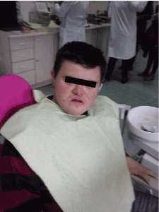
The changes in body of the children with Down syndrome are manifest trough the short stature, short legs and arms, slow skeletal maturation, poor muscle development, always with the presence of obesity, poorly developed male genitalia, short and wide neck, dry and rough skin, straight and smooth hair. The changes in the head and face are characterized by brachycephaly , microcephaly, undeveloped vertex bone (os parientale) of the skull that gives the appearance as if the back of the skull is flattened. Also nasal bones are hypoplastic, with flat nasal bridge. The combination of minor and major abnormalities characterize this syndrome that give typical phenotypic appearance of the head and face ː hypertelorism, small and oblique eyes, spots on the iris, strabismus in 30% of patients, deformed and small ears. The limbs are short, the typical appearance of the hands with ruffle and short fingers and transverse crease on the palms present in about 80% of cases. Figure 1 (own material)

Figure 1. Boy with Down syndrome (own material)
Oral manifestation
Open mouth with protrusion of the hypotonic and fissured tongue, macroglossia, hypoplastic upper jaw, short and hard palate, pseudoprogenic bite, delay eruption of the teeth [15], persistence of some deciduous tooth up to 15 years, the order of eruption of teeth is disturbed [16], hypodontia , microdontia, atypical form of dental crowns and taurodontism [17]. Also patients with Down syndrome can present periodontal disease, premature tooth loss, reduced salivary flow, crowding of teeth in both arches, and decreased occlusal vertical dimension. Increased frequency of periodontal disease, reduced incidence of dental caries [18], often spilling of saliva from the mouth. In these patients, cleft lip, cleft lip and palate or palate only occurs 3 times more often than in the general population. The findings by van der Linden MS et al showed that dental development in children with Down syndrome is similar to the development of control children and that a relationship exists between hypodontia and dental development [18,19].
Altintas NY et al. in their case report article emphasized that implant-retained overdentures with Locator attachment system could be good therapeutic option even for patients with Down syndrome. This therapy can be implemented only for patients with mild mental impairment. The prosthodontist, first of all have to educate patients and caregivers about importance of optimum oral hygiene and also carefully to select patients, which is essential for successful and safe implant treatment of Down syndrome patients [20]. Alqahtani NM et al presented 44-year-old male patient with Down syndrome with moderate intellectual disability. The treatment approach of this patient with congenital and acquired tooth loss and significant occlusal discrepancies is used of a single dental implant [21].
Orthodontic treatment
Before starting of the implementation of the orthodontic treatment within the patients with Down syndrome, it is necessary to restore all decay teeth. If the patient is unable for clinical work, orthodontic treatment can be initiated with a mobile device [22], which usually carried extension of the upper jaw and, if necessary, '' pull '' of the front teeth due to the presence of the pseudoprogenia [23]. Treatment can be continued with a fixed appliance that compensates dental chains, regulates the placement of the teeth in the dental chains and adjust dental bite [24]. Of course, given that there is some degree of disability in these patients, we must often make trade-offs in the orthodontic therapy [25]. Miyazaki H et al. describe orthodontic treatment in a patient with Down’s syndrome with unilateral cleft lip and alveolus. After surgical intervention and further mental and physical growth of the patient, the orthodontic treatment was indicated with multi-bracket. Reverse occlusion was corrected by labial inclination of the incisors [26].
Fakhruddin KS et al. from their clinical study involving 22 children with Down syndrome concluded that routine psychological (Tell-Show-Do) intervention along with visual distraction using video eyewear and use of CDS-IS (computerized delivery system-intrasulcular) system for anesthetic delivery is effective behavior management technique during invasive dental treatment [27].
Considering the great emotional, psychological and material goods that require such member of a family and the modest effects of the expensive therapy, enormous efforts to the prenatal diagnosis are invested nowadays [28-31]. Prenatal diagnosis of a mother who previously gave birth to a child with trisomy, and in women who give birth after the age of 35 years, significantly reduces the number of children born with the disease. However most of children with Down syndrome remain undetected prenatally.
Resently it’s believed that with the analysis of certain ultrasonic parameters associated with the combination of determining α fetal-protein, estriol and β HCG (beta-chorionic gonadotropin) in the mother's blood at the 14th week of pregnancy can be assign degrees of urgency to certain group of pregnant women with the increased risk of delivering a child with trisomy 21. Kochova and its associates [32] considered that this group should undergo prenatal diagnosis by amniocentesis. In this manner 75-80% of pregnancies with a Down syndrome fetus can be detected.
Treatment
In the treatment of children with Down syndrome, the following health professionals are included: pediatricians, family medicine physicians, internists, pediatric dentists, but also the following specialists: a speech-language pathologist [33,34], a physical therapist, an occupational therapist and special education teachers play important role. The main priority of all centers for children with Down syndrome in the world is to provide adequate rehabilitation to these children and their training for independent survival.
Adults with disabilities in our country are employed at a significantly lower rate than adults without disabilities. The situation of the unemployed persons with disabilities is also similar in other countries of the world, even in the United States [33].
Today in many research centers all over the world, the potential pathological mechanisms that cause altered cell physiology in Down syndrome are still under exploration. We hope that they will contribute to the design of better therapeutic strategies that will improve the quality of life [35] of people with Downs syndrome.
References
- Lefter S, Costello DJ, McNamara B, Sweeney B (2011) Clinical and EEG features of seizures in adults with down syndrome. J Clin Neurophysiol 28: 469-473. [Crossref]
- Schwartzman O, Savino AM, Gombert M, Palmi C, Cario G, et al. (2017) Suppressors and activators of JAK-STAT signaling at diagnosis and relapse of acute lymphoblastic leukemia in Down syndrome. Proc Natl Acad Sci USA. 114: E4030-E4039. [Crossref]
- Jastaniah W, Alsultan A, Daama AS, Ballourah W, Bayoumy M, et al. (2017) Treatment results in children with myeloid leukemia of Down syndrome in Saudi Arabia: A multicenter SAPHOS leukemia group study. Leuk Res 58: 48-54. [Crossref]
- Percy ME, Potyomkina Z, Dalton AJ, Fedor B, Mehta P, et al. (2003) Relation between apolipoprotein E genotype, hepatitis B virus status, and thyroid status in a sample of older persons with Down syndrome. Am J Med Genet A. 120A: 191-8. [Crossref]
- Colvin KL, Yeager ME (2017) What people with Down Syndrome can teach us about cardiopulmonary disease. Eur Respir Rev 26. [Crossref]
- Dekker AD, Fortea J, Blesa R, De Deyn PP (2017) Cerebrospinal fluid biomarkers for Alzheimer's disease in Down syndrome. Alzheimers Dement (Amst). 8: 1-10. [Crossref]
- Foerste T, Sabin M, Reid S, Reddihough D (2016) Understanding the causes of obesity in children with trisomy 21: hyperphagia vs physical inactivity. J Intellect Disabil Res 60: 856-864. [Crossref]
- Aversa T, Valenzise M, Corrias A, Salerno M, Iughetti L, et al. (2016) In children with autoimmune thyroid diseases the association with Down syndrome can modify the clustering of extra-thyroidal autoimmune disorders. J Pediatr Endocrinol Metab 29: 1041-6. [Crossref]
- Benhaourech S, Drighil A, Hammiri AE (2016) Congenital heart disease and Down syndrome: various aspects of a confirmed association. Cardiovasc J Afr 27(5): 287-290. [Crossref]
- Hill CM, Evans HJ, Elphick H, Farquhar M, Pickering RM, et al. (2017) Corrigendum to "Prevalence and predictors of obstructive sleep apnea in young children with Down syndrome". Sleep Med. [Crossref]
- Skotko BG, Macklin EA, Muselli M (2017) A predictive model for obstructive sleep apnea and Down syndrome. Am J Med Genet A 173: 889-896. [Crossref]
- Aivazidis S, Coughlan CM, Rauniyar AK, Jiang H, Liggett LA, et al. (2017) The burden of trisomy 21 disrupts the proteostasis network in Down syndrome. PLoS One 12: e0176307. [Crossref]
- Ishihara K, Akiba S (2017) A Comprehensive Diverse '-omics' Approach to Better Understanding the Molecular Pathomechanisms of Down Syndrome. Brain Sci 7. [Crossref]
- Saghazadeh A, Mahmoudi M, Dehghani Ashkezari A, Oliaie Rezaie N, Rezaei N (2017) Systematic review and meta-analysis shows a specific micronutrient profile in people with Down Syndrome: Lower blood calcium, selenium and zinc, higher red blood cell copper and zinc, and higher salivary calcium and sodium. PLoS One 12: e0175437. [Crossref]
- Ondarza A, Jara L, Muñoz P, Blanco R (1997) Sequence of eruption of deciduous dentition in a Chilean sample with Down's syndrome. Arch Oral Biol 42: 401-406. [Crossref]
- Jara L, Ondarza A, Blanco R, Valenzuela C (1993) The sequence of eruption of the permanent dentition in a Chilean sample with Down's syndrome. Arch Oral Biol 38: 85-89. [Crossref]
- Alpöz AR, Eronat C (1997) Taurodontism in children associated with trisomy 21 syndrome. J Clin Pediatr Dent 22: 37-39. [Crossref]
- Hashizume LN, Schwertner C, Moreira MJS, Coitinho AS, Faccini LS (2017) Salivary secretory IgA concentration and dental caries in children with Down syndrome. Spec Care Dentist 37: 115-119. [Crossref]
- van der Linden MS, Vucic S, van Marrewijk DJF, Ongkosuwito EM (2017) Dental development in Down syndrome and healthy children: a comparative study using the Demirjian method. Orthod Craniofac Res 20: 65-70. [Crossref]
- Altintas NY, Kilic S, Altintas SH (2017) Oral Rehabilitation with Implant-Retained Overdenture in a Patient with Down Syndrome. J Prosthodont. [Crossref]
- Alqahtani NM, Alsayed HD, Levon JA, Brown DT (2017) Prosthodontic Rehabilitation for a Patient with Down Syndrome: A Clinical Report. J Prosthodont. [Crossref]
- Andersson EM, Axelsson S, Katsaris KP (2016) Malocclusion and the need for orthodontic treatment in 8-year-old children with Down syndrome: a cross-sectional population-based study. Spec Care Dentist 36: 194-200. [Crossref]
- Abdul Rahim FS, Mohamed AM, Nor MM, Saub R (2014) Malocclusion and orthodontic treatment need evaluated among subjects with Down syndrome using the Dental Aesthetic Index (DAI). Angle Orthod 84: 600-6. [Crossref]
- Abeleira MT, Pazos E, Limeres J, Outumuro M, Diniz M, et al. (2016) Fixed multibracket dental therapy has challenges but can be successfully performed in young persons with Down syndrome. Disabil Rehabil 38: 1391-1396. [Crossref]
- Rey D, Campuzano A, Ngan P (2015) Modified Alt-RAMEC treatment of Class III malocclusion in young patients with Down syndrome. J Clin Orthod 49: 113-120. [Crossref]
- Miyazaki H, Ohtawa Y, Sueishi K (2014) Orthodontic treatment in Down's syndrome patient with unilateral cleft lip and alveolus. Bull Tokyo Dent Coll 55: 199-206. [Crossref]
- Fakhruddin KS, Batawi H, Gorduysus MO (2017) Effectiveness of audiovisual distraction with computerized delivery of anesthesia during the placement of stainless steel crowns in children with Down syndrome. Eur J Dent 11: 1-5. [Crossref]
- Gilboa Y (2017) Second-Trimester Ultrasound for Adjusting Patient's Risk for Down Syndrome. Isr Med Assoc J 19: 55-56. [Crossref]
- Spaggiari E, Dreux S, Stirnemann JJ, Czerkiewicz I, Houfflin-Debarge V, et al. (2017) Impact on spina bifida screening of shifting prenatal Down syndrome maternal serum screening from the second trimester to the first. Prenat Diagn. [Crossref]
- Oxenford K, Daley R, Lewis C, Hill M, Chitty LS (2017) Development and evaluation of training resources to prepare health professionals for counselling pregnant women about non-invasive prenatal testing for Down syndrome: a mixed methods study. BMC Pregnancy Childbirth 17: 132. [Crossref]
- Guseh SH, Little SE, Bennett K, Silva V, Wilkins-Haug LE (2017) Antepartum management and obstetric outcomes among pregnancies with Down syndrome from diagnosis to delivery. Prenat Diagn [Crossref]
- Kochova, Sukarova-Angelovska E, Anastasovska V, Koceva S, Ilieva G. Medical Genetics. University Ss. Cyril and Methodius, Faculty of Medicine, 2013 Skopje.
- Bush KL, Tassé MJ (2017) Employment and choice-making for adults with intellectual disability, autism, and down syndrome. Res Dev Disabil 65: 23-34. [Crossref]
- Deckers SRJM, Van Zaalen Y, Van Balkom H, Verhoeven L (2017) Core vocabulary of young children with Down syndrome. Augment Altern Commun 33: 77-86. [Crossref]
- van den Driessen Mareeuw FA, Hollegien MI, Coppus AMW, Delnoij DMJ, de Vries E (2017) In search of quality indicators for Down syndrome healthcare: a scoping review. BMC Health Serv Res 17: 284. [Crossref]

