Purpose: Chest radiography (CXR) and CT scan are the preferred method for lung imaging in diagnosing pneumonia in intensive care unit, in spite of their limitations. The aim of this study was to assess the role of bedside lung ultrasound examination by a critical care physician compared to CXR and chest CT in the diagnosis of acute pneumonia in the ICU.
Materials and methods: This was an observational, prospective, single-center study conducted in the intensive care unit of Ahamdi General Hospital. Lung ultrasound examinations (LUSs) were performed by trained critical care physicians, and a chest radiograph was interpreted by another critical care physician blinded to the LUS results. CT scans were obtained when considered clinically indicated by the senior physician.
Results: Out of 92 patients, 73 were confirmed to have a diagnosis of pneumonia (79.3%). Of the 73 patients, 31 (42.5%) were male and 42 (57.5%) were female, with a mean age of 68.3 years (SD, 13.56). Eleven (15%) patients had community-acquired pneumonia, and 62 (85%) had hospital-acquired pneumonia. In the group of patients with confirmed pneumonia, 72 (98.6%) had LUSs positive for consolidation (sensitivity 98.6%, 95% CI 92.60% to 99.97%) and in group without pneumonia ,16 (85%) had LUS negative for consolidation (specificity 84.2%, 95% CI 60.42% to 96.62%) compared to 40 (55%) with CXRs positive for consolidation (sensitivity 54.8%, 95% CI 42.70% to 66.48%) and 33 (45%) with CXRs negative for consolidation (specificity 63.16%, 95% CI 38.36% to 83.71%).
A chest CT was performed in 38 of the 92 enrolled patients and was diagnostic for pneumonia in 32 cases. LUSs were positive in 31 of 32 patients with CT-confirmed pneumonia (sensitivity 96%), and CXR was positive in 5 of 32 patients with CT-confirmed pneumonia (sensitivity 15.6%).
Conclusion: Bedside lung ultrasound is a reliable and accurate tool that is superior to CXR for diagnosing pneumonia in the ICU setting. LUS allows for a faster, non-invasive, and radiation-free method to diagnose pneumonia in the ICU.
Lung ultrasound, pneumonia
Pneumonia is still a major healthcare problem with an important effect on mortality and morbidity worldwide [1]. Pneumonia affects 3-5 people per 1000 person-years, with same incidence for the past few decades , affecting mainly the young and the elderly [2]. Pneumonia is the second-most common type of nosocomial infection of high mortality and considered an important healthcare-related complication [3]. Diagnosing pneumonia is constantly challenging to physicians as there is wide variety of differential diagnosis needs to be ruled out.
The diagnosis of pneumonia is made by a constellation of suggestive clinical features such as tachypnea, fever, and respiratory rales or reduced breath sounds on auscultation, along with the presence of consolidation or opacification on Chest Radiograph (CXR) or Computerized Tomography (CT) scan of the chest [4,5]. As medical history and physical examinations cannot provide certainty in diagnosing pneumonia, currently, a CXR is recommended for the routine evaluation of a patient with suspected pneumonia [6]. For hospitalized patients with suspected pneumonia and a negative chest radiograph, the 2007 IDSA/ATS consensus guidelines consider it reasonable to initiate empiric presumptive antibiotic therapy and repeat the chest radiograph in 24 to 48 hours [4].
Alternatively, a CT scan can be performed in patients with a negative chest radiograph when there is a clinical suspicion for pneumonia. A CT scan, especially a high-resolution CT (HRCT), is more sensitive than plain films for the evaluation of interstitial lung disease, bilateral disease, cavitation, empyema, and hilar lymphadenopathy [7]. A CT scan is not generally recommended for routine use because the data for its use in CAP are limited, the cost is high, and without affecting the outcomes.
There is growing interest for the use of lung ultrasound to diagnose pneumonia, particularly in unstable patients in the emergency department or intensive care unit where it is difficult to obtain a quality chest radiograph. In a meta-analysis of 12 trials, lung ultrasound had a sensitivity of 88 percent and a specificity of 86 percent when compared to chest radiography or computed tomography [8]. It is reasonable for those with experience in performing lung ultrasounds to use this modality when a quality chest radiograph cannot be obtained.
The absence of radiation, real-time imaging, better portability, and the ability to perform dynamic imaging, are advantages of lung ultrasound over traditional radiographic imaging of the pleura.
The aim of this study was to assess the ability of bedside lung US as a first line diagnostic test to confirm the diagnosis of pneumonia in patients with clinical suspicion for pneumonia in the ICU compared to traditional standard radiological tests.
The study was conducted in a medical–surgical ICU. The study was approved by the Hospital Ethics Committee, and informed consent was obtained from each patient or next of kin. Patients consecutively admitted to our ICU from the ED and general wards, and patient on ventilators with signs and symptoms of pneumonia were included.
In patients with clinical histories and physical examinations suggestive of pneumonia, Lung Ultrasounds (LUSs) were performed by a trained intensivist. Chest radiography was consistently carried out after LUS. A positive CXR was considered sufficient for the diagnosis of pneumonia, irrespective of US results. On the other hand, cases with a positive LUS and a negative CXR were evaluated by CT according to preexisting protocols. Chest radiography and CT scans were read by the senior radiologist on duty, who was aware of the clinical suspicion for pneumonia but not the US findings. In each patient with confirmed pneumonia, a 10-day follow-up was conducted to verify clinical and laboratory improvement following antibiotic therapy. We obtained an additional separate informed consent only from patients undergoing CT scan because an US examination and CXR are considered routine procedures, while a CT scan is not.
The diagnosis of pneumonia is made by a constellation of suggestive clinical features such as tachypnea, fever, and respiratory rales or reduced breath sounds on auscultation, along with the presence of consolidation or opacification on a chest radiograph (CXR) or computerized tomography (CT) scan of the chest [4,5]. We excluded children (<16 years old) and pregnant women because of the restrictions for the use of CT in these patients.
The following four pathologic entities were evaluated using each imaging method: (1) consolidation, (2) interstitial syndrome, (3) pneumothorax, and (4) pleural effusion.
A convex 5-MHz ultrasound probe (GE Vivid S6N, N-3191 Horten, Norway) was used. At the bedside, the probe was set perpendicular, oblique, and parallel to the ribs in the anterior, lateral, and posterior (lower and upper) thorax, respectively. The sitting and lateral decubitus positions were used to scan the posterior chest wall.
In accordance with the literature [9-11], each hemithorax was divided into the following 5 areas: 2 anterior, 2 laterals, and 1 posterior. The anterior chest wall was defined from the parasternal line to the anterior axillary line. This zone was divided into an upper region (from the collar bone to the second-third intercostal space) and a lower region (from the third intercostal space to the diaphragm). The lateral area from the anterior to the posterior axillary line was divided into the upper and lower halves. The posterior zone was identified from the posterior axillary line to the paravertebral line.
The normal lung displays lung sliding and A-lines (repetitive lines parallel to the pleural line) on ultrasound [12]. In interstitial syndrome, there are multiple (more than 3 lines in one region) B lines, defined as discrete laser-like vertical hyperechoic reverberation artifacts that arise from the pleural line extending to the bottom of the screen that fade and move synchronously with lung sliding [12]. The lung consolidation sign is the presence of a subpleural echo-poor region or tissue-like echotexture. The signs present in almost all free pleural effusions are a space (usually anechoic) between the parietal and visceral pleura and respiratory movement of the lung within the effusion [12].
Sensitivity, specificity and positive and negative likelihood ratios of lung ultrasound and CXR for the diagnosis of pneumonia were calculated. The McNemar test was used for dicotomic variables when appropriate.
Out of 92 patients, 73 were confirmed to have a diagnosis of pneumonia (79.3%). There were 31 (42.5%) males and 42 (57.5%) females with a mean age of 68.3 years (SD, 13.56). Eleven (15%) patients had community-acquired pneumonia and 64 (85%) had hospital-acquired pneumonia. In the group of patients with confirmed pneumonia, 72 (98.6%) and 40 (54.8%) cases were positive for consolidation on LUS and CXR, respectively (Table 1). The specificity and sensitivity of CXR and LUS are shown in Table 2. There was overall concordance between the findings of consolidation on lung US and CXR (k statistic = 0.63; 95% confidence interval, 0.43-0.84), but the number of cases with positive LUS and negative CXR was significantly greater than the number of patients with negative LUS and positive CXR (McNemar P = .0196).
Table 1. Lung ultrasound and CXR profiles based on diagnosis of pneumonia.
Diagnostic tool |
Bronchopneumonia |
Sensitivity (%) |
Specificity (%) |
PPV (%) |
NPP (%) |
PLR (%) |
NLR (%) |
present |
Absent |
LU + |
72 |
3 |
98.63 |
84.21 |
96 |
94 |
6.25 |
0.02 |
LU- |
1 |
16 |
CXR + |
40 |
7 |
54.79 |
63.16 |
85 |
26.67 |
1.49 |
0.72 |
CXR - |
33 |
12 |
LU: Lung Ultrasound; CXR: Chest X-Ray; positive (+) or negative (-) for the abnormality; TP true positive; TN: True Negative; FP: False Positive; FN: False Negative; PPV: Positive Predictive Value; NPV: Negative Predictive Value; PRL: Positive Likelihood Ratio; NLR: Negative Likelihood Ratio.
Table 2. Ultrasound and CXR compared to CT
Diagnostic tool |
CT chest |
Sensitivity (%) |
Specificity (%) |
PPV (%) |
NPP (%) |
PLR (%) |
NLR (%) |
positive |
Negative |
LU + |
31 |
1 |
96.9 |
83 |
97 |
83 |
5.81 |
0.04 |
LU- |
1 |
5 |
CXR + |
5 |
5 |
15.6 |
16.67 |
50 |
3 |
0.19 |
5 |
CXR - |
27 |
1 |
LU: Lung Ultrasound; CXR: Chest X-Ray; positive (+) or negative (-) for the abnormality; TP true positive; TN: True Negative; FP: False Positive; FN: False Negative; PPV: Positive Predictive Value; NPV: Negative Predictive Value; PRL: Positive Likelihood Ratio; NLR: Negative Likelihood Ratio
A chest CT was performed in 38 of the 92 enrolled patients and was diagnostic for pneumonia in 32 cases. LUS was positive in 31 of 32 patients with CT-confirmed pneumonia, and CXR was positive in 5 of 32 patients with CT-confirmed pneumonia. The sensitivity and specificity of LUS and CXR compared to those of the CT scan are shown (Table 3). The low sensitivity of the CXR may be attributed to the lack of lateral view CXRs and repeat CXRs after 48 hours as recommended in the guidelines for non-diagnostic radiology [13].
Complete LUSs (scanning the anterior, lateral and posterior chest walls) were performed in all patients and took 5 minutes on average. The ultrasound findings are summarized in Table 3. Examples of consolidation on ultrasound are shown in figures. 1 and 2. Complicated effusions were detected in 3 cases (Figure 3). In these cases, empyema was suspected early on LUS, and a pigtail catheter was inserted under ultrasound guidance. One case showed multiple encysted effusions on ultrasound and 2 pigtail catheters were inserted.
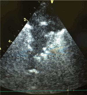
Figure 1. A consolidated right lung on ultrasound in a case of pneumonia.
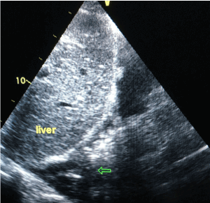
Figure 2. Alveolar consolidation with a frank tissue pattern arising from the pleural line with an irregular, shredded border (arrows). The shred sign is seen because the consolidation is in contact with the aerated lung.
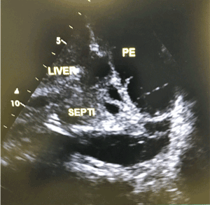
Figure 3. Complex pleural effusion with multiple septations. PE pleural effusion.
Table 3. US findings in patients with pneumonia and positive LUS
US findings in pneumonia |
Frequency (%) |
interstitial syndrome |
11(15) |
consolidation |
61(85) |
effusion |
23(31.5)
|
complicated effusion |
3 (4) |
dynamic air bronchogram |
58 (79) |
In all cases, portable antero-posterior CXRs were performed without lateral views. Portable lateral view CXRs are difficult to obtain and are not routinely practiced in the ICU.
Diagnosing pneumonia in the ICU is challenging. Pneumonia is a diverse disease consisting of both community-acquired and nosocomial pneumonias, most commonly ventilator-associated pneumonia, which have common radiological findings. As a rule, community-acquired pneumonia must be present on hospital admission or occur within the first 48 hours after admission, indicating that there was already incubation at the time of admission. In contrast, according to the American Thoracic Society (ATS) guidelines, Healthcare-Associated Pneumonias (HCAPs) are defined as those occurring after 48 hours or more following admission to a healthcare facility, implying that there was no incubation at the time of admission. Ventilator-Associated Pneumonia (VAP) develops in Intensive Care Unit (ICU) patients who have been mechanically ventilated for at least 48 hours.
Lung ultrasound has been shown to be a very useful tool for critical care physicians for the diagnosis of pneumothorax, pleural effusions and other thoracic conditions. Its use in the diagnosis of pneumonia has also been investigated in view of the limitations of CXR. The limitation of CXR will be clear when performed in ICU and ED, where many patients are critically ill and can be examined only in the supine position, often with bedside equipment [14]. In the emergency department, it was found that a bedside chest ultrasound is a reliable tool for the diagnosis of pneumonia and is superior to CXR in this setting. The study concluded that broader use of lung ultrasound will allow for more timely diagnosis and implementation of appropriate therapy [15]. A meta-analysis of published data has also shown that lung ultrasound is an accurate bedside tool to diagnose and monitor Ventilator-Associated Pneumonia (VAP), especially in the intensive care setting, and reduces patients’ exposure to radiation [16].
The diagnostic sign of pneumonia on ultrasound is the presence of consolidation and interstitial syndrome, which is nonspecific. Consolidation, identified in most of our cases, can also be present in obstructive atelectasis. Dynamic air bronchograms observed within the consolidation due to pneumonia can be easily detected by ultrasound and help differentiate pneumonia from atelectasis with high sensitivity and specificity [17]. This dynamic sign on LUS is an advantage over CXR and even CT images. There was dynamic air bronchograms in 58 of 73 patients. The presence of static air bronchograms within a large pneumonic consolidation in association with the “lung pulse” sign and decreased pleural sliding, is an indication for bronchoscopic examination [18]. The minority of patients with pneumonia in our study had the ultrasound pattern of interstitial syndrome. Focal interstitial pattern is suggestive of pneumonia, but the finding of a bilateral interstitial syndrome of acute onset suggests pulmonary edema or other causes of ALI/ARDS [12]. In our study, we found four such cases, and we used the presence of small subpleural consolidations, the absence or reduction of pleural gliding, and the irregularity and thickening of the pleural lines to rule out pulmonary edema. The ultrasound features in these four patients were like those described by Volpicelli et al. [12] in ALI/ARDS; microbiological results supported the final diagnosis of lung infection as the cause of the primary ALI.
Detecting the nature of effusions by chest US was an advantage over CXR, as detecting echoes within the effusion, whether mobile particles or septa, is highly suggestive of exudate [12]. In our study, complicated effusions were detected by chest US and followed by ultrasound-guided chest tube insertions.
The findings of our study are consistent with those of others showing that lung ultrasound is superior to CXR in diagnosing pneumonia. In this study, the gold standard chest CT was performed in a greater number of patients compared to other studies [19,20]. In this study, the decision to obtain a chest CT was made by the treating critical care physician. The inferiority of CXR observed in this study may be due in part to the inability to perform lateral view CXRs. Chest CT was performed in a limited number of nonrandomized patients, in our study. The advantage of ultrasound over CXR was therefore not confirmed with reference to the gold standard. In agreement with other studies [6], we agreed that performing CT scans in all patients would not be ethically justified; therefore, we decided to use the hospital discharge diagnosis as a surrogate for the CT diagnosis in a large number of patients with suspected pneumonia who had no clear clinical indication for further imaging.
2021 Copyright OAT. All rights reserv
In conclusion, bedside lung ultrasound is a reliable and accurate tool for diagnosing pneumonia in the critical care setting and is superior to CXR. It is a timely, non-invasive, and radiation-free modality for the diagnosis of pneumonia in the ICU.
This study was funded by Kuwait Oil Company, Ahmadi hospital. The funders had no role in study design, data collection and analysis, interpretation of data, or preparation of the manuscript.
- File TM, Jr., Marrie TJ (2010) Burden of community-acquired pneumonia in North American adults. Postgrad Med 122: 130-141. [Crossref]
- Welte T, Torres A, Nathwani D (2012) Clinical and economic burden of community-acquired pneumonia among adults in Europe. Thorax 67: 71-9. [Crossref]
- Almirall J, Bolibar I, Vidal J, Sauca G, Coll P, et al. (2000) Epidemiology of community-acquired pneumonia in adults: a population-based study. Eur Respir J 15: 757-763.
- Vincent JL, Rello J, Marshall J, Silva E, Anzueto A, et al. (2009) International study of the prevalence and outcomes of infection in intensive care units. JAMA 302: 2323-2329. [Crossref]
- Mandell LA, Wunderink RG, Anzueto A, Bartlett JG, Campbell GD, et al. (2007) Infectious Diseases Society of America/American Thoracic Society consensus guidelines on the management of community-acquired pneumonia in adults. Clin Infect Dis 44: S27-72. [Crossref]
- Woodhead M, Blasi F, Ewig S, Garau J, Huchon G, et al. (2011) Guidelines for the management of adult lower respiratory tract infections - full version. Clin Microbiol Infect 17: E1-59. [Crossref]
- Metlay JP, Fine MJ (2003) Testing strategies in the initial management of patients with community-acquired pneumonia. Ann Intern Med 138: 109-118. [Crossref]
- Syrjala H, Broas M, Suramo I, Ojala A, Lahde S (1998) High-resolution computed tomography for the diagnosis of community-acquired pneumonia. Clin Infect Dis 27: 358-363.
- Long L, Zhao H-T, Zhang Z-Y, Wang G-Y, Zhao H-L (2017) Lung ultrasound for the diagnosis of pneumonia in adults. Medicine 96: e5713. [Crossref]
- Lichtenstein DA, Lascols N, Meziere G, Gepner A (2004) Ultrasound diagnosis of alveolar consolidation in the critically ill. Intensive Care Med 30: 276-381. [Crossref]
- Lichtenstein DA, Mezière G, Lascols N, Biderman P, Courret J-P, et al. (2005) Ultrasound diagnosis of occult pneumothorax. Crit Care Med 33: 1231-1238. [Crossref]
- Volpicelli G, Mussa A, Garofalo G, Cardinale L, Casoli G, et al. (2006) Bedside lung ultrasound in the assessment of alveolar-interstitial syndrome. Am J Emerg Med 24: 689-696. [Crossref]
- Volpicelli G, Elbarbary M, Blaivas M, Lichtenstein DA, Mathis G, et al. (2012) International evidence-based recommendations for point-of-care lung ultrasound. Intensive Care Med 38: 577-591. [Crossref]
- Zagli G, Cozzolino M, Terreni A, Biagioli T, Caldini AL, et al. (2014) Diagnosis of ventilator-associated pneumonia. Chest 146: 1578-1585.
- Esayag Y, Nikitin I, Bar-Ziv J, Cytter R, Hadas-Halpern I, et al. (2010) Diagnostic value of chest radiographs in bedridden patients suspected of having pneumonia. Am J Med 123: 88.e1-5. [Crossref]
- Cortellaro F, Colombo S, Coen D, Duca PG (2012) Lung ultrasound is an accurate diagnostic tool for the diagnosis of pneumonia in the emergency department. Emerg Med J 29: 19-23. [Crossref]
- Wang G, Ji X, Xu Y, Xiang X (2016) Lung ultrasound: a promising tool to monitor ventilator-associated pneumonia in critically ill patients. Crit Care 20: 320.
- Lichtenstein D, Mezière G, Seitz J (2009) The dynamic air bronchogram. A lung ultrasound sign of alveolar consolidation ruling out atelectasis. Chest 135: 1421-1425. [Crossref]
- Lichtenstein DA, Lascols N, Prin S, Mezière G (2003) The "lung pulse": an early ultrasound sign of complete atelectasis. Intensive Care Med 29: 2187-2192. [Crossref]
- Parlamento S, Copetti R, Di Bartolomeo S (2009) Evaluation of lung ultrasound for the diagnosis of pneumonia in the ED. Am J Emerg Med 27: 379-384. [Crossref]