Carotid-subclavian bypass and endovascular aortic repair of Kommerell’sdiverticulum with aberrant left subclavian artery: A case report and literature review
Akilu Wajeehullahi
Cardiothoracic Surgical Center, Tongji Hospital, Huazhong University of Science and Technology, Wuhan, China
Yi Feng
Cardiothoracic Surgical Center, Tongji Hospital, Huazhong University of Science and Technology, Wuhan, China
Zhangqiang Zhu
Cardiothoracic Surgical Center, Tongji Hospital, Huazhong University of Science and Technology, Wuhan, China
Li Shi Liang
Cardiothoracic Surgical Center, Tongji Hospital, Huazhong University of Science and Technology, Wuhan, China
XianTao Ma
Cardiothoracic Surgical Center, Tongji Hospital, Huazhong University of Science and Technology, Wuhan, China
Wei Xiang
Cardiothoracic Surgical Center, Tongji Hospital, Huazhong University of Science and Technology, Wuhan, China
Cheng Cai
Cardiothoracic Surgical Center, Tongji Hospital, Huazhong University of Science and Technology, Wuhan, China
Congenital deformities of the aortic arch are very uncommon. We present a case report ofKommerell’s Diverticulum with aberrant left subclavian artery in a 50 years old man who has no past medical history and is non-smoker and non-alcoholic. Patient presented with shortness of breath, chest pain and dizziness for six months. Blood tests were done and CT angiogram confirmed the diagnosis. Surgery was planned as the treatment modality.Carotid-Subclavian artery bypass and endovascular aortic repair was conducted. Being a rare disease, little literature is found about the surgical treatment. The procedure we conducted is innovative and will bring focus and better treatment options in managing KD with ASCA.
Keywords
Kommerell’sdiverticulum, left common carotid artery, left subclavian artery, medtronic stent catheter endovascular repair, case report
Introduction
Originally described in 1936, by a German Radiologist, named Kommerell[1], Kommerell’s Diverticulumis also known as “lusoria diverticulum”, “lusoria root” or “remnant diverticulum”[1].This was seen in a patient with left sided aortic arch, where a pulsatile mass seen to the posterior of the oesophagus, leading to the compression of that part of gut, detected in barium swallow[1].It is defined as the aneurysmal dilatation of the descending aorta at the origin of an aberrant subclavian artery, that can be located in both right and left sided aortic arches[2]. Kommerell’s diverticulum (KD) is due to persistence remnant of the fourth primitive dorsal arch, which failed to retrogress [3].
As per the classification of Salomonowitz et al, there are three types of aortic arch diverticulum namely: 1. diverticulum in left aortic arch with right aberrant subclavian artery (ASCA), 2.diverticulum in right aortic arch with left aberrant subclavian artery (ASCA), 3.aortic diverticulum without ASCA (at the aortic-ductal junction) [4].
Clinical history and examination
A 50-year-old Chinese man was admitted to our hospital, presented with difficulty in breathing, sudden onset chest painand dizziness on and off for 6 months. There was no dysphagia, syncopal attacks, abdominal pain, palpitations or other symptoms.
His past medical history, past surgical history, smoking- drinking habits, family history and drug allergy were not significant. The vital signs were: blood pressure was 120/70 mmHg, temperature was 36.5 degrees Celsius, pulse was 73bpm, regular (bilateral radial, brachial sides were normal), no pallor, no cyanosis, no clubbing, no pedal oedema, no peripheral vascular signs. The systemic examinations were normal. The blood investigations such as the haematological and biochemical investigations were within normal limits. A Computed Tomography Aortagraphy (CTA) was performed on him as diagnostic assessment and findings as shown below.
Imaging findings
At the local provincial hospital, on day 1 admission, CT Aortography of the brain and neck region identified aortic arch Kommerell’s diverticulum with oesophageal compression, left subclavian was lost and proximally occluded, cold and left subclavian artery steal syndrome was seen. The patient was referred to our Cardiothoracic Surgery Department. On day 5, Another CT Aortagraphywas performed and showed mild dilatation of the thoracic aorta, adjacent oesophagus and trachea was compressed and displaced. The right upper lobe tip was streaked, and was mostly inflammatory lesions, several small round low-density foci in the liver(Figures 1-3).
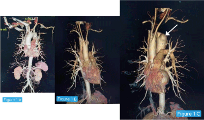
Figure 1A:Normal shape of the aorta of this patient; 1B: Shape of the patient's aorta, 1C: White arrow showing Kommerell’s Diverticulum in the arch of aorta
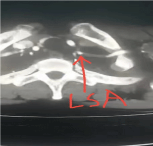
Figure 2:Location of Left Subclavian Artery (LSA)
Figure 3: 3D video of the CTA
The treatment options were then discussed with the patient including that of a surgical treatment along with all the risks and benefits involved. The patient agreed for surgical intervention.
Surgical open repair
Pre-operative CT angiography film of the chest,showing a 3.5cm Kommerell’s Diverticulum(KD). After placing the patient in supine position, general anaesthesia was administered successfully. The anterior aspect of the chest wall and groin area wasroutinely disinfected. We began a transverse supraclavicular 5 cm incision. We separated the platysma muscle and proceeded to the 2 heads of the sternocleidomastoid muscle(Figure 4).
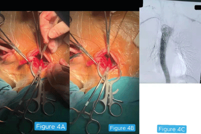
Figure 4A:Transverse supraclavicular 5cm incision exposing the surgical site
Figure 4B:Clamping and anastomosis of the Left Subclavian Artery (LSA) to the left common carotid artery (LCCA)
Figure 4C: Angiography picture showing the insertion of the large imported aortic stent (Medtronic Stent Catheter)
Firstly, a dissection was done to reach the internal jugular vein. We applied deep wound spreaders that help reveal the area. Medial to this vein, we cautiously liberated the vagus nerve, which lies between the internal jugular vein and the left common carotid artery (LCCA) and we open the carotid sheath. This way allowed us the chance to do anastomosis proximally on the left common carotid artery at the left lateral side.
The next procedure was exposing the left subclavian artery (LSA), usually 1 cm deeper in the neck and its lateral away from the LCC. There was subcutaneous fatty tissue with lymph nodes that obstructs LSA dissection, hence these lymph nodes were resected. We also exposed and looped the left internal thoracic artery using a vessel loop.
We then finally revealed the deep LSA in the neck and the association between the LCCA and LSA was obviously seen. Heparin was administered and there was activated clotting time, then we clamped the LSA. The LSA was incised to make round-shaped incised artery. We usedprolene 5-0 C1 sutures then meticulously anastomosed 6-mm Dacron prosthesis to the LSA round-shape incised artery and haemostasis was checked afterwards. We made a good size length of our Dacron prosthesis and it was able to stretch from the left subclavian artery to the lateral side of LCCA. It was made to be under the crossing of vagus nerve.
We clamped the LCCA while at thesame time monitoring the Transcranial Doppler(TCD) signals. We ensured that there was enough cerebral perfusion pressure by letting the mean blood pressure stable at 90 mmHg. We made a tinny incision on the LCCA at the lateral side and a mild punch to create a round-hole. We then started our anastomosis using 5-0 C1 sutures on a 6-mm Dacron prosthesis end to side with LCCA. A clamp was used clamping the distal LCCA and then was flushed. Afterwards, we now unclamped the LSA and the first clamp on the LCCA to de-air them. Haemostasis was checked from the vessels by unclamping the vessels.
Total endovascular repair
The groin region was opened by an incision. After the right femoral artery angiography, the left common carotid artery (LCCA), the right common carotid artery (RCCA), right subclavian artery, the proximal end of the left subclavian artery was occluded, and the descending aorta was obviously bulged. The bridge vessel is unobstructed (from the left common carotid artery to the left subclavian artery). The large imported aortic stent (Medtronic Stent Catheter) was placed through the femoral artery, positioned by the right subclavian artery angiography catheter, and released at the proximal end of the right subclavian artery and sutured. The internal aortic rupture was now closed and there was no leakage of contrast agent after re-angiography. We withdrew the stent conveyor, bleeding stopped thoroughly. Afterwards we closed all incisions and drainages inserted layer by layer after checking. Patient was now transferred to the ward.
Post-operative care
Patient was discharged on Tablet Plavix 75 mg once a day and later shifted to enteric coated Aspirin tablet 100 mg once a day and its side-effects (such as bleeding gums, bleeding from any orifices, gastrointestinal bleeding, black stools) have been explained to the patient and to attend hospital immediately once any of it happens.
Strict bed rest for one month should be taken. Dietary advice such as low salt, low fat, low sugar diets and to perform moderate exercise, keep weight control and proper heart activity, maintain peace of mind, avoid stress, smoking, alcohol, avoid infections, fatigue and take proper rest are given to the patient.
We suggest the patient for regular monitoring of blood pressure, heart rate, blood routine examination including renal function, liver function tests, cardiac enzymes, electrocardiogram and cardiac echography and to have regular follow-up after 1, 3, 6,12-month.
Discussion
Kommerell’s diverticulum (KD) in brief, is a rare abnormal congenital condition that occurs in either right or left sided aortic arch and seen to be associated with an aberrant subclavian artery (ASCA)[2].Kommerell’s diverticulum is aneurysmal dilated change in the aortic walls and has the tendency of dissection[5].In 1936, initially described in a patient with left sided aortic arch, German Radiologist Kommerell saw on barium swallow: a pulsatile mass compressing the oesophagus posteriorly[1].The incidence of KD with a right-sided aortic arch; seen in radiological studies is 0.05-0.1%[ 6,7].
KD symptoms of oesophageal or trachealorigins,are indicative factors for a surgical treatment. A KD with a size greater than 30 mm in diameter should be considered for operation[5]. It is known that Kommerell’s diverticulum has tendency of activating aortic aneurysm, or dissection or even rupture. Open repair and revascularization of the left arm was the surgical approach of preference. Using the techniques of minimally invasive and endovascular techniques for surgical repairs, have recently helped to reduce anguish, pain when compared to open sternotomy[8-10].This approach of surgical treatment, we performed; is unique, safe and reliable strategy for the Kommerell’s Diverticulum management. Again, our technique was able to give a full comprehensive insight of how the stent replacement and left handrevascularization can be performed in an orderly way.
3. Edwards JE (1984)Anomalies of the derivatives of the aortic arch system.Med Clin North Am32: 925-949. [Crossref]
4. Salomonowitz E,Edwards JE, Hunter DW, Castaneda-Zuniga WR, Lund G, et al. (1984)The three types of aortic diverticula. AJR Am J Roentgenol142: 673-679. [Crossref]
5. Cinà CS,Althani H, Pasenau J, Abouzahr L (2004)Kommerell's diverticulum and right-sided aortic arch: a cohort study and review of the literature.J Vasc Surg39: 131-139. [Crossref]
6. Tsukube T, Ataka K, Sakata M, Wakita N, Okita Y (2001)Surgical treatment of an aneurysm in the right aortic arch with aberrant left subclavian artery.Ann Thorac Surg71: 1710-1711. [Crossref]
7. Svensson LG, Crawford ES (1997) Cardiovascular and vascular disease of the aorta. Saunders.
9. Hayakawa M,Nagano T, Nishijima I, Shinzato K, Ikemura R, et al. (2020)Successful Endovascular Repair of a Kommerell's Diverticulum and a Right-Sided Aortic Arch.Heart Surg Forum23: E860-E862. [Crossref]
10. Ghincea CV,Ikeno Y, Weyant MJ, Mitchell JD, Aftab M, et al. (2020)Right Thoracoscopic Aberrant Right Subclavian Artery Division and Subclavian-Carotid Transposition.Ann Thorac Surg110: e431-e433. [Crossref]
Editorial Information
Editor-in-Chief
Andy Goren
University of Studies Guglielmo Marconi
Article Type
Case Report
Publication history
Received: May 23, 2023
Accepted: June 24, 2023
Published: June 27, 2023
Wajeehullahi A, Feng Y, Zhu Z, Liang LS, Ma XT, et al. (2023) Carotid-subclavian bypass and endovascular aortic repair of Kommerell’s diverticulum with aberrant left subclavian artery: A case report and literature review. Clin Case Rep Rev 9: DOI: 10.15761/CCRR.1000521.
Corresponding author
Dr. Cheng Cai, M.D
Professor of Surgery, Cardiothoracic Surgical Center, Tongji Hospital, Huazhong University of Science and Technology 1095 Jie Fang Da Dao, Wuhan, China, 430030.
Figure 1A:Normal shape of the aorta of this patient; 1B: Shape of the patient's aorta, 1C: White arrow showing Kommerell’s Diverticulum in the arch of aorta
Figure 2:Location of Left Subclavian Artery (LSA)
Figure 3: 3D video of the CTA
Figure 4A:Transverse supraclavicular 5cm incision exposing the surgical site
Figure 4B:Clamping and anastomosis of the Left Subclavian Artery (LSA) to the left common carotid artery (LCCA)
Figure 4C: Angiography picture showing the insertion of the large imported aortic stent (Medtronic Stent Catheter)