Aim: To define the differential hepatic artery variations in two cadavers during routine dissections.
Materials and methods: Hepatic artery dissections, in two 10% formalin fixed cadavers from de Ege University School of Medicine Department of Anatomy, were performed. Photos were taken with digital camera and measurements were made photogrammetrically.
Results: In the first case; at the end of the common hepatic artery, the left hepatic artery was branched out as a pair. It was observed that both left hepatic arteries supply the left lobe of the liver. The common hepatic artery continues as the right hepatic artery. The length of the left hepatic artery in first case was measured 15.72 mm and 18.95 mm in the second case.
In the second case; a thick right hepatic artery arises from the superior mesenteric artery and acting as a proper hepatic artery. Although it acts like a proper hepatic artery, it seems to supply the right lobe of the liver. The length of the right hepatic artery was measured as 46.68 mm, the diameter was 4 mm. The left hepatic artery originates from the common hepatic artery and divided into three branches. Two branches supply the left lobe and one branch supplies the quadrate lobe of the liver.
Conclusion: These defined variations are useful for the planning and guide for surgical and radiological procedures of the liver transplantation, pancreas or biliary surgery.
Proper hepatic artery, common hepatic artery, arterial supply of liver
The coeliac trunk passes almost horizontally forwards and slightly right above the pancreas and splenic vein and divided into left gastric, common hepatic and splenic arteries. In adults the common hepatic artery (CHA) is intermediate in size when compared with the left gastric and splenic arteries; but in later fetal and early postnatal life, it is the largest branch of the coeliac trunk [1]. Accompanied by the hepatic autonomic plexus it first passes forwards and right, below the epiploic foramen to the upper aspect of the superior part of the duodenum [1]. When it reaches to the superior part of the duodenum, it divides into right gastroduodenal (RGDA) and proper hepatic artery. Proper hepatic artery enters to the hepatoduodenal ligament and extends to the hepatic porta [2–4]. It divides into right and left branches to supply the corresponding lobes of the liver at the hepatic porta [5]. Right hepatic artery (RHA) supplies the right lobe and the half of the caudate lobe of the liver. It also gives cystic artery, which supplies the gall bladder. Left hepatic artery (LHA) supplies the left lobe, quadrate lobe and the other half of the caudate lobe of the liver [2,3].
While the classical branching pattern in these arteries is 52-80% in the literature, variations about this branching pattern are not uncommon [6]. The most common variations are, LHA originating from the left gastric artery and RHA originating from the superior mesenteric artery [3,6]. In addition to different branching patterns of the hepatic arteries, defined accessory arteries are also common. The term accessory is used for arteries concomitant to the main artery that leads to the same region. Accessory LHA (11%) is mainly originating from the left gastric artery or coeliac trunk [6]. However, CHA originated accessory LHA instance is relatively rare (4%) [6,7]. Due to growing interventional radiological procedures, transplantation, laparotomy or laparoscopic surgery procedures, variations of the CHA and its branches are becoming more important.
During a routine cadaver dissection program for anatomy training in Ege University, School of Medicine, Department of Anatomy, some vascular variations of the liver were noticed in two male cadavers. The cadavers were obtained in accordance with the laws of the country and embalmed with 10% formalin solution. The variations were identified according the literature and photographs were taken with a digital camera. Shootings were done at right angle to the objects with a millimetric scale next to the targeted structure. All measurements were made photogrammetrically using the "Image J" program.
Case reports
Case 1
During the abdominal dissection of a male cadaver, although the coeliac trunk was in classical pattern (left gastric artery, CHA and splenic artery); the LHA was branched out as a pair at the end of the CHA, with the same level of the RGDA but to the opposite side. Both LHAs were headed parallel to the left hepatic lobe through the left sagittal fissure (Figure 1). The CHA continues as the RHA and give off the cystic artery at the hepatic porta. Some autonomic nerve fibers to the liver were situated and run between the paired LHAs. There wasn’t an artery that could be defined as proper hepatic artery (Figure 1).
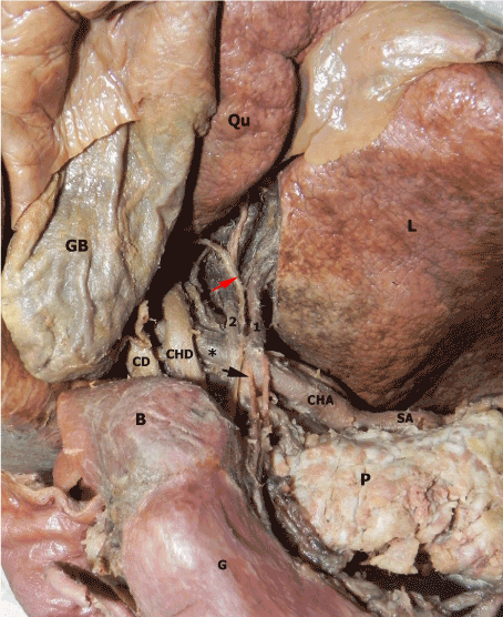
Figure 1. Case 1, antero-inferior aspect of porta hepatis. Gaster (G) and the pancreas (P) removed antero-inferiorly, the gall bladder (GB) superiorly. Right inferior corner of the left lobe (L) was excised minimally to show the arteries. After giving two branches (1 and 2) the common hepatic artery (CHA) proceeds as right hepatic artery (*) instead of proper hepatic artery behind the common hepatic duct (CHD). SA: splenic artery, Qu: quadrate lobe of the liver, B: bulbus duodeni, CD: cystic duct, red and black arrows: Ascending autonomic nerve fibers.
The diameter of the CHA before giving off the LHAs is 8.33 mm. The diameters of the first and second LHAs were 5.20 mm and 3.11 mm, respectively. The diameter of the right hepatic artery was 6.81 mm. The lengths of the LHAs from the branching point to the entrance to the liver were also measured. The length of the first LHA was 15.72 mm and the second one was 18.95 mm.
Case 2
In another routine abdominal dissection of a male cadaver, a noticeable thick right hepatic artery (RHA) that arises from the superior mesenteric artery as a short common trunk together with inferior pancreaticoduodenal artery was detected (Figure 2). After a careful dissection, it was noticed that the RHA is acting as proper hepatic artery and accompanies the portal vein, ductus choledochus and autonomic nerve bundles through the hepatic porta within the hepatoduodenal ligament. Although it acts like a proper hepatic artery, it seems to supply the right lobe of liver. The diameter of the right hepatic artery was 4mm. The length was measured as 46.68 mm.
The left hepatic artery originates from the common hepatic artery and divided into three branches: the anterior, intermediate and posterior (Figure 2). It is observed that the intermediate and posterior branches supply the left lobe and the anterior branch supplies the quadrat lobe of the liver (Figure 3). The length of the anterior branch was 26.02 mm, the intermediate branch was 20.61 mm and the posterior branch was 17.24 mm.

Figure 2. Case 2, antero-inferior aspect of porta hepatis. Right hepatic artery (**) arising as a branch of superior mesenteric artery (SMA). The arteries (1, 2 and 3) to the left lobe of the liver (L) are branches of the left hepatic artery (*) originated from common hepatic artery (CHA). CT: coeliac trunk, SA: splenic artery, LGA: left gastric artery, IVC: inferior vena cava, Pv: portal vein, GB: gall bladder, BD: biliary duct, Qu: quadrate lobe of the liver.
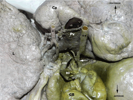
Figure 3: Case 2, porta hepatis, inferior aspect. The intermediate (2) and posterior (1) branches supply the left lobe (L) and the anterior branch (3) supplies the quadrat lobe of the liver. Ca: caudate lobe, Pv: portal vein, *: left hepatic artery **: right hepatic artery, white arrow: cystic artery, CHD: common hepatic duct, BD: biliary duct, CD: cystic duct, GB: gall bladder.
Variations of the arteries supplying the liver are caused by an abnormal persistence or regression of embryonic arteries. The origin of these variant arteries varies according to which lobes they supply. Accordingly, LHA that supplies the left lobe of the liver may originate from RGDA, left gastric artery or coeliac trunk. RHA that supplies the right lobe of the liver may originated from superior mesenteric, RGDA or CHA [3,8,9]. These variations are very common. Uğurel et al. [10] determined the ratio of hepatic artery variations to 48% as a result of multidetector CT (MDCT) angiography of the abdominal aorta applied in 100 patients. However, accessory left hepatic artery duplication is an uncommon but a significant variation. Generally, double hepatic artery is originated from the common hepatic artery at the distal level of the RGDA [3,8]. In the case of duplication, one or both of them can originate directly from the coeliac trunk or abdominal aorta. Tharao et al. [11] had 102 cadaveric dissections and there was one case in which the LHA divided into two branches before entering the liver.
In our first case, the LHAs were branching as a pair from the CHA with the same level of the RGDA and directed to the left hepatic lobe. The CHA continues as the right hepatic artery and give off the cystic artery at the hepatic porta. The proximal one of the paired LHA was evaluated as an accessory branch. Some autonomic fibers to the liver were situated and run between them.
Kastamoni et al. [3] reported a LHA duplication originated from the CHA at the distal level of RGDA. Covey et al. described the hepatic artery variations in the study performed on 600 patients between 1999 and 2002. According to this study; one hundred nineteen patients (19,8%) had variant left hepatic arteries, eighty-nine patients (14.8%) had variant right hepatic arteries. Double hepatic arteries were seen in twenty-two patients (3.7%) [8]. Another “double hepatic artery” cases described by Winston et al. They examined 371 patients’ computerized tomography angiographies, between 2001–2003 years and they reported this rate as 4% [12]. Futara et al. found the accessory LHA originated from CHA in 5 of 110 cadavers [7].
When the current literatures are reviewed, a number of variations related to the arterial branching patterns of the liver are observed. Therefore, it is very important to know well the common patterns and variations of the CHA and its branches both preoperative and intraoperative phases. Unawareness or ignorance of these variations especially on liver transplantation, pancreas or biliary surgery; can cause vital complications. The possibility of encountering such variations must always be considered for the safety of surgeries and interventional radiological procedures.
- Williams PL (1995) Gray’s Anatomy: the anatomical basis of medicine and surgery. 38th Ed. New York: Churchill Livingstone, p. 1548-1552, 1802.
2021 Copyright OAT. All rights reserv
- Arinci K, Elhan E (2014) Anatomi 2. cilt. 5th ed. Ankara, p. 54-57.
- Kastamoni Y, Albay S (2012) Arteria hepatica sinistra duplikasyonu ve arteria mesenterica superior’dan çikan “replaced” arteria hepatica dextra: Olgu Sunumu. S.D.Ü. Tip Fak. Derg 19: 148-150.
- Ozan H (2014) Ozan Anatomi, 3th ed. Ankara, p. 302-403.
- Snell R (2007) Clinical anatomy by systems. 1st ed. Philadelphia: Lippincott Williams & Wilkins, Pp. 220-223.
- Pai RS, Hunnargi AS, Srinivasan M (2008) Accessory left hepatic artery arising from common hepatic artery. Indian J Surg 70: 80-82. [Crossref]
- Futara G, Ali A, Kinfu Y (2001) Variations of the hepatic and cystic arteries among Ethiopians. Ethiop Med J 39: 133-142. [Crossref]
- Covey AM, Brody LA, Maluccio MA, Getrajdman GI, Brown KT (2002) Variant Hepatic Arterial Anatomy Revisited: Digital Subtraction Angiography Performed in 600 Patients. Radiology 224: 542-547. [Crossref]
- Çiçekçibasi AE, Dogan NÜ, Fazliogullari Z, Sanli ÖÖ, Büyükmumcu M, et al. (2011) Arteria mesenterica superior’dan orijin alan arteria hepatica dextra. S.D.Ü. Tip Fak. Derg 18: 21-24.
- Ugurel MS, Battal B, Bozlar U, Nural M, Tasar M, et al. (2010) Anatomical variations of hepatic arterial system, coeliac trunk and renal arteries: an analysis with multidetector CT angiography. Br J Radiol 83: 661-667. [Crossref]
- Tharao MK, Saidi H, Kitunguu P, Julius OA (2007) Variant anatomy of the hepatic artery in adult Kenyans. Eur J Anat 11: 155-161.
- Winston CB, Lee NA, Jarnagin WR, Teitcher J, DeMatteo RP, et al. (2007) CT Angiography for Delineation of Celiac and Superior Mesenteric Artery Variants in Patients Undergoing Hepatobiliary and Pancreatic Surgery. AJR Am J Roentgenol 189: W13-W19. [Crossref]