Objectives: The purpose of this prospective study was to characterize the clinical appearance of oral squamous cell carcinoma (OSCC) in a population of South African patients attending the Medunsa Oral Health Centre (MOHC).
Patients and methods: Thirty-two random cases of OSCC seen and diagnosed at the MOHC during the period July 2011 to June 2015 were evaluated. Data as to age, gender, use of tobacco, alcohol and snuff, oral site affected and clinical appearance of the lesion (leukoplakia, erythroplakia, exophytic, ulcer) were recorded.
Results: The mean age of the patients at the time of diagnosis of OSCC was 60 years and the male:female ratio was 3:1. The tongue was the most frequently affected site (30%) followed by the floor of the mouth (19%). Seventy-eight percent of the patients both smoked tobacco and drank alcohol. Only 13% of OSCCs occurred in patients younger than 45 years of age. OSCC most frequently manifested as ulcers (59%) or as exophytic lesions (30%). Leukoplakic changes next to the carcinomata were present in 13% of patients. None of the patients had observed oral leukoplakia in their mouths before the OSCC appeared.
Conclusion: In Black South Africans in the Ga-Rankuwa area, OSCC affects most frequently male smokers manifesting either as ulcers or as exophytic lesions.
oral squamous cell carcinoma, leukoplakia, erythroplakia, exophytic, ulcer
Abbreviations:
OSCC: oral squamous cell carcinoma
About 8% of all malignancies worldwide are squamous cell carcinomas of the head and neck (Posner et al., 2007). Oral squamous cell carcinoma (OSCC) is predominantly a disease of elderly males who have a long history of smoking tobacco and drinking alcohol [1]. Other risk factors for OSCC include the use of betel nut and a diet poor in fresh vegetables and fruits [2]. OSCC most commonly affects the tongue followed by the floor of the mouth, the gingiva and alveolar mucosa [3]. After treatment, local recurrence or distant metastasis occurs in 40-60% of these subjects; only 30-50% survive for longer than three years [2,4].
Carcinogenesis is a multi-step process involving cellular epigenetic modifications and sequential genetic changes giving the altered cells growth dominance over normal neighboring cells, resulting in clonal expansion and uncontrolled proliferation of the transformed cells [5]. It is suggested that between five and ten cytogenetic events in different critical genes are required to transform a normal cell into a cell with a malignant phenotype [5]. However, a number of non-genetic factors also play important roles in carcinogenesis: this include repetitive trauma, chronic inflammation, increased intrinsic mechanical stresses within the extracellular matrix, and dysregulation in epithelial-mesenchymal interactions, in cell-to-cell communication, in cell-to-extracellular interactions and in mechanotransduction pathways [6,7].
Most, if not all OSCCs arise within fields of precancerized oral epithelium with genetically altered keratinocytes. Stem/progenitor cells in the basal cell layer of oral epithelium which have acquired one or more genetic changes, and have subsequently undergone initial transformation may evolve into an expanding clones of precancerized cells which may manifest either as clinically normal fields of precancerized epithelium, as leukoplakia or as erythroplakia [8]. An altered cell upon acquiring additional genetic lesions, may undergo clonal divergence, develop a malignant phenotype and manifest clinically and histologically as OSCC [9-11]. Thus, OSCC can evolve from leukoplakia/erythroplakia or from normal looking epithelium with genetically altered keratinocytes. It is estimated that between 17 and 35 percent of OSCCs arise from pre-existing leukoplakic lesions, while the remainder arise de novo from apparently normal oral epithelium [12].
Oral squamous cell carcinoma may present clinically as leukoplakia or as erythroplakia that in fact has already become malignant, as a necrotic ulcer with irregular raised indurated borders or as a broad-based exophytic mass with a surface texture that can be relatively smooth, verrucous or pebbled [13]. In South Africa the prevalence of OSCC is higher in Blacks than in Whites [14], but the prevalence of potentially malignant oral leukoplakia appears to be substantially lower in Blacks than in Whites [15,16]. This may be because, in general, many South African Blacks neglect to seek prompt medical treatment for non-painful oral lesions, and therefore, by the time of diagnosis, premalignant leukoplakia may already have progressed to frank OSCC [13,17]; or because, in Black South Africans, for reasons unknown, many cases of OSCC arise de-novo and not from premalignant leukoplakia [16].
Worldwide, about one-third of cases of de-novo OSSC develop in close proximity to pre-existing leukoplakias, and many other cases of apparently de-novo OSCC show tell-tale signs of leukoplakic origin [13]. As there are no data from South Africa about how OSCC evolves in Black persons, the aims of this prospective research were to determine the proportion of cases of oral leukoplakia that on histopathological examination turn out to be OSCCs, the proportion of frank OSSCs that show tell-tale signs of a leukoplakic origin, and to characterize the clinical appearance of OSCC in a population of South African patients attending the Medunsa Oral Health Centre.
This prospective study is based on random cases seen and diagnosed by a single clinician (MB) in the Department of Maxillofacial and Oral surgery at Medunsa Oral Health Centre, Ga-Rankuwa, South Africa, during the period July 2011 to June 2015 and not on the total number of OSCCs that were diagnosed in the centre during this period. Randomization was achieved not according to any predetermined plan, but according to the times that the clinician that was conducting the research (MB) was present in the clinic. Relevant personal information including age, gender, oral site affected and the clinical appearance of the lesion (leukoplakia, erythroplakia, ulcer, exophytic), and information as to alcohol consumption, tobacco and snuff use were collected.
Thirty-two Black patients histopathologically confirmed as having OSCC were included in this study. Of these, 24 (75%) were males and 8 (25%) were females (Table 1), with a male to female ratio of 3:1. Twenty-five patients (78%) smoked cigarettes and all of these patients also drank alcohol. Thirteen percent used snuff and six percent of patients used alcohol, tobacco and snuff. Thirteen percent were younger than 45 years,
In this study of 32 patients, 70 oral sites were affected: eight patients had one site affected, 14 had two sites affected, six had three sites affected and four had four sites affected. In every case with more than one site affected, there was a single lesion that extended to one of the neighbouring oral sites. There were no multifocal anatomical lesions (Table 1) and there were no cases of new second cancers.
Table 1. Gender in relation to age, alcohol, smoking and snuff use
|
Males |
Females |
Total |
Number |
24 (75%) |
8 (25%) |
32 (100%) |
Age (in years)
Mean
Median
Std. deviation |
60.25
59.5
10.05 |
57.25
54
20.06 |
59.5
58
12.74 |
Alcohol alone
Yes
No |
23
1 |
2
6 |
25 (78%)
7 (22%) |
Smoking alone
Yes
No |
23
1 |
2
6 |
25 (78%)
7 (22%) |
Snuff alone
Yes
No |
2
22 |
2
6 |
4 (13%)
28 (88%) |
Combination use
Smoking + alcohol
Smoking + alcohol+ snuff |
23
2 |
2
0 |
25 (78%)
2 (6%) |
The sites most commonly affected were the tongue (30%) followed by the floor of the mouth (19%) (Table 2). Oral squamous cell carcinoma most frequently manifested as ulcers (59%) (Figure 1) or as exophytic lesions (30%) (Figure 2). Leukoplakic (Figure 3) or erythroplakic (Figure 4) changes next to the carcinoma were present in 11% of lesions (Table 2). None of the patients had observed oral leukoplakia before the OSCC appeared, but on examination four patients (13%) exhibited tell-tale signs of leukoplakia in relation to their OSCC.
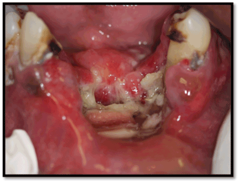
Figure 1. OSCC manifesting as an ulcer on the floor of the mouth and ventral surface of the tongue
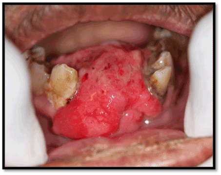
Figure 2. OSCC manifesting as an exophytic lesion of the floor of the mouth and anterior mandibular buccal gingiva
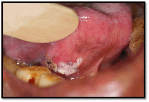
Figure 3. OSCC manifesting as a leukoplakia on the ventral surface of the tongue
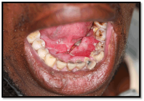
Figure 4. OSCC manifesting as erythroleukoplakia of the anterior floor of the mouth
According to self-reports of the 32 patients, none had had any pre-existing leukoplakia before they developed OSCC, but on examination four patients (13%) exhibited tell-tale signs of leukoplakia (Figure 3) in relation to their OSCC [7,15,18]. This reinforces the notion that in Black South Africans the vast majority of OSCCs arise de-novo.
However, one cannot exclude the possibility that some patients were not aware of having had pre-existing leukoplakia at the site of OSCC while others may have been treated by traditional healers for some kind of unidentified oral mucosal lesions which in fact was premalignant leukoplakias or early OSCCs which by the time of diagnosis had already progressed to frank OSCC manifesting clinically as an ulcer, or as an exophytic lesion [7,17].
The distribution of carcinomatous lesions by clinical type is shown in Table 2. It has been suggested that if the growth of the OSCC is very rapid so that the mass of malignant cells out-paces its blood supply, then the lesion will become necrotic and breakdown into an ulcer (Figure 1), but if the mass of malignant keratinocytes has grown more slowly and has sufficient blood supply, an exophytic (Figure 2) OSCC will develop.
Table 2. Clinical appearance of lesion in relation to site affected
|
Number of lesions |
Clinical appearance of lesion |
Leukoplakia |
Erythroplakia |
Ulcer |
Exophytic |
Mixed
(L+E) |
Tongue |
21 (30%) |
1 |
|
14 |
6 |
|
Floor of mouth |
13 (19%) |
1 |
|
9 |
3 |
|
Lower alveolar mucosa |
7 (10%) |
|
1 |
3 |
3 |
|
Lower gingiva |
7 (10%) |
|
1 |
2 |
2 |
2 |
Lower retromolar |
5 (7%) |
|
|
2 |
2 |
1 |
Soft palate |
4 (6%) |
|
|
4 |
|
|
Buccal mucosa |
3 (4%) |
|
|
|
3 |
|
Hard palate |
2 (3%) |
|
|
1 |
1 |
|
Upper retromolar |
2 (3%) |
|
|
|
1 |
|
Lower labial mucosa |
2 (3%) |
|
|
2 |
|
|
Upper lip |
1 (1%) |
|
|
1 |
|
|
Lower lip |
1 (1%) |
|
|
1 |
|
|
Upper labial mucosa |
1 (1%) |
|
|
1 |
|
|
Upper alveolar mucosa |
1 (1%) |
|
|
|
|
1 |
Total |
70 (100%) |
2 (3%) |
2 (3%) |
41 (59%) |
21 (30%) |
4 (6%) |
The most important risk factors for OSCC are tobacco smoking, heavy drinking of alcoholic beverages, a diet low on fresh fruits and vegetables, and the use of betel quid and/or areca nut in those populations that practice it. The greater the amount, the greater the frequency and the longer the duration of tobacco smoking and alcohol consumption, the greater is the risk of carcinoma, and the risk is increased by the concurrent use of these agents. However, OSCC can be idiopathic; and it is clear that subject-specific genetic factors and environmental factors also play important roles either predisposing to or affording protection against OSCC [13]. In this study of patients with OSCC, 78% of patients both smoked tobacco and drank alcohol: two of these patients also used snuff. Two affected patients use snuff only. There were five patients that didn’t use any of the aetiological agents considered in this study and these were classified as having idiopathic OSCC (Table 1). Therefore, it is that it is likely the incidence of OSCC could be substantially reduced by abstinence from tobacco smoking or exercising moderation in alcohol consumption.
OSCC can affect any part of the oral mucosa [7], but in Western countries the tongue is affected most frequently (20%-40%), followed by the floor of the mouth (15%-20%), and together these two sites account for about 50% of all OSCCs [13]. In our study, the tongue was affected in 30% of cases followed by the floor of the mouth (19%) (Table 2).
It is probable that the thin non-keratinized epithelium of both the ventral surface of the tongue and the floor of the mouth allow penetration of carcinogens, particularly constituents of tobacco smoke, and alcohol to a greater extent than thicker and more keratinized parts of the oral epithelium so that these agents can reach the progenitor cell compartment in the basal cell layer of the epithelium, mediating malignant transformation. This may explain why the ventral surface of the tongue and the floor of the mouth are the sites most commonly affected by OSCC [13].
It has been reported that OSCC affects males more frequently than females, probably because males more often indulge in high-risk habits such as tobacco smoking and drinking alcoholic beverages than do females [13,19]. Similarly, in this study OSCC affected males (75%) more frequently than females (25%); and tobacco smoking and alcohol consumption were more prevalent in males than in females (Table 1). The mean age of the patients at the time of diagnosis of OSCC was 59.5 years, similar to the mean age of 57 years previously reported in Black South Africans [7].
OSCC more frequently affects Blacks than Whites, and at the time of diagnosis is significantly more advanced in the former than in the latter [7,13]. Furthermore, in general, the average 5-year survival rate is lower for Blacks than for Whites most probably because Blacks have more limited access to health care services, and because, for educational and socioeconomic reasons Blacks delay seeking medical advice and treatment. Although these factors play no direct role in the development of OSCC, they indirectly influence morbidity and mortality [13].
Eighty-eight percent of the patients in this study had more than one site affected. This probably reflects extension of a single lesion to one or more contiguous oral sites; but one cannot rule out the possibility that some cases of widespread OSCCs developed by conjugation of lesions that arose from multiple subclones of cells in fields of precancerised oral epithelium in which the precancerised keratinocytes were already committed to the pathway of cancerization [20].
In Black South Africans in the Ga-Rankuwa area, OSCC affects most frequently male smokers manifesting either as ulcers or as exophytic lesions.
2021 Copyright OAT. All rights reserv
We certify that we have participated adequately in the intellectual content, conception and design of this work or the analysis and interpretation of the data as well as writing of the manuscript.
- Poeta ML, Manola J, Goldwasser MA, Forastiere A, Benoit N, et al. (2007) TP53 mutations and survival in squamous-cell carcinoma of the head and neck. N Engl J Med 357: 2552-2561. [Crossref]
- Scully C, Porter S (2000) ABC of oral health. Oral cancer. BMJ 321: 97-100. [Crossref]
- Neville BW, Day TA (2002) Oral cancer and precancerous lesions. CA Cancer J Clin 52: 195-215. [Crossref]
- Posner MR, Hershock DM, Blajman CR, Mickiewicz E, Winquist E, et al. (2007) Cisplatin and fluorouracil alone or with docetaxel in head and neck cancer. N Engl J Med 357: 1705-1715. [Crossref]
- Feller L, Wood NH, Khammissa RA, Lemmer J (2010) Human papillomavirus-mediated carcinogenesis and HPV-associated oral and oropharyngeal squamous cell carcinoma. Part 1: human papillomavirus-mediated carcinogenesis. Head Face Med 6: 14. [Crossref]
- Feller L, Altini M, Lemmer J (2013) Inflammation in the context of oral cancer. Oral Oncol 49: 887-892. [Crossref]
- Khammissa RA, Meer S, Lemmer J, Feller L (2014) Oral squamous cell carcinoma in a South African sample: Race/ethnicity, age, gender, and degree of histopathological differentiation. J Cancer Res Ther 10: 908-914. [Crossref]
- Feller L, Bouckaert M, Chikte UM, Wood NH, Khammissa RA, et al. (2010) A short account of cancer--specifically in relation to squamous cell carcinoma. SADJ 65: 322-324. [Crossref]
- Dakubo GD, Jakupciak JP, Birch-Machin MA, Parr RL (2007) Clinical implications and utility of field cancerization. Cancer Cell Int 7: 2. [Crossref]
- Rhiner C, Moreno E (2009) Super competition as a possible mechanism to pioneer precancerous fields. Carcinogenesis 30: 723-728. [Crossref]
- Mao, L., El-Naggar, A. K., Papadimitrakopoulou, V., Shin, D. M., Shin, H. C., Fan, Y., Zhou, X., Clayman, G., Lee, J. J., Lee, J. S. et al: (1998) Phenotype and genotype of advanced premalignant head and neck lesions after chemopreventive therapy. J Natl Cancer Inst 90: 1545-1551. [Crossref]
- Petti S (2003) Pooled estimate of world leukoplakia prevalence: a systematic review. Oral Oncol 39: 770-780. [Crossref]
- Feller L, Lemmer J (2012) Oral squamous cell carcinoma: Epidemiology, Clinical Presentation and Treatment. J Cancer Ther 3: 263-268.
- Steyn K, Fourie J, Temple N (2006) Chronic diseases of life-style in South Africa: 1995-2005. Cape Town: Medical research council.
- Feller L, Altini M, Slabbert H. (1991) Pre-malignant lesions of the oral mucosa in a South African sample--a clinicopathological study. J Dental Assoc S Afr 46: 261-265.
- Chandran R, Meer S, Feller L (2013) Oral leukoplakia in a South African sample: a clinicopathological study. Oral Dis 19: 592-597. [Crossref]
- Feller L, Lemmer J (2011) Field Cancerization and Oral Leukoplakia. In: Field Cancerization: Basic Science and Clinical Applications. Editor: Ontario GDD. Canada: Nova Science: 95-111.
- Chandran R, Feller L, Lemmer J, Khammissa RAG (2014) HIV associated oral melanin hyperpigmentation. SADJ 69: 370-371.
- Warnakulasuriya S (2009) Global epidemiology of oral and oropharyngeal cancer. Oral Oncol 45: 309-316. [Crossref]
- Feller LL, Khammissa RR, Kramer BB, Lemmer JJ (2013) Oral squamous cell carcinoma in relation to field precancerisation: pathobiology. Cancer Cell Int 13: 31. [Crossref]