Key words
rosary, neurofibromatosis, subcutaneous nodules
Abstract
Segmental neurofibromatosis is a rare genodermatosis secondary to post zygotic somatic mutation of the NF1 gene. It may be accompanied by neurological and skeletal complications. We report the caseof a 13 years old child with bilateral segmental neurofibromatosis on the thighs and legs. Clinicalmonitoring was proposed each year and genetic counseling was lavished.
Introduction
Segmental neurofibromatosis a rare genodermatosis characterized by segmental symptoms. We report a case who presented painful and bilateral nodules arranged in series.
Observation
The child HJ, 15 years old, consulting for non pruritic painless swelling localized at gradually lower members that increasein size since 13 years old. The patient had no notion of consanguinity or similar cases in the family. Clinical examination found multiple subcutaneous bilateral nodules with a facing skin flesh-colored, grouped rosary on the thighs and medial sides of the legs. The rest of examination shows both lentigines and coffee with milk spots localized in the left flank without axillaire location (Figures 1-3).There was no plexiform tumors or osteo-articular abnormalities or ophthalmic neurologiques signs. Ocular examination didn't found Lisch nodules. We discussed the diagnosis of schwannoma, a rare lipomatosis or variant neurofibromatosis. Ultrasound soft tissue revealed multiple tissue pictures dented measuring 20mm to 36mm in diameter, resembling to a nervous origin. The Histopathological exam of the nodule was in favor of a neurofibroma (Figure 4). The brain scan andX-rays of long bones have not objectified anomalies. Segmental neurofibromatosis was detention. Clinical monitoring has been proposed every year.
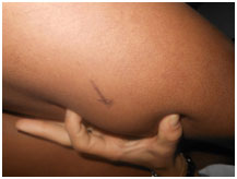
Figure 1. Nodule of the thigh
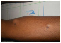
Figure 2. Nodules rosary at the leg (2 cm/2 cm)
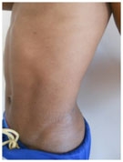
Figure 3. Spots latte2021 Copyright OAT. All rights reserv
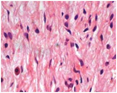
Figure 4. Proliferation of nerve fibers entangled in the connective tissue
Discussion
Segmental neurofibromatosis is extremely rare: 0.0014 to 0.002%, with female predominanceat average 30 years old [1]. This is a somatic mutation postzygotic the NF1 gene. The risk of transmission is very low (1 in 18 cases) [1,2]. Segmental localization is due to a phenomenon of late somatic mosaicism.Lesions are usually unilateral. The particularityof our observation is represented by the following atypical clinical appearance of bilateral lesions in a stringnerve pathway. Serious complications such as scoliosis, learning disorders, hematologicalhave been reported [3]. The care of patients is not codified. In absence of a study regarding therisks involved in the case of segmental neurofibromatosis, it seems justified to provide an initial assessment andclinical monitoring. Genetic counseling should be done.
References
- Saurat JH, Lachapelle JM, Lipsker D, Thomas L (2009) Saurat (5thedn), Masson France. Allanore L, P. Wolkenstein P (Eds.), Neurofibromatosi: 10,4.
- Galhotra V, Sheikh S, Jindal S, Singla A (2014) Segmental neurofibromatosis. Indian J Dent 5: 166-169. [Crossref]
- Sobjanek M, Dobosz-Kawałko M, Michajłowski I, Peksa R, Nowicki R (2014) Segmental neurofibromatosis. Postepy Dermatol Alergol 31: 410-412.
Editorial Information
Editor-in-Chief
Andy Goren
University of Rome "G.Marconi"
Article Type
Case Report
Publication history
Received date: June 21, 2015
Accepted date: July 25, 2015
Published date: July 28, 2015
Copyright
©2015 Abdou A. This is an open-access article distributed under the terms of the Creative Commons Attribution License, which permits unrestricted use, distribution, and reproduction in any medium, provided the original author and source are credited.
Citation
Abdou A, Meknassi IE, Ismaili N, Benzekri L, Senouci K, et al. (2015) Rosary nodules of the legs. Clin Case Rep Rev 1: doi: 10.15761/CCRR.1000150
Corresponding author
Ahlam Abdou
Department of Dermatology, Ibn Sina Hospital University, 1 Bir Rami Ouest Kénitra, 14090, Maroc, Tel: 00212671568343,
E-mail : sophialondon9@hotmail.fr ahlam.siham.abdou@gmail.com