Purpose: To present a new phenomenon with color objects and its principle hypothetical interpretation.
Methods: Twenty-five volunteers with normal color vision were included in the study. Red and blue objects placed on different color fields under condition of varied illumination were given to determine whether fixated by glance object seems different as compared with adjustment ones. Visual perception was studied during the prolong fixation of the red and blue objects on their blue and red backgrounds respectively.
Results: Under condition of the mesopic illumination the reds objects projection on the fovea was perceived as desaturated light spot, whereas blue objects seemed more saturated. A zone of the brightness altering of the fixated objects subtended an area corresponding to the fovea diameter.
After the prolong fixation of the blue and red objects faded and disappeared one by one. The red field became dark-gray, the blue field sustained its hue.
Conclusion: The changed and distorted visual perception in mesopic vision may be explained by mutual antagonistic relationship between the rod and cone systems regulated two competing parts of the vegetative nervous system.
color perception, saturation, rods, cones, mesopic vision, vegetative
An explanation of the nature of phenomenons like bubbles in a boiling water, an appearance of the rainbow or thunder in a cloudy sky provided us an opportunity for more deep understanding of the surrounding world. In visual physiology such a role played, for instance, a blind spot in the visual field or the Purkinje phenomenon with color pictures.
It has been our lucky opportunity to find the new phenomenon with color objects [1]. The report on the issue had been presented at the international meeting (Regional symposium of the international research group on color vision deficiencies) in Dresden in 1978 [2]. The description of the phenomenon appeared to be unknown for the audience. A core of the phenomenon occurring only under the mesopic illumination is as follows. When viewing several red objects on the blue background the fixated by the glance object, in other words, projected on the fovea, immediately becomes substantially brighter. And inversely, the fixated by a glance the blue object on the red background turns darker (Figure 1 and 2).
Within later years’ new additional features of the phenomenon requiring an explanation were noticed. A purpose of this study is the deeper examination of the phenomenon and elaboration of a hypothesis for its interpretation.
Altogether 35 volunteers with the normal visual acuity and color perception participated in the study. All tests were conducted monocularly with one of eyes. The research was selected in 3 sections:
Illumination measurement
Determination of the illumination range when fixating objects alter their brightness and the influence of a relationship between a color of the objects and the background. A set of sheets of a different color paper (red, rose, yellow, green blue, white and black) 120-200 mm in size were prepared. Six round pieces of the red or blue paper about 10 mm in diameter were glued to these paper sheets. Each sheet served as a target picture. Two of them are shown in Figure 1a and Figure 2a. The Photometer ТЕС 06-93 (Ukraine) was used for the photometric dimensions.
Initially the blue sheet with red objects and red sheet with blue objects were selected for the examination. One of the sheets was placed at distance of 15-20 cm from one of an eye of the person examined, who was asked to match the color objects in course of shifting his/her glance successively from one of them to other and compare the fixating object with all the adjacent ones. An illumination in the room had been slowly diminishing from bright to very low. When the subject reported the change of a brightness of the fixating object, the illumination level was registered. Then this procedure was repeated with other sheets: yellow, green, white and black.
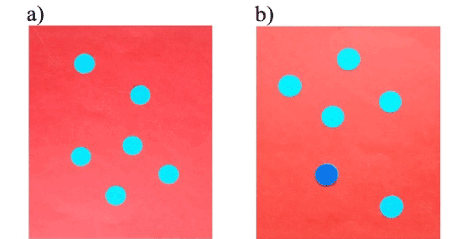
Figure 1. The target with red field and blue objects in a) photopic and b) mesopic vision. Dark blue object is fixated by glance.
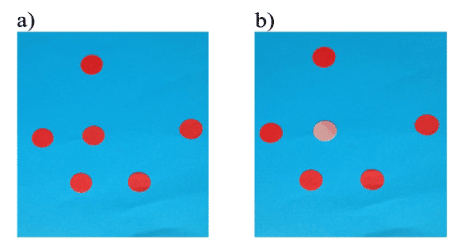
Figure 2. The target with blue field and red objects in a) photopic and b) mesopic vision. Light rose object is fixated by glance.
Zone of the color perception change
Measurement of a size of the central retina area, where the change of object brightness occurs. The new targets with blue and red fields were prepared. Each picture consists one central and 4 peripheral objects located at a circle with diameter of 30 mm (Figure 3a). At the beginning under illumination around of 10 lux the target picture was held at about 10-15 cm from the examined eye. The central blue object clearly seemed darker and the red object brighter as compared with 4 objects around them. Then the target picture was slowly moved away until the brightness of the central and peripheral objects were perceived as similar (Figure 3b). This distance was registered.
Prolong fixation
Examination of the color and brightness perception during prolonged stable viewing of the targets: red objects on blue background and blue objects on red background. Subject was asked to sustain stable gaze on either of objects of the target picture during prolong time, 7-15 seconds, and to report on a possible alteration in the visual perception .
Range of illumination
All 35 subjects noted a change of brightness of the color objects as follows: a) the red objects became brighter on the blue and green background (Figure 1b), b) blue objects were darker on red and orange background (Figure 2b). Altering of the brightness of the color objects, red and blue as well, did not observed on the yellow, black and white background.
A range of illumination was measured on 7 subjects: a change of brightness of the fixating objects began under illumination of 23 ± 3.4 lux. A disappearance of the phenomenon appeared to be at illumination below 1.0 lux. It was some technical difficulty because a change of the object brightness occurred not instantly. A discrimination of the initial little altering of brightness depended on a varying individual ability. It is likely, the volunteers noticed the phenomenon more easily with blue objects on red field.
Size of the central retinal area
While the target was moving away from distance 10 cm from the eye examined initially the central object was different in brightness in respect the peripheral ones (Figure 3a). Then at distance of 31 ± 4.8 cm all 5 objects became equal in brightness, though not all immediately (Figure 3b). A calculation was conducted on a basis of the schematic eye. The area where color objects perceive as similar in brightness subtended 5° 30' angular arc, that corresponds to an area of 1.61 mm in diameter.
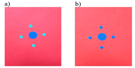
Figure 3. a) The target placed near with an eye examined, b) change of the target perception by its viewing at distant position.
Transformation of visual perception
Initially, the fixating red object on blue background entirely lost its hue and looked as a gray spot. Then peripheral red objects also are loosing their hue, became dark-gray and disappear one by one, though the blue field sustained its hue.
The fixating blue object in the red field turned in dark-blue while peripheral objects are perceived as achromatic bright spots, which then were also faded and disappeared. After 3-5 seconds the red background grew being dark-gray and then almost black.
In an event of the slight fixation shift all disappeared objects reappeared instantly, however, their saturation and brightness were different. Near them at points where the objects were located initially the positive post- images appeared: the rose ones on the gray field and the light blue ones on the blue field. It is a problem to show this picture with quickly varying details, and Fig.4 only approximately express a visual perceiving of the target (Figure 4).
Prior to formulation of a physiological nature of the phenomenon it is reasonable to indicate basic questions which appeared in a course of the study:
- Why does the functional discrepancy between the center area and periphery of the retina develop?
- What is a size of the specific center area?
- Why the phenomenon is developed under in the mesopic illumination?
- Why the fixated red object on the blue background was loosing its color, and on the contrary the blue object on the red background became dark blue?
- Why during the prolong fixation of the target the red field lost its hue and became gray? Why the peripheral objects disappear?
Rods and cones function
The photo-receptor cells rods and cones are characterized by specific distribution over the retina causing the functional discrepancy. Around 90 millions rods being extremely sensitive to light energy and active at very low illumination provide so called the scotopic vision. They are located at the periphery and para-central area of the retina. Cones, near 4.5 millions, are operating under bright light are securing the photopic vision [3]. The fovea area of 1.5 mm horizontal diameter is occupied mostly by cones. The exclusively densely packed cones are presented within the tiny fovea centralis region (foveola, 0.4 mm in size) [4-6].
This specific feature creates a ground for a distinguished condition of the light perception. Mostly cones are responsible for analyzing the light image located in the fovea, whereas both cones and rodes are producing signals, which are carrying to the brain from images at the retinal periphery. Its would be more accurate to speak about not only the red and blue colors but in respect the long and short light wavelengths, because the phenomenon took place, for instance, with green objects on the red field and rose objects of the blue field.
Fovea function
The central c retinal area where the phenomenon did not manifest appeared to be 1.61 mm (5° 30' angular arc). This value coincides closely with classical data in regard a size of the fovea [5-6]. Therefore, we can conclude that the phenomenon with color objects does not occur if solely cones are responsible for analyzing images in the fovea. Understandably, the only rod system can not produce the color phenomenon because rods are unable to distinguish colors.
Link between photo-receptive cells and vegetative nervous system
The mutual operation of both cones and rods occur under condition of an illumination between 50 and 0.05 lux presenting the mesopic vision. In our study the phenomenon took place within the illumination range between 23 ± 3.4 and 1.0 lux, though subjects noticed the difference between viewing color objects more easily and clearly by illumination approximately within 7-13 lux.
The retina is characterized by evident structural and functional differences in terms of rods and cones, which are managing by different competing parts of the vegetative nervous system. The rods have links with the sympathetic system, whereas cones with the parasympathetic system [7]. In post-synaptic fibers the noradrenalin and acetylcholine are acting in sympathetic and parasympathetic systems.
Two competing regions are organized in the retina: the fovea is represented by parasympatheticophyling cones and its periphery settled predominantly by sympatheticophyling rods [8]. Stimulation of the retinal periphery induces functional hampering of the fovea, and vice versa Kravkov,1950. This fact may serve as argument in favor of an explanation why perceptive ability at the retinal periphery and at the fovea is distinguished. In the single photopic or scotopic vision when solely cone or rods are actively working there are not conditions for a competition. Therefore the phenomenon is appeared only with mesopic vision.
2021 Copyright OAT. All rights reserv
Explanation of the phenomenon
The vegetative nervous system is bound up not only with rods and cones, but regulate actions within the cones system. It became known that the red light belongs to the parasympathetic, and the blue light to sympathetic system [4-9]. Careful investigations discovered the cones heterogeneity, and their devision into 3 types: red-catching (2.9 millions), green-catching (1.4 millions) and blue-catching (0.2 millions) ones [10-12]. It may be assumed that the red-catching rods are parasympatheticophylic, and blue-catching cones sympatheticophylic. On a ground of mentioned above arguments it is an ground to conceive a hypothetical interpretation of the phenomenon. The red and blue fields of the used target are affecting the retinal periphery in opposed ways: the red color suppresses, but the blue color stimulates the sympatheticophylic rod system. In the first case, the suppressed retinal periphery activates the fovea cones and stimulates the hue sensation. As a consequence, the saturation of the blue object is increased which subjectively perceived as less bright. In another case, the blue background stimulates the retinal periphery which in its turn inhibits the parasympatheticophylic fovea, where the cones appeared to be suspended. The red object loses its red hue and seems like white-gray spot.
Originally, when the phenomenon had been discovered, the stimulating effect of red light on the cone system and fovea had originated an idea to treat with the red light the impaired vision of eyes suffered of the amblyopia [1].
Prolong gazing
The third stage of the study was associated with a problem because only 4 from 11 volunteers were able to meet the mandatory requirement of the procedure, namely, to stare steadily for several seconds on one of the color objects. Really, it a hard task. Our finding in regard a prolong viewing the color targets revealed the dramatical impact on a visual perception. In respect the backgrounds, the profound changes occurred only with red one. Our interpretation is as follows. In the mesopic illumination an active state of the rod system and sympathetic part of the vegetative nervous system generates an antagonistic impact on the parasympatheticophyling red-catching cones causing a hampering of the red light sensation and eventually the full desaturation. In its turn the red field making suppression of the rod system diminishes the rod light perception. As a result the red field became dark-gray, if not black.
The sympatheticophyling blue field falls on a positive conditions of the retinal periphery. A perception of the blue field remains practically not distorted.
As regards a behavior of the objects red and blue alike on their color fields, the two mutually antagonistic components of the vegetative nervous system in mesopic illumination develops a fatigue in the color perception both red and blue objects. They gradually disappear from the fields.
The glance shift during viewing of the targets displaces the color objects on neighboring areas, where slight functional ability remains. As a consequence, a visual perception of the objects reappears for a short period. At the same time at the spots of initial objects projections the positive post-images appear (Figure 4). Their development may be explained by an existence of an active processes in the cone system although the brain does not “see” the objects.
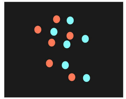
Figure 4. View of the target with the red field during the prolong fixation of one of the blue objects and a shift of the fixation to the right.
Main intention of this paper was a presentation of the new phenomenon on color perception with its interpretation. This paper should be regarded as invitation to verify the core of the phenomenon. Now we present a preliminary and principle hypothesis for its explanation. Its detailed understanding requires further investigations. We do not exclude any possible corrections or additions, moreover, would be pleased to meet them. Certainly, the deep comprehending of this phenomenon will contribute our knowledge of the super complex mechanism of the visual function.
Changed and distorted visual perception in mesopic vision may be explained by mutual antagonistic relationship between the rod and cone systems regulated by two competing parts of the vegetative nervous system.
- Sergienko NM (1966) New possibility in treatment of an amblyopia with eccentric fixation. Ophthalmol J : 403-407.
- Sergienko NM (1978) New color phenomenon. Regional symposium of the international research group on color vision deficiencies.
- Curcio CA, Sloan KR, Kalina RE, Hendrickson A (1990) Human photoreceptor topography. J Comparative Neurol 2929: 497-523.
- Kravkov SV (1950) Eye and its work. Academy of Science of USSR.
- Kozart DM (1989) Anatomic correlates of the retina. Duane's Clin Ophthalmol 3:1-18.
- Benson WE (1989) An introduction to color vision. Duane's Clin Ophthalmol 3:1-18.
- Babsky EB (1947) Significance of sympathetic nervous system in regulation the vision analyzer function. Problem of Physiol Optics 4: 13-30.
- Lebedinsky AV, Pressman JM, Fadeeva AA (1948) New data in regard relationship between centre and periphery of the retina. Problem of Physiol Optics 6: 104-111.
- Kravkov SV (1948) About nature of color vision. Problem of Physiol Optics 6:17-27.
- Baker GE (2009) Anatomy of vision (2009) In: Optometry, Elsevier.
- Gouras P, Charles S (1989) Physiology of the retina. Duane's Clini Ophthalmol 3:1-18.
- Oyster CW (1999) The human eye:structure and function. 1st edn, Sinaeur, Massachusetts.