A 35-year-old male was referred to our hospital complaining for bleeding of per rectum. Colonoscopy from anus to the terminal ileum revealed only a solitary pedunculated polyp in the sigmoid colon and polypectomy was performed without complications (Figure 1). . He had no mucocutaneous pigmentation or family history of Peutz-Jeghers syndrome (PJS). Histopathological examination revealed polypoid structure with hyperplastic epithelial covering. And, the underlying core showed branching smooth muscle fibers (Figure 2a). The presence of muscle fibers confirmed by Desmin & alpha smooth muscle actin (SMA) immunehistochemical staining (Figure 3). Epithelial covering showed moderate pleomorphism with moderate nuclear atypia and atypical mitotic figures (Figure 2b). The polyp was diagnosed as a hamartomatous polyp with dysplastic foci.

Figure 1: Solitary pedunculated polyp in the sigmoid colon during snare polypectomy.
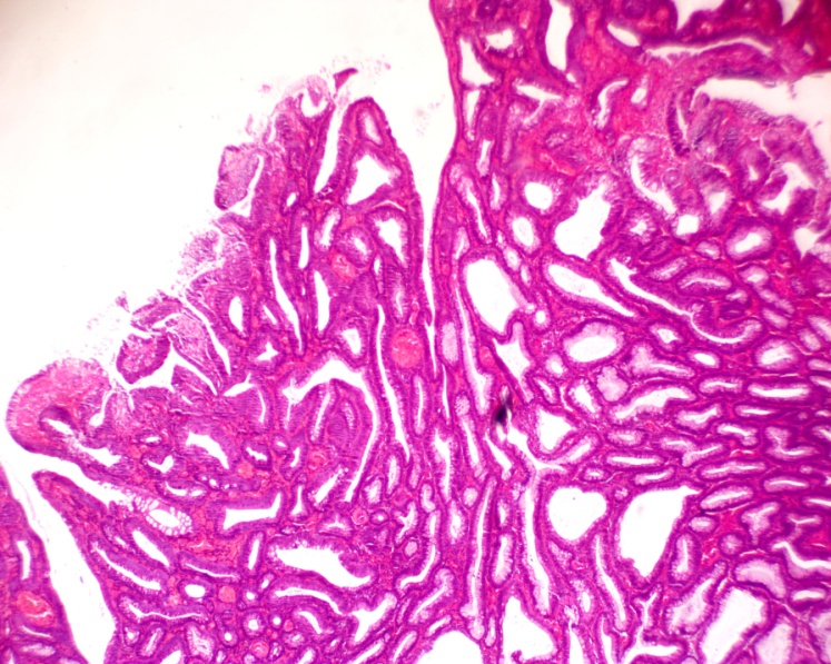
Figure 2:[a]Polypid mass with irregular hamartomatous glands (H&E x40), [b]epithelial covering shows moderate dysplasia with little mitosis (H&E x400).
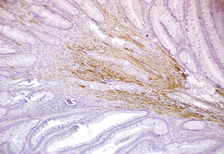
Figure 3:[a]Positive Desmin staining in smooth muscle fibers within the core (100x), [b]positive α SMA staining in smooth muscle fibers within the core (100x).
Discussion
A Peutz-Jeghers polyp without either mucocutaneous pigmentation or a positive family history was described as a solitary Peutz-Jeghers (P-J) type hamartomatous polyp. which has been suggested to be a clinical entity different from PJS [1]. The malignant potential of solitary P-J type polyps is unclear; however, the hypothesis of hamartoma-adenoma-carcinoma sequence has been suggested [2]. Only 28 previous case reports of solitary P-J type polyps were found. One polyp was located in the stomach, 21 in the small intestine, and 6 in the colon. Only 3 cases of malignant transformation in a solitary P-J-type polyp have been reported to date; one polyp was located in the duodenum, and the others were colorectal [3]. The present case is the 3rd case report of malignant transformation in a colorectal P-J type polyp, supporting the hypothesis of hamartoma-adenoma-carcinoma sequence.
Abbreviations
PJS: Peutz-Jeghers syndrome; SMA: smooth muscle actin
References
- Nebril BA, Filgueira LT, Calvo PA, García RG, Rodríguez GD, et al. (1993) Solitary hamartomatous duodenal polyp; a different entity: report of a case and review of the literature. Surgery Today 23: 1074-1077.
- Bosman FT (1999) The hamartoma-adenoma-carcinoma-sequence. J Pathol 188: 9-13.
- Arima S, Kunimura T, Matsuo K, Hobo T, Nogaki K, et al. (2010) Solitary Peutz-Jeghers type colorectal polyp with hamartoma-adenoma-carcinoma sequence in a non-Peutz-Jeghers syndrome patient. Showa Univ J Med Sci 22: 69-72.
Editorial Information
Editor-in-Chief
Marcel C C Machado
Department of Clinical Emergencies,
University of São Paulo School of Medicine,
Brazil
Article Type
Case Report
Publication history
Received date: March 01, 2016
Accepted date: April 20, 2016
Published date: April 23, 2016
Copyright
©2016 Altonbary AY. This is an open-access article distributed under the terms of the Creative Commons Attribution License, which permits unrestricted use, distribution, and reproduction in any medium, provided the original author and source are credited.
Citation
Altonbary AY, Bahgat MH, Elkashef WF(2016) Hamartoma-adenoma-carcinoma sequence in a solitaryPeutz-Jeghers type sigmoid polyp.Gastroenterol Hepatol Endosc1: doi:10.15761/GHE.1000104
Corresponding author
Dr. Ahmed Youssef Altonbary
Department of hepatology and gastroenterology, Mansoura specialized medical hospital, Mansoura, Egypt
E-mail : tomiki@juntendo.ac.jp
Figure 1: Solitary pedunculated polyp in the sigmoid colon during snare polypectomy.
Figure 2:[a]Polypid mass with irregular hamartomatous glands (H&E x40), [b]epithelial covering shows moderate dysplasia with little mitosis (H&E x400).
Figure 3:[a]Positive Desmin staining in smooth muscle fibers within the core (100x), [b]positive α SMA staining in smooth muscle fibers within the core (100x).