Objective: To evaluate and compare the error in cephalometric measurements by manual and computerized methods using the softwares Dolphin Imaging version 11.0 and Dentofacial Planner version 7.02.
Methods: The sample comprised 45 lateral cephalograms. A calibrated operator conducted 90 manual and 180 digital cephalometric tracings, using 8 angular measurements (SNA, SNB, ANB, FMA, 1.NA, 1.NB, SN-Occlusal, SN-GoGn) and 5 linear measurements (1-NA, 1-NB, Co-Gn, Co-A e LAFH). For analysis of results, the dependent t test was used to compare the error of each method (intragroup error); comparison of measurements between the three methods was performed by the one-way ANOVA and Tukey test in the presence of significant result.
Results: The results showed statistically significant differences, especially between measurements evaluated on the Dolphin software and by manual tracing (p>0.05).
Conclusions: According to the present results, the three cephalometric methods have proven to be accurate, since they presented few systematic errors. The computerized method using the Dentofacial Planner software showed the highest reliability, followed by the manual method, while the Dolphin Imaging software was the least effective and more likely to produce systematic errors in the identification of points.
Since the advent of the first dental X-ray machine in 1919, radiographies have been used for several purposes, including diagnosis, evaluation of pathologies, periapical lesions, tooth resorptions and evaluation of existing endodontic and restorative treatments.
Cephalometrics appeared as an evolution of craniometry from the standardization o radiographic techniques and development of the cephalostat proposed by Broadbent [1] in 1931.
The radiographic image is an important aid in the diagnosis and evaluation of skeletal and dental relationships. Since 1931 [1], bidimensional images have been used to identify the anatomical landmarks with which the cephalometric measurements are obtained [2].
For a long time, manual tracing was the only method available for cephalometric tracing and achievement of angular and linear measurements required for interpretation [3].
For cephalometric analysis, it is necessary to accurately define how to identify the different specific points used. The radiographs must be technically perfect, without distortions, to allow good visualization of bony anatomical structures and the soft tissue profile.
With the increasing presence of informatics in the clinical routine, it has been a great aid to speed up the work, optimize tasks, simplify procedures and increase the productivity and refinement of diagnosis. The constant technological advances in the field of computing, combined to scientific advances in dental radiology, led to the development of computerized softwares for cephalometric tracings and measurements, as well as accomplishment of different types of analysis [4]. Digital images are currently routinely used in the orthodontic practice. Several systems and formats are available to produce, store, recover, visualize and share these images [5].
This study aimed to compare the error in cephalometric measurements with manual and computerized methods using the softwares Dolphin Imaging and Dentofacial Planner, aiming to evaluate the reliability of cephalometrics techniques analyzed.
This study aimed to evaluate the reliability of manual cephalometrics compared to two specific softwares and quantified the degree of reliability of identification of cephalometric points manually and digitally, evaluating and comparing the outcomes achieved and comparing them to each other, to aid the clinician in selecting the best option for cephalometrics, either manual or computerized.
Material
To evaluate the reliability of manual tracings compared to specific softwares for this purpose, the sample was composed of 45 lateral cephalograms (Figure 1) of individuals submitted to orthodontic treatment.
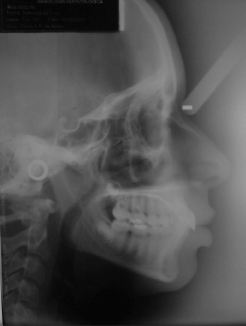
Figure 1. Initial lateral cephalogram.
Only the initial cephalograms available from the initial orthodontic records of individuals were used, therefore no additional examination was necessary.
The study included lateral cephalograms of young and young adult individuals, aged 12 to 35 years. The basic criterion for sample selection was presence of the permanent maxillary and mandibular first molars. Individuals presenting hypodontia or missing permanent teeth, deciduous teeth, impacted or unerupted teeth or any previous orthodontic treatment were excluded from the sample.
The radiographs were selected by observing the quality of image (contrast/density) and sharpness of points to be marked. The study included only lateral cephalograms with optimal quality, since the goal was to analyze the accuracy of identification of points, which is directly influenced by the quality of radiographs.
Methods
Manual method: The following instruments were used for manual tracing: film viewer, protractor, set square, ruler, 0.5mm mechanical pencil, eraser and 80 sheets of Ultraphan paper (G&H Wire Company). One sheet of Ultraphan paper (G&H Wire Company) with 0.07-mm thickness was adapted on each radiograph. The cephalogram (Figure 2) was traced in a darkened room. The cephalometric points were identified following a sequence pre-established by the examiner, using 0.5mm mechanical pencil with the aid of ruler, protractor and setsquare for achievement of cephalometric measurements. After adaptation of an Ultraphan sheet (G&H Wire Company), manual tracing (Figure 3) of each radiograph was performed following a minimum interval of one month for repetition.
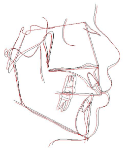
Figure 2. Schematic drawing of cephalometric tracing.
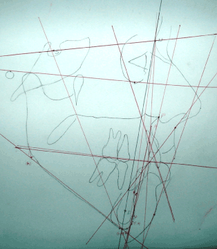
Figure 3. Manual tracing on ultraphan paper G&H Wi.
Computerized method: The computerized analysis used the Dentofacial Planner software version 7.02 (Figure 4), which allows transferring the points marked on the manual tracing to the computer, using a digitizing table, following points previously marked on the Ultraphan paper (G&H Wire Company). The study also used the Dolphin Imaging software version 11.0, which performs analyses on previously digitized lateral cephalograms.
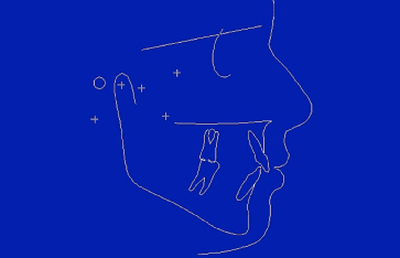
Figure 4. Tracing on the dentofacial planner software.
The cephalometric points required for ____ standard analysis were identified on each tracing. The tracings were performed twice on each cephalogram, with a one-month interval between the first and second measurements to assure reliability.
The following 13 measurements were selected: SNA, SNB, ANB, Co-Gn, FMA, SN.GoGn, LAFH, 1.NA, 1-NA,1.NB, 1-NB, SN.OCCL, Co-A, considering their great importance to aid the diagnosis and orthodontic treatment planning.
For utilization on the Dolphin Imaging software, the cephalograms were digitized using a scanner Microtek, model ScanMaker i800 (9600 x 4800 dpi, Microtek International, Inc., Carson, CA, USA) with minimum resolution of 300 dpi and connected to a microcomputer.
Cephalometrics was performed using the Orthodontics software. The identification of points using the Dolphin Imaging software was also performed in a darkened room, following the sequence established by the software.
Concerning the Dentofacial Planner software, initially the points were identified on a sheet of Ultraphan paper (G&H Wire Company), similar to the manual method, marking the points on the sequence previously established by the software, and then digitized using a digitizing table.
The manual and digital tracings on the Dolphin Imaging software version 11.0 (Figure 5) were performed in the ______________________________________ while tracings on the Dentofacial Planner software version 7.02 were performed in ______________________________________.
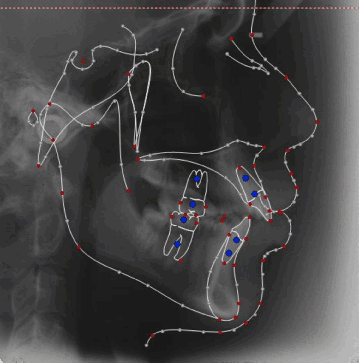
Figure 5. Tracing on the dolphin Imaging software.
Statistical analysis
All radiographs were evaluated and traced by a single examiner, previously calibrated using 10 lateral cephalograms for each measurement technique, both the manual method and the two computerized softwares used in this study.
For evaluation of intra-examiner error, all points were identified twice on all 45 cephalograms. After tracings, the data were plotted and statistically analyzed by the dependent t test to evaluate the intragroup error.
Intergroup comparison
The values obtained in the three groups related to the means of initial and final values and the difference between them were compared by analysis of variance ANOVA followed by the Tukey test for intergroup comparison.
Statistical analysis was performed with software Statistica for Windows (Statistica for Windows – Release 7.0 - Copyright Statsoft, Inc. 2005). Results were considered statistically significant at p<0.05.
Results
Results are presented in Tables 1-4. The three cephalometric methods presented to be properly accurate, since they presented few systematic errors.
Table 1. Results of error of manual tracing, comparing the first and second measurements (dependent t test) (N=45).
|
Variables
|
1st measurement
|
2nd measurement
|
P
|
|
Mean
|
SD
|
Mean
|
SD
|
|
SNA (º)
|
81.48
|
3.83
|
81.23
|
3.90
|
0.056
|
|
SNB (º)
|
77.74
|
3.45
|
77.63
|
3.47
|
0.390
|
|
ANB (º)
|
3.74
|
2.45
|
3.62
|
2.24
|
0.439
|
|
Co-A (mm)
|
95.73
|
4.72
|
95.66
|
5.04
|
0.755
|
|
Co-Gn (mm)
|
125.04
|
6.34
|
124.72
|
6.76
|
0.102
|
|
FMA (º)
|
23.01
|
4.92
|
22.87
|
5.09
|
0.387
|
|
SN.GoGn (º)
|
29.85
|
4.90
|
30.75
|
4.99
|
0.000*
|
|
SN.Occl (º)
|
14.26
|
3.69
|
14.28
|
3.68
|
0.894
|
|
LAFH (mm)
|
74.35
|
5.24
|
73.97
|
5.45
|
0.020*
|
|
1-NA (mm)
|
6.55
|
4.08
|
6.24
|
3.90
|
0.017*
|
|
1.NA (º)
|
23.25
|
7.25
|
23.11
|
7.10
|
0.379
|
|
1-NB (mm)
|
7.00
|
3.09
|
6.65
|
3.01
|
0.000*
|
|
1.NB (º)
|
28.64
|
6.36
|
28.26
|
6.32
|
0.007*
|
*Statistically significant at P<0.05.
Table 2. Results of error of tracing on the Dentofacial Planner software, comparing the first and second measurements (dependent t test) (N=45).
|
Variables
|
1st measurement
|
2nd measurement
|
P
|
|
Mean
|
SD
|
Mean
|
SD
|
|
SNA (º)
|
79.92
|
5.22
|
80.11
|
5.54
|
0.292
|
|
SNB (º)
|
74.82
|
4.35
|
74.93
|
4.58
|
0.318
|
|
ANB (º)
|
5.09
|
2.36
|
5.17
|
2.42
|
0.303
|
|
Co-A (mm)
|
78.88
|
3.47
|
78.81
|
3.52
|
0.248
|
|
Co-Gn (mm)
|
127.64
|
5.81
|
127.97
|
6.07
|
0.020*
|
|
FMA (º)
|
28.98
|
4.97
|
28.99
|
4.98
|
0.869
|
|
SN.GoGn (º)
|
40.02
|
6.20
|
39.85
|
6.67
|
0.454
|
|
SN.Occl (º)
|
19.66
|
4.97
|
19.56
|
4.96
|
0.554
|
|
LAFH (mm)
|
75.30
|
4.93
|
75.30
|
5.04
|
0.976
|
|
1-NA (mm)
|
2.04
|
2.98
|
2.02
|
3.06
|
0.809
|
|
1.NA (º)
|
23.11
|
5.55
|
22.96
|
5.53
|
0.271
|
|
1-NB (mm)
|
5.33
|
2.83
|
5.46
|
2.95
|
0.086
|
|
1.NB (º)
|
30.69
|
7.32
|
30.59
|
7.26
|
0.419
|
*Statistically significant at P<0.05.
Table 3. Results of error of tracing on the Dolphin Imaging software, comparing the first and second measurements (dependent t test) (N=45).
|
Variables
|
1st measurement
|
2nd measurement
|
P
|
|
Mean
|
SD
|
Mean
|
SD
|
|
SNA (º)
|
87.03
|
4.47
|
86.36
|
4.28
|
0.002*
|
|
SNB (º)
|
80.59
|
3.39
|
79.69
|
3.37
|
0.000*
|
|
ANB (º)
|
6.44
|
3.12
|
6.65
|
2.97
|
0.230
|
|
Co-A (mm)
|
101.88
|
5.17
|
102.03
|
5.75
|
0.756
|
|
Co-Gn (mm)
|
128.71
|
6.76
|
129.06
|
7.46
|
0.326
|
|
FMA (º)
|
26.00
|
5.12
|
26.40
|
5.30
|
0.102
|
|
SN.GoGn (º)
|
30.52
|
5.05
|
30.81
|
5.09
|
0.005*
|
|
SN.Occl (º)
|
13.92
|
3.62
|
14.60
|
3.37
|
0.001*
|
|
LAFH (mm)
|
73.78
|
5.12
|
73.87
|
4.97
|
0.693
|
|
1-NA (mm)
|
1.50
|
3.91
|
1.40
|
3.68
|
0.513
|
|
1.NA (º)
|
21.17
|
6.85
|
21.83
|
6.25
|
0.158
|
|
1-NB (mm)
|
6.39
|
3.09
|
6.44
|
3.19
|
0.791
|
|
1.NB (º)
|
28.60
|
6.07
|
29.32
|
6.51
|
0.114
|
*Statistically significant at P<0.05.
Table 4. Results of the one-way ANOVA and Tukey test, when needed, for comparison of mean error of the three types of tracing: manual, Dentofacial Planner and Dolphin Imaging (N=45).
|
Variables
|
Manual
|
Dentofacial
|
Dolphin
|
P
|
|
Mean (SD)
|
Mean (SD)
|
Mean (SD)
|
|
SNA (º)
|
-0.25 (0.87)AB
|
0.19 (1.20)A
|
-0.67 (1.42)B
|
0.003*
|
|
SNB (º)
|
-0.11 (0.85)A
|
0.11 (0.75)A
|
-0.90 (1.15)B
|
0.000*
|
|
ANB (º)
|
-0.12 (1.50)A
|
0.08 (0.54)A
|
0.20 (1.14)A
|
0.254
|
|
Co-A (mm)
|
-0.06 (1.42)A
|
-0.06 (0.35)A
|
0.15 (3.24)A
|
0.848
|
|
Co-Gn (mm)
|
-0.32 (1.29)A
|
0.32 (0.91)A
|
0.35 (2.40)A
|
0.094
|
|
FMA (º)
|
-0.13 (1.02)A
|
0.01 (0.45)A
|
0.39 (1.60)A
|
0.073
|
|
SN.GoGn (º)
|
0.90 (1.69)A
|
-0.16 (1.50)B
|
0.56 (1.29)AB
|
0.003*
|
|
SN.Occl (º)
|
0.02 (1.11)A
|
-0.09 (1.07)A
|
0.67 (1.32)B
|
0.004*
|
|
LAFH (mm)
|
-0.37 (1.05)A
|
0.00 (0.51)A
|
0.08 (1.46)A
|
0.099
|
|
1-NA (mm)
|
-0.31 (0.84)A
|
-0.01 (0.49)A
|
-0.18 (1.83)A
|
0.510
|
|
1.NA (º)
|
-0.14 (1.90)A
|
-0.15 (0.93)A
|
0.65 (3.07)A
|
0.082
|
|
1-NB (mm)
|
-0.34 (0.55)A
|
0.13 (0.51)B
|
0.04 (1.11)B
|
0.009*
|
|
1.NB (º)
|
-0.37 (0.90)A
|
-0.10 (0.87)AB
|
0.71 (2.99)B
|
0.017*
|
*Statistically significant at P<0.05.
Radiographs
Since standardization is fundamental in comparative studies, the procedures were performed by a single examiner. Manual tracing of all 45 radiographs was randomly performed as suggested by Houston [6], as well as the sequence of computerized methods, in which care was taken so that each selected point would represent the manual counterpart.
Considering the great acceptance of the computerized method, studies are necessary to compare the efficiency of this method compared to conventional cephalometric tracing. Emphasis should also be given to the reduced time necessary for measurement of cephalometric dimensions when using computerized cephalometrics [7].
Statistical analysis and error
Systematic errors occurred in this study, which are expected in any cephalometric measurement, even performed by experienced examiners. Therefore, a series of measurements may be systematically different from a series obtained in another moment. This may have occurred between the manual and computerized methods, which may have contributed to the several significant differences between means [6].
When analyzing the advantages and disadvantages of different methods of cephalometric tracing, it may be inferred that manual or conventional tracing is among the most investigated concerning the reliability of identification of cephalometric points and measurements [8,9].
Errors in the projection and identification of points may be included among the great errors of conventional cephalometrics.
Errors are common in cephalometrics and may occur even among experienced operators. Comparative measurements are five times more accurate than the individual identification of cephalometric points [10,11].
The analyses employed are extremely important for statistical analysis, because they primarily aim to perform an isolated analysis of the examiner’s error in each group (dependent T test), followed by comparison between groups (ANOVA and Tukey).
Data
Data were obtained using three different methods, namely manual tracing and computerized analyses using the softwares Dolphin Imaging and Dentofacial Planner. Manual tracing was selected because so far it has been the most employed by most orthodontists for diagnosis and treatment planning. Conversely, the softwares Dolphin and Dentofacial were selected because they are currently the most used and known, even though several cephalometrics softwares are available. Both softwares have good acceptance by many orthodontists and, to our knowledge, have been presenting good reliability of data.
Studies in the literature on the same subject and with similar purpose as the present investigation reveal the lack of criteria concerning the selection of cephalometric points and the ideal angular and linear measurements for such studies [12-15].
The manual method demanded longer time to trace, yet this was similar to the Dentofacial Planner and shorter than for the Dolphin software, due to the stage of identification of points on the Ultraphan paper. The manual method is the most common for achievement of tracings, identification of points, measurement of distances and angles between points, yet it also presents high probability of error [16].
The errors found in the present methods demonstrate that the apparent lack of accuracy of measurements is not related to the method employed, but rather to the likely uncertainty at the moment of identification of points, which occurs in any technique, thus making computerized cephalometrics as reproducible and uncertain as the conventional manual tracing [7].
Several studies have been performed to evaluate the accuracy of computerized methods compared to the manual technique, revealing variable results. Some studies did not reveal significant differences [7], while others indicated fewer errors in the computerized method [17,18] and suggested their utilization due to the reduced time spent for cephalometric analysis and the reliability of data achieved.
Albuquerque, et al. [17], stated that the examiner presents great interference in the incorporation of systematic errors, which depend on several factors such as the experience of examiners, calibration of equipment and points and measurements analyzed.
Errors may be incorporated in cephalometric tracings by any method, either conventional, manual or computerized. According to several studies [6,19-22] these errors may occur due to failures involving the projection of cephalometric points, poor image quality, differences between examiners (e.g. during achievement of the radiograph, including incorrect patient positioning).
Some cephalometric points are normally subject to error because their location is more difficult, making them less reliable [7,12,22,23].
In modern Orthodontics, quantitative, systematic and objective measurements based on reference points located in hard and soft tissues in some cephalograms are daily used in the clinical practice. Accuracy and reproducibility in data achieved by cephalometrics are important for the orthodontist. Errors in conventional methods occur from the identification and measurement of cephalometric points. The advances in computer technology in Orthodontics allowed easy storage, handling and transmission of images, besides the possibility of highlighting, as well as questions on the validity of digital cephalometrics [20,24,25].
In the present study, some measurements presented statistically significant differences when the first and second measurements were compared.
Table 1 presents the results of manual tracings when the first and second measurements were compared, which reveal statistically significant outcomes for measurements as SNGoGn, LAFH, 1-NA, 1-NB and 1.NB. A relatively high error was observed, in among 13 measurements analyzed, corresponding to 38.46%, which demonstrates some difficulty in the identification of points for correct measurement of cephalometric dimensions. This difficulty in the identification of points in manual tracing should be considered, because it may impair the correct orthodontic diagnosis.
Table 2 displays the outcomes of the Dentofacial Planner software, evidencing nearly no systematic error except for one cephalometric dimension, namely the CoGn, or 7.69%.
Table 3 presents the outcomes for the Dolphin Imaging software, which revealed similar systematic error as the manual tracing, yet only 4 cephalometric dimensions presented statistical significance, including SNA, SNB, SNGoGn and SNOccl, or 30.76%. This error may have occurred due to the software’s sensitivity in the identification of points.
Concerning the angular measurement SNGoGn, even though this dimension involves the point N, which is difficult to identify in both methods, it was not statistically different compared to the Dentofacial Planner software and presented statistically significant result when manual tracing was compared to the Dolphin Imaging software [10].
Concerning the linear measurements 1-NA and 1-NB, which require identification of points A and B – which are also difficult to identify, statistically significant differences were observed between the first and second measurements. However, no statistically significant differences were observed between initial and final measurements for the computerized method using both softwares [10].
Similar to the linear measurements, the angular measurement 1.NB only presented statistically significant difference between first and second measurements in the manual tracing, which presented to be less accurate than the computerized softwares [10].
The main difficulty was observed for the identification of points on the apices of incisors, in which the digitized image involves gray shades that may be confounded at this region.
The study conducted by Gravely [26] revealed that the computerized cephalometric analysis does not cause error in measurements, especially when the reference points are identified by a human examiner. However, other study revealed statistically significant differences in the identification of cephalometric points between original and digitized cephalograms [12].
The three cephalometric methods presented to be accurate, since they presented few systematic errors. The computerized method using the Dentofacial Planner software showed the highest reliability, followed by the manual method, while the Dolphin Imaging software was the least effective and more likely to produce systematic errors in the identification of points.
- Broadbent BH (1931) A new x-ray technique and its application to orthodontia. Angle Orthod 1:45-66.
- Moshiri M, Scarfe WC, Hilgers ML, Scheetz JP, Silveira AM, et al. (2007) Accuracy of linear measurements from imaging plate and lateral cephalometric images derived from cone-beam computed tomography. Am J Orthod Dentofacial Orthop132: 550-560. [Crossref]
- Chen SK, Chen YJ, Yao CC, Chang HF (2004) Enhanced speed and precision of measurement in a computer-assisted digital cephalometric analysis system. Angle Orthod 74: 501-507.
- Hazey MA, 3rd, Ngan P, Reed H, Razmus T, Crout R, et al. (2009) Comparison of computer-generated, enhanced and conventional 2-dimensional radiographic imaging. Am J Orthod Dentofacial Orthop 135: 463-467.
- Decker JD, Bollen AM, Chen CS (2007) A model for digital archiving of radiographs into a searchable database. Am J Orthod Dentofacial Orthop 132: 856-859. [Crossref]
- Houston WJ (1983) The analysis of errors in orthodontic measurements. Am J Orthod 83: 382-390. [Crossref]
- Brangeli LAM, Henriques JFC, Vasconcelos MHF, Janson G (2000) Comparative study of cephalometric analysis through manual and computerized methods. Rev APCD 54: 234-241.
- Adenwalla ST, Kronman JH, Attarzadeh F (1988) Porion and condyle as cephalometric landmarks--an error study. Am J Orthod Dentofacial Orthop 94: 411-415. [Crossref]
- Barrett MJ, Brown T, McNulty EC (1968) A computer-based system of dental and cranio-facial measurement and analysis. Aust Dent J 13: 207-212. [Crossref]
- Ferreira JT1, Telles Cde S (2002) Evaluation of the reliability of computerized profile cephalometric analysis. Braz Dent J 13: 201-204. [Crossref]
- Gonçalves FA, Schiavon L, Pereira Neto JS, Nouer DF (2006) Comparison of cephalometric measurements from three radiological clinics. Braz Oral Res 20: 162-166. [Crossref]
- Chen YJ, Chen SK, Chang HF, Chen KC (2000) Comparison of landmark identification in traditional versus computer-aided digital cephalometry. Angle Orthod 70: 387-392.
- Chen YJ, Chen SK, Yao JC, Chang HF (2004) The effects of differences in landmark identification on the cephalometric measurements in traditional versus digitized cephalometry. Angle Orthod 74: 155-161.
- Davis DN, Mackay F (1991) Reliability of cephalometric analysis using manual and interactive computer methods. Br J Orthod 18: 105-109. [Crossref]
- Nimkarn Y, Miles PG (1995) Reliability of computer-generated cephalometrics. Int J Adult Orthodon Orthognath Surg 10: 43-52. [Crossref]
- Guedes PA, Souza JÉN, Tuji FM, Nery EM (2010) Comparative study of manual and computerized cephalometric analysis. Dental Press J Orthod 15: 44-51.
- Albuquerque HR, Almeida HC (1998) Evaluation of error in reproducibility of cephalometric values applied to Tweed-Marrifield philosophy by methods computerized and conventional. Orthodontics 31: 18-30.
- Dana JM, Goldstein M, Burch JG, Hardigan PC (2004) Comparative study of manual and computerized cephalometric analyses. J Clin Orthod 38: 293-296. [Crossref]
- Cohen AM (1984) Uncertainty in cephalometrics. Br J Orthod 11: 44-48. [Crossref]
- Houston WJ, Maher RE, McElroy D, Sherriff M (1986) Sources of error in measurements from cephalometric radiographs. Eur J Orthod 8: 149-151. [Crossref]
- Kamoen A, Dermaut L, Verbeeck R (2001) The clinical significance of error measurement in the interpretation of treatment results. Eur J Orthod 23: 569-578. [Crossref]
- Midtgård J, Björk G, Linder-Aronson S (1974) Reproducibility of cephalometric landmarks and errors of measurements of cephalometric cranial distances. Angle Orthod 44: 56-61. [Crossref]
- Sandler PJ (1988) Reproducibility of cephalometric measurements. Br J Orthod 15: 105-110. [Crossref]
- Forsyth DB, Shaw WC, Richmond S (1996) Digital imaging of cephalometric radiography, Part 1: Advantages and limitations of digital imaging. Angle Orthod 66: 37-42. [Crossref]
- Forsyth DB, Shaw WC, Richmond S, Roberts CT (1996) Digital imaging of cephalometric radiographs, Part 2: Image quality. Angle Orthod 66: 43-50. [Crossref]
- Gravely JF, Benzies PM (1974) The clinical significance of tracing error in cephalometry. Br J Orthod 1: 95-101. [Crossref]