Abstract
Breast engorgement is, the swelling and distension of breasts, usually in the early days of initiation of lactation, due to vascular dilatation as well as the arrival of the milk. Breast engorgement occurs in 72% to 82% of lactating women. Various physiotherapeutic interventions for treatment of breast engorgement have been documented such as, Hot Moist Pack, Massage, Ultrasound. Although Ultrasound is used in the treatment of breast engorgement but literature is scarce on use of it. The objective of the present study was to comparative effectiveness of ultrasound therapy with conventional therapy on breast engorgement in immediate post-partum mothers. In randomized controlled trial, 80 post-partum mothers with breast engorgement were randomly assigned to Group A (Ultrasound therapy) and Group B (Conventional therapy) during the study period from April, 2010 to December 2010. The outcome measures used were Visual Analogue Scale (VAS), Hardness scale and Six-point engorgement scale (SPES). The pre-treatment and post-treatment values of outcome measures were noted on for all the four days.
The intra group comparison results showed that pain on VAS, hardness score and SPES score was statistically significant in both groups. Whereas inter-group comparison results showed that VAS was statistically significant on 2nd and 3rd day post-intervention. Pre-intervention and post-intervention at 4th day were statistically significant in both groups by the hardness and SPES scores. The study concluded that ultrasound therapy added with conventional therapy helps in reduction of pain with non-tender breasts which further helps the post-partum mothers to recover better from discomforts of breast engorgement. This in turn can facilitate better breast feeding.
Key words
breast engorgement, ultrasound, massage, hot moist pack, visual analogue scale (vas), hardness scale and six-point engorgement scale (spes)
Introduction
Breast engorgement is defined as the swelling and distension of breasts, usually in the early days of initiation of lactation, due to vascular dilatation as well as the arrival of the milk. It is the painful overfilling of the breasts with milk. This is usually caused by an imbalance between milk supply and infant demand. Early breast fullness occurs as milk supply develops and while newborn has an irregular breast-feeding routine. The normal fullness is caused by the milk and extra-blood and fluids in the breasts as body uses the extra fluids to make breast milk and if baby is not breast-fed for several days then breast engorgement can occur. Breast engorgement occurs in 72% to 82% of lactating women [1]. Engorgement is most common during the first week of breast-feeding and occurs as a result of delayed, infrequent or interrupted removal of milk from the breast [2]. The factors which may place a mother at a higher risk of engorgement are, failure to prevent or resolve milk stasis resulting from infrequent or inadequate drainage of the breasts [3,4].
When milk production increases rapidly, the volume of milk in the breast can exceed the capacity of the alveoli to store it. If the milk is not removed, over-distention of the alveoli can cause the milk-secreting cells to become flattened and drawn out, even to rupture [3]. Accumulation of milk and the resulting engorgement causes programmed cell death, thus resulting in involution of the milk-secreting gland, milk reabsorption, collapse of the alveolar structures, and the cessation of milk production [5]. A mother, who is experiencing normal breast fullness, should be encouraged to nurse frequently, at least eight to twelve times in 24 hours, waking the baby if necessary [6]. The most common cause of sore nipples in the first few days of feeding is the incorrect position of the baby at the breast meaning that the baby sucks only at the "nipple" [7].
Engorgement of the breast can be prevented by keeping the baby on mother's milk both in hospital and home, unrestricted and exclusive breastfeeding on demand.3 Mother's milk is undoubtedly the best food for babies. It is a living fluid and contains exactly the right amount of nutrients required by a baby [8].
It is important to distinguish between physiological and pathological engorgement [3,9]. Treatment for breast engorgement include conservative, medical, and surgical [9]. Though various interventions are available few studies support that ultrasound is effective in relieving symptoms of breast engorgement [9,10]. Heat application in the form of hot moist heat is a comfort measure to activate the milk ejection reflex [11]. Gentle massage can be used as conventional intervention for relieving breast engorgement. Massage prior to feedings is helpful [9]. Studies have evaluated the effect of hot moist pack and massage but literature is scarce on the use of ultrasound therapy for the same. Hence the present study has been undertaken with the intention to compare the effect of ultrasound therapy with conventional therapy on breast engorgement in immediate post-partum mothers. The objective of this study was to evaluate and compare the effectiveness of ultrasound, hot moist pack and massage on breast engorgement in immediate post-partum mothers.
Materials and methods
Source of data
The data will be taken from KLE’S Dr.Prabhakar Kore Hospital and Medical Research Centre, Belgaum from April 2010 to December 2010.
Study design: randomized controlled trial
Participants: Immediate post-partum mothers (primipara & multipara) having breast engorgement, were referred to us from The Department of Obstetrics and Gynaecology, KLE’S Dr.Prabhakar Kore Hospital and MRC, Belgaum.
Sample size: Total of 80 participants were recruited out of which 40 were in group A receiving ultrasound, hot moist pack and massage and 40 in group B receiving conventional therapy (hot moist pack and massage).
Sampling design: Non-probability sampling.
Sampling method: Convenience method of sampling.
Sample allocation: Participants were randomly allocated into two groups i.e., group A and group B as stated above.
Statistical analysis
Statistical analysis was done both manually as well as using statistics software SPSS 18 version so as to verify the results obtained. Various statistical measures such as Mean, Standard Deviation (SD) and Mann-Whitney U test and Wilcoxon Signed Rank test were utilized for this purpose.
Results
The results of this study were analyzed in terms of pain relief indicated by decrease in Visual Analog Scale (VAS) scores, decrease in hardness score indicated by hardness scale and decrease in engorgement score indicated by Six-point engorgement scale (SPES). Comparison was made between the pre-intervention 1st and post-intervention 4th day readings. Intra group and inter group differences were compared so as to evaluate the effectiveness of physiotherapy intervention under consideration in the present study.
Age distribution
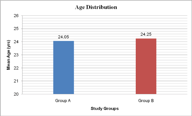
The average age of the participants in Group A (experimental) was 24.05 ± 2.04 years and in Group B (control) was 24.25 ± 1.82 years. There was no significant difference between the mean ages of the participants in both the groups. (Table1 & Figure1)
|
Groups
|
Mean Age(Yrs) + SD
|
|
Group A (Experimental)
|
24.05 (± 2.04)
|
|
Group B (Control)
|
24.25 (±1.82)
|
Table 1. Mean Age distribution in study groups
Outcome measures
VAS SCORE (in centimeters): The VAS score pre- and post-intervention in Group A were 6.29 ± 1.14 and 5.23 ± 1.12 on Day 1; 4.65 (± 0.94) and 3.64 (± 0.80) on Day 2; 3.08 (± 0.87) and 2.13 (± 0.74) on Day 3; 1.66 (± 0.71) and 0.58 (± 0.49) on Day 4. The VAS score pre- and post- intervention in Group B were 6.51 (± 1.10) and 5.65 (± 1.04) on Day 1; 4.80 (± 1.04) and 4.15 (± 1.01) on Day 2; 3.37 (± 0.96) and 2.80 (± 0.84) on Day 3; 2.25 (± 0.74) and 1.46 (± 0.44) on Day 4. VAS score was statistically significant post-intervention day 2 and day 3 in both groups and also pre-intervention and post-intervention day 4 in both groups as shown in Table 2 & Figures 2 and 3.
|
Visit
|
Intervention
|
Group A
|
Group B
|
p- Value
|
Inference
|
|
Day 1
|
Pre
|
6.29 (± 1.14)
|
6.51 (± 1.10)
|
0.488
|
Statistically not significant
|
|
Post
|
5.23 (± 1.12)
|
5.65 (± 1.04)
|
0.156
|
Statistically not significant
|
|
Day 2
|
Pre
|
4.65 (± 0.94)
|
4.80 (± 1.04)
|
0.721
|
Statistically not significant
|
|
Post
|
3.64 (± 0.80)
|
4.15 (± 1.01)
|
0.032
|
Statistically significant
|
|
Day 3
|
Pre
|
3.08 (± 0.87)
|
3.37 (± 0.96)
|
0.236
|
Statistically not significant
|
|
Post
|
2.13 (± 0.74)
|
2.80 (± 0.84)
|
0.002
|
Statistically significant
|
|
Day 4
|
Pre
|
1.66 (± 0.71)
|
2.25 (± 0.74)
|
0.001
|
Statistically significant
|
|
Post
|
0.58 (± 0.49)
|
1.46 (± 0.44)
|
0.000
|
Statistically significant
|
Table 2. Mean and SD values of VAS score in Experimental and control groups
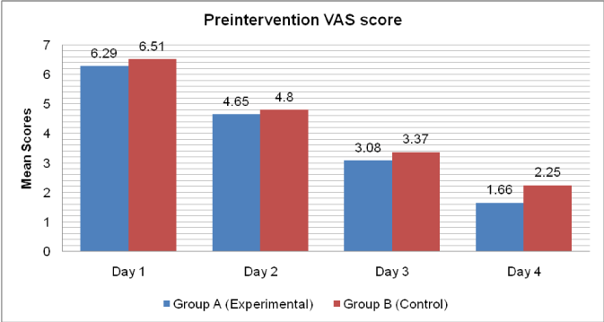
Figure 2. Pre-intervention VAS score between Group A and Group B at Day 1, Day 2, Day 3 and Day 4
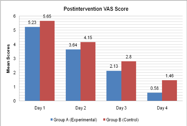
Figure 3. Post-intervention VAS score between Group A and Group B at Day 1, Day 2, Day 3 and Day 4
Hardness score (in centimeters)
The hardness score pre-intervention and post-intervention in Group A were 5.79 ± 1.22 and 4.76 ± 1.09 on Day 1; 4.19 (± 0.92) and 3.23 (± 0.88) on Day 2; 2.93 (± 0.91) and 1.93 (± 0.71) on Day 3; 1.93 (± 0.81) and 0.55 (± 0.43) on Day 4. The hardness score pre-intervention and post- intervention in Group B were 6.58 (± 0.90) and 5.72 (± 0.80) on Day 1; 4.97 (± 0.72) and 4.27 (± 0.61) on Day 2; 3.62 (± 0.59) and 3.03 (± 0.59) on Day 3; 2.42 (± 0.54) and 1.58 (± 0.44) on Day 4. Hardness score was statistically significant for all the four days in both groups as shown in Table 3 & Figures 4 and 5.
|
Visit
|
Intervention
|
Group A (Experimental)
|
Group B (Control)
|
p-Value
|
Inference
|
|
Day 1
|
Pre
|
5.79 (± 1.22)
|
6.58 (± 0.90)
|
0.0001
|
Statistically significant
|
|
Post
|
4.76 (± 1.09)
|
5.72 (± 0.80)
|
0.000
|
Statistically significant
|
|
Day 2
|
Pre
|
4.19 (± 0.92)
|
4.97 (± 0.72)
|
0.000
|
Statistically significant
|
|
Post
|
3.23 (± 0.88)
|
4.27 (± 0.61)
|
0.000
|
Statistically significant
|
|
Day 3
|
Pre
|
2.93 (± 0.91)
|
3.62 (± 0.59)
|
0.000
|
Statistically significant
|
|
Post
|
1.93 (± 0.71)
|
3.03 (± 0.59)
|
0.000
|
Statistically significant
|
|
Day 4
|
Pre
|
1.93 (± 0.81)
|
2.42 (± 0.54)
|
0.000
|
Statistically significant
|
|
Post
|
0.55 (± 0.43)
|
1.58 (± 0.44)
|
0.000
|
Statistically significant
|
Table 3. Mean and SD values of Hardness scale in Experimental and control groups
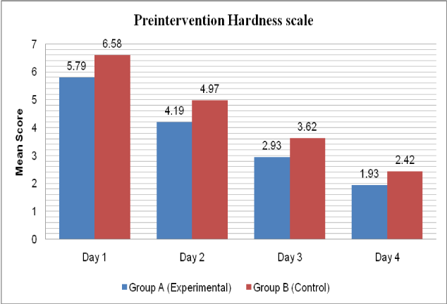
Figure 4. Pre-intervention Hardness scale between Group A and Group B at Day 1, Day 2, Day 3 and Day 4
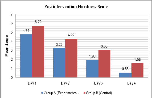
Figure 5. Post-intervention Hardness scale between Group A and Group B at Day 1, Day 2, Day 3 and Day 4
Six point engorgement scale (SPES):
The SPES score pre- and post-intervention in Group A were 5.52 ± 1.21 and 4.47 ± 1.19 on Day 1; 4.00 (± 1.08) and 3.12 (± 0.94) on Day 2; 2.75 (± 0.93) and 2.11 (± 0.75) on Day 3; 1.65 (± 0.75) and 0.54 (± 0.45) on Day 4. The SPES score pre-intervention and post intervention in Group B were 6.33 (± 0.98) and 5.51 (± 0.78) on Day 1, 4.79 (± 0.84) and 4.27 (± 1.01) on Day 2, 3.52 (± 0.84) and 2.91 (± 0.73) on Day 3, 2.31 (± 0.66) and 1.52 (± 0.46) on Day 4. SPES score was statistically significant for all the four days in both groups as shown in Table 4 & Figure 6 and 7.
|
Visit
|
Intervention
|
Group A (Experimental)
|
Group B (Control)
|
p Value
|
Inference
|
|
Day 1
|
Pre
|
5.52 (± 0.21)
|
5.33 (± 0.18)
|
0.002
|
Statistically significant
|
|
Post
|
4.47 (± 0.19)
|
4.51 (± 0.78)
|
0.000
|
Statistically significant
|
|
Day 2
|
Pre
|
4.00 (± 1.08)
|
4.79 (± 0.84)
|
0.000
|
Statistically significant
|
|
Post
|
3.12 (± 0.94)
|
4.27 (± 1.01)
|
0.000
|
Statistically significant
|
|
Day 3
|
Pre
|
2.75 (± 0.93)
|
3.52 (± 0.84)
|
0.000
|
Statistically significant
|
|
Post
|
2.11 (± 0.75)
|
2.91 (± 0.73)
|
0.000
|
Statistically significant
|
|
Day 4
|
Pre
|
1.65 (± 0.75)
|
2.31 (± 0.66)
|
0.002
|
Statistically significant
|
|
Post
|
1.24 (± 0.14)
|
1.52 (± 0.46)
|
0.000
|
Statistically significant
|
Table 4. Mean and SD values of SPES in experimental and control groups
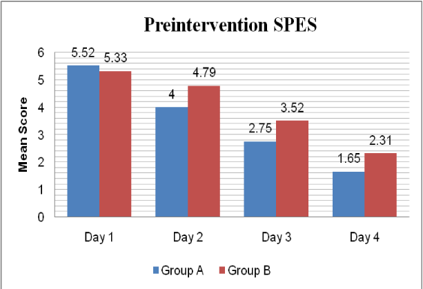
Figure 6. Pre-intervention SPES between Group A and Group B at Day 1, Day 2, Day 3 and Day 4
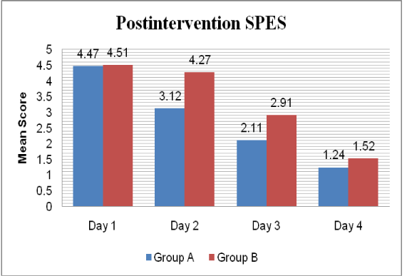
Figure 7. Post-intervention SPES between Group A and Group B at Day 1, Day 2, Day 3 and Day 4
The inter-group analysis for VAS change from Day1 to Day 4 pre-intervention for group A and B was 4.62 (± 1.11) and 4.25 (± 0.80). The change from Day1 post- to Day 4 post-intervention for group A and B was 4.65 (± 1.01) and 4.19 (± 0.81). For hardness scale change from Day1 to Day 4 pre-intervention for group A and B was 4.06 (± 1.33) and 4.16 (± 0.76). Change from Day 1 post- to Day 4 post-intervention for group A and B was 4.21 (± 1.05) and 4.14 (± 0.73). For Six-point engorgement scale change from Day1 to Day 4 pre-intervention for group A and B was 3.87 (± 1.09) and 4.01 (± 0.77). The change from Day 1 post- to Day 4 post-intervention for group A and B was 3.93 (± 1.07) and 3.99 (± 0.61). The change in VAS from Day 1 to Day 4 pre-intervention and Day 1 to Day 4 post-intervention was statistically significant for both groups, whereas for the hardness scale and six-point engorgement scale (SPES) it was not significant as shown in Table 5 & Figure 8.
|
Parameters
|
Comparison
|
Group
|
Mean (± SD)
|
p value
|
Inference
|
|
VAS
|
Change from Day 1(Pre) to Day 4 (Pre)
|
A
|
4.62 (± 1.11)
|
0.036
|
Statistically significant
|
|
B
|
4.25 (± 0.80)
|
|
Change from Day 1(Post) to Day 4 (Post)
|
A
|
4.65 (± 1.01)
|
0.022
|
Statistically significant
|
|
B
|
4.19 (± 0.81)
|
|
Hardness scale
|
Change from Day 1(Pre) to Day 4 (Pre)
|
A
|
4.06 (± 1.33)
|
0.893
|
Statistically not significant
|
|
B
|
4.16 (± 0.76)
|
|
Change from Day 1(Post) to Day 4 (Post)
|
A
|
4.21 (± 1.05)
|
0.771
|
Statistically not significant
|
|
B
|
4.14 (± 0.73)
|
|
SPES
|
Change from Day 1(Pre) to Day 4 (Pre)
|
A
|
3.87 (± 1.09)
|
0.524
|
Statistically not significant
|
|
B
|
4.01 (± 0.77)
|
|
Change from Day 1(Post) to Day 4 (Post)
|
A
|
3.93 (± 1.07)
|
0.552
|
Statistically not significant
|
|
B
|
3.99 (± 0.61)
|
Table 5. Comparison of Groups with change in VAS, Hardness scale, SPES from Day 1 to Day 4 by Mann-Whitney U test
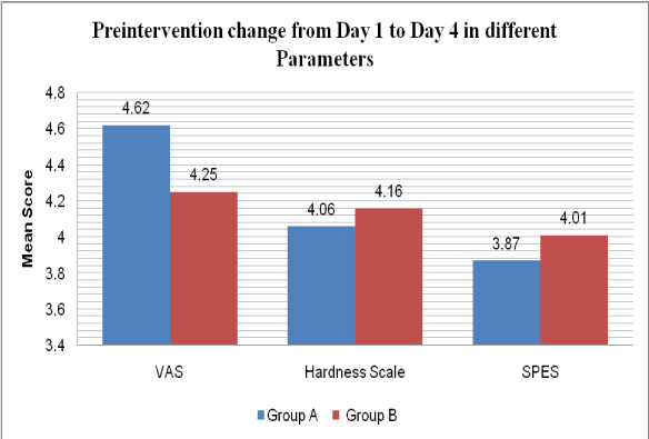
Figure 8. Comparison of Pre-intervention change in parameters from Day 1 to Day 4
The intra group analysis for VAS change from Day 1 pre-intervention to Day 4 post-intervention was 6.29 (± 1.14) and 0.58 (± 0.49) for group A and 6.51 (± 1.10) and 1.46 (± 0.44) for group B. For hardness scale change from Day 1 pre-intervention to Day 4 post-intervention was 5.79 (± 1.22) and 0.55 (± 0.43) for group A and 6.58 (± 0.90) and 1.58 (± 0.44) for group B. For Six-point engorgement scale (SPES) change from Day 1 pre-intervention to Day 4 post-intervention was 5.52 (± 1.21) and 0.54 (± 0.45) for group A and 6.33 (± 0.98) and 1.52 (± 0.46) for group B. Change in VAS, hardness scale, and six-point engorgement scale (SPES) was statistically significant for both groups as shown in Tables 6-8 & Figure 9-11).
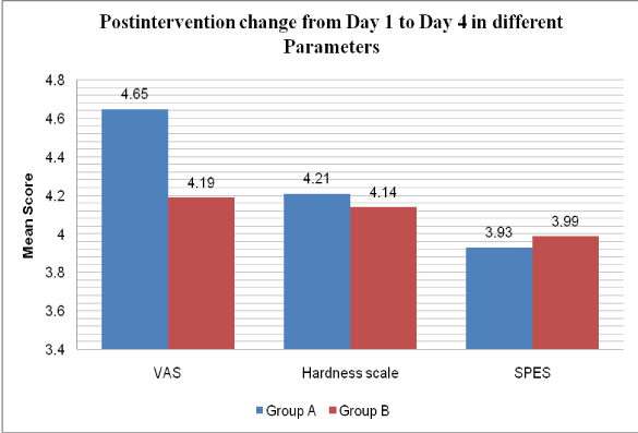
Figure 9. Comparison of Post-intervention change in parameters from Day 1 to Day 4

Figure 10. Intra-group comparison in group A
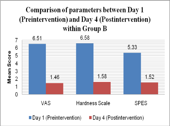
Figure 11. Intra-group comparison in group B
|
Parameters
|
Pre Day 1
|
Post Day 4
|
P-value
|
Inference
|
|
VAS
|
6.29 (± 1.14)
|
0.58 (± 0.49)
|
0.000
|
Statistically significant
|
|
Hardness Scale
|
5.79 (± 1.22)
|
0.55 (± 0.43)
|
0.000
|
Statistically significant
|
|
SPES
|
5.52 (± 0.21)
|
1.24 (± 0.14)
|
0.000
|
Statistically significant
|
Table 6. Comparison of Visual Analogue scale, Hardness scale and SPES between Day 1(Pre- intervention) and Day 4 (Post-intervention) within Group A using Wilcoxon Signed Rank test
|
Parameters
|
Pre Day 1
|
Post Day 4
|
P-value
|
Inference
|
|
VAS
|
6.51 (± 1.10)
|
1.46 (± 0.44)
|
0.000
|
Statistically significant
|
|
Hardness Scale
|
6.58 (± 0.90)
|
1.58 (± 0.44)
|
0.000
|
Statistically significant
|
|
SPES
|
5.33 (± 0.18)
|
1.52 (± 0.46)
|
0.000
|
Statistically significant
|
Table 7. Comparison of Visual Analogue scale, Hardness scale and SPES between Day 1(Pre- intervention) and Day 4 (Post-intervention) within Group B using Wilcoxon Signed Rank test
|
Parameters
|
Groups
|
Day 1
(Pre intervention)
|
Day 4
(Post intervention)
|
p-value
|
Inference
|
|
VAS
|
A
|
6.29 (± 1.14)
|
0.58 (± 0.49)
|
0.000
|
Statistically significant
|
|
B
|
6.51 (± 1.10)
|
1.46 (± 0.44)
|
0.000
|
Statistically significant
|
|
Hardness Scale
|
A
|
5.79 (± 1.22)
|
0.55 (± 0.43)
|
0.000
|
Statistically significant
|
|
B
|
6.58 (± 0.90)
|
1.58 (± 0.44)
|
0.000
|
Statistically significant
|
|
SPES
|
A
|
5.52 (± 0.21)
|
1.24 (± 0.14)
|
2021 Copyright OAT. All rights reserv
0.000
|
Statistically significant
|
|
B
|
5.33 (± 0.18)
|
1.52 (± 0.46)
|
0.000
|
Statistically significant
|
Table 8. Comparison of Visual Analogue scale, Hardness scale and SPES between Day 1(Pre- intervention) and Day 4 (Post-intervention) within groups using Wilcoxon Signed Rank test
Discussion
The present controlled trial was conducted to study the effectiveness of ultrasound therapy and conventional therapy in the treatment of breast engorgement in immediate post-partum mothers in terms of reduction of pain using VAS, Hardness scale and Six-point engorgement scale (SPES).
The results from of the present study supported alternative hypothesis which stated that there will be significant difference in terms of pain reduction using VAS, Hardness scale or Six-point engorgement scale with Ultrasound therapy added to conventional therapy.
In the present study age distribution showed no statistical difference in both the groups which suggests that the subjects mean age in both groups were same. The modality used in Group A was ultrasound machine. The sound waves produced by US cause cavitations and acoustic micro-streaming in the tissue, which essentially is a micro-massage for the individual cells. This helps the tissue to heal more efficiently. Both of these are non-thermal effects, which pose no safety concerns. The result of the combined effect of stable cavitations and acoustic streaming is that the cell membrane becomes excited (up regulated), this increases the activity levels of the whole cell. The US energy acts as a trigger for this process, but it is the increased cellular activity which is in effect responsible for the therapeutic benefits of the modality (Dinno et al. 1989) [10].
In the present research reduction in pain level, as quantified by the VAS, with ultrasound is consistent with the findings of McLachlan and collaborators, who conducted a study using two ultrasound machines one working normally, the other having crystal with a resistor producing superficial heat were used. Assessment of effectiveness was subjective, using visual analogue scale and hardness scale was measured pre-intervention and post-intervention and objective using tonometry. Results indicated that both true and sham machines were effective in reducing subjective perceived pain and hardness. However, the effect was attributed to the heat and massage effect and not to the ultrasound component. In this pre-intervention and pos-intervention scores after a single session of treatment helped in reduction of pain and non tender breast. The inter group comparison of VAS scores and hardness scores in both the studies shows significant reduction [11].
It is possible that lower or higher dosages of ultrasound, different durations of treatment or the use of pulsed instead of continuous wave might have a therapeutic effect beyond the placebo effects detected in this trial. More controlled trials are needed to test modifications of dosage and its effect on the condition.
The findings of the present study are in contrast with the earlier study conducted by Shellshear et al. on breast engorgement after delivery using continuous ultrasound for 10 min at 2.75 W/cm2 was given for primipara and continuous ultrasound for 6 minutes at 2.0 W/cm2 was given for multipara women for 1 days. Continuous US were given prior to feeding the baby and the breasts were comfortable and soft after feeding. Two sessions of US was given in a day. In this study the tenderness was reduced after the 2nd session of the treatment and breasts were non-tender and mother had no difficulty in feeding. In present study pulsed US was given with treatment a single session in a day. With the use of pulsed ultrasound the mothers showed significant improvement whereas with pulsed US the significance was not achieved after 1st day of treatment though the values were significant on 2nd day post-intervention [12].
The results of the present study are in consistent with recent study conducted by Chiu at al. in 54 women, who received Gua-Sha therapy on selected appropriate acupoint positions. Each position was lightly scraped seven times in two cycles. For the control group, hot packs and massage for 10 min each was given. Results concluded that, Gua-Sha therapy may be used as an effective technique in the management of breast engorgement [13].
Recent trials conducted by Snowden et al. involving on breast engorgement during lactation indicated that ultrasound is equally effective with or without ultra-violet emitting crystals; same is for the cabbage leaves and hot packs, and pharmacological treatment for breast engorgement. Duration of treatment in each group was different as the form of treatment is varied in this study. The results obtained showed that the engorgement and tenderness was reduced with all the treatment the same with hardly any difference in between the groups [14].
Heat promotes the opening or widening of the milk ducts. This can dislodge the ducts paving way for the release of the breast milk. Pain is reduced by massage by the stimulation of sensory nerve endings which blocks the pathway of pain in accordance with the Melzack and Wall’s theory of pain gate. The mechanical movement of massage stretches the individual fibers of soft tissue reduces their tension. Removal of metabolic wastes results due to increased drainage of the massaged area. This results in the reduction of pain, as these substances are noxious to the tissue and irritate the free nerve endings. Increased blood flow following massage reduces the anoxic condition present in the tissue due to compression of the blood vessels produced by the sustained muscular contraction. Thus, it reduces the danger of increased damage. In case of breast engorgement, there is edema due to vascular and lymphatic stasis. If no relief is obtained, milk production is interrupted, with later reabsorption of residual milk. The increase in intraductal pressure causes the residual milk to undergo an intermolecular transformation and to become thicker [5,15].
The present study is consistent with the findings of Jones et al., who studied either sequential pumping with no massage or sequential pumping with massage, simultaneous pumping with no-massage, or simultaneous pumping with massage in various receiving groups. The results indicated that simultaneous pumping is more effective than sequential pumping and breast massage has an additive effect, improving milk production in both. Gentle massage from the outer edge of the breast inward toward the outer edge of the nipple in small, soft circles was given in this study which is comparable with present study as same gentle massage was used [16].
Hence, the present study can provide evidence for the use of both US along with conventional therapy including hot moist pack and massage in the management of pain and improving lactation in breast engorgement in immediate post-partum mothers.
Conclusion
The study concluded that ultrasound therapy added with conventional therapy helps in reduction of pain with non-tender breasts which further helps the post-partum mothers to recover better from discomforts of breast engorgement. This in turn can facilitate better breast feeding.
Ethics committee approval
This study was approved by KIPT institutional ethics committee on Human Subjects Research.
References
- Humenick SS, Hill PD, Anderson MA (1994) Breast engorgement: patterns and selected outcomes. J Hum Lact 10: 87-93. [Crossref]
- Lee WT, Lui SS, Chan V, Wong E, Lau J (2006) A population-based survey on infant feeding practice (0-2 years) in Hong Kong: Breastfeeding rate and patterns among 3,161 infants below 6 months old. Asia Pac J Clin Nutr 15: 377-387. [Crossref]
- Arora S, Vatsa M, Dadhwal V (2008) A Comparison of Cabbage Leaves vs. Hot and Cold Compresses in the Treatment of Breast Engorgement. Indian J Community Med 33: 160-162. [Crossref]
- de Sousa L, Haddad ML, Nakano AM, Gomes FA (2012) [A non-pharmacologic treatment to relieve breast engorgement during lactation: an integrative literature review]. Rev Esc Enferm USP 46: 472-479. [Crossref]
- Dawson EK (1935) Histological study of normal mamma in relation to tumor growth; mature gland in lactation and pregnancy. Edinburgh Med J 42: 569.
- Marti A, Feng Z, Altermatt HJ, Jaggi R (1997) Milk accumulation triggers apoptosis of mammary epithelial cells. Eur J Cell Biol 73: 158-165. [Crossref]
- Hill PD, Humenick SS (1994) The occurrence of breast engorgement. J Hum Lact 10: 79-86. [Crossref]
- Academy of Breastfeeding Medicine Protocol Committee, Eglash A (2010) ABM clinical protocol #8: human milk storage information for home use for full-term infants (original protocol March 2004; revision #1 March 2010). Breastfeed Med 5: 127-130. [Crossref]
- Robson BA (1990) Breast engorgement in breast feeding mothers. Doctoral dissertation, Case Western Reserve University, Cleveland, Ohio.
- Dinno MA, Crum LA, Wu J (1989) The effect of therapeutic ultrasound on electrophysiological parameters of frog skin. Ultrasound Med Biol 15: 461-470. [Crossref]
- McLachlan Z, Milne EJ, Lumley J, Walker BL (1991) Ultrasound treatment for breast engorgement: A randomised double blind trial. Aust J Physiother 37: 23-28. [Crossref]
- Shellshear M (1981) Therapeutic ultrasound in post-partum breast engorgement. Aust J Physiother 27: 15-16. [Crossref]
- Chiu JY, Gau ML, Kuo SY, Chang YH, Kuo SC, et al. (2010) Effects of Gua-Sha therapy on breast engorgement: a randomized controlled trial. J Nurs Res 18: 1-10. [Crossref]
- Snowden HM, Renfrew MJ, Woolridge MW (2001) Treatments for breast engorgement during lactation. Cochrane Database Syst Rev 2: CD000046. [Crossref]
- Smillie C M (2004) The Prevention and Treatment of plugged Ducts. Clinical Handouts. Stratford, CT: Breast Feeding Resource.
- Jones E, Dimmock PW, Spencer SA (2001) A randomised controlled trial to compare methods of milk expression after preterm delivery. Arch Dis Child Fetal Neonatal Ed 85: F91-F95. [Crossref]