Expanded or small organizing hematoma is found everywhere in the body and frequently simulates a neoplasm. It has a central mass of blood, a wall of granulation tissue, and a dense, fibrous tissue at the periphery. We report a case of an enormous organizing hematoma of the left carotid neck triangle, in a patient without a history of trauma in the neck or blood disorders and also review the literature.
organizing hematoma; neck; carotid triangle
Most hematomas resolve without causing notable clinical problems, but some may persist and appear as slowly expanding lesions that simulate neoplasms. The last lesions occurring have been described as chronic expanding organizing hematoma. The term “organizing” explains the histologic findings such as fibrous tissue, neovascularization and extravasated red blood cells. The formation of a fibrous capsule around the hematoma prevents its absorption and allows for recurrent intracapsular bleeding and progressive expansion [1]. The organizing hematoma was first described by Tadokoro in 1917 [2]. Sporadic cases have been reported in the head and neck.
The carotid triangle is the most important anatomic space of the neck, comprising carotid artery, jugular vein and vagus nerve. Most tumors of this region are benign and are mainly from the carotid body. Other lesions of this region include carotid artery aneurysm, branchial cleft cyst, thyroid tumor, enlarged lymph nodes and parotid gland tumor [3].
In this paper, was port the first case of an enormous organizing hematoma appearing as a carotid triangle tumor that displaced the trachea in the precisely opposite side of the neck and also review the literature.
A man aged 60 years was admitted to the outpatient department of Maxillofacial Surgery Clinic complaining of discomfort in breathing because of a mass in the neck, without any history of blunt trauma or operation. The mass appeared on the neck 25 years ago, gradually increased in the first ten years and remained stable in size. Physical examination showed an enormous sub-hard mass in the left carotid triangle area (Figure 1A).Computed tomography (CT) ,CT angiography and 3D CT showed a well-defined mass lesion, about 8 × 8 × 7 cm in size, manifesting calcification and heterogeneous enhancement involving the left carotid triangle with extension to the larynx, trachea and left clavicle region, displacing the trachea to opposite right neck carotid triangle (Figure 2).The relationship between the mass and the thyroid gland was unclear, but the SPECT showed that the left thyroid lobe was atrophic. Laboratory data for thyroid gland function were within normal limits. At laryngoscopy the movement of the vocal cords was normal, and showed no lesions. The coagulation tests showed normal platelet number, prothrombin time (PT) and activated partial thromboplastin time (aPTT). Open biopsy was performed and the histology showed necrosis. The patient was advised to have undergone a total excision of the mass. The anesthesia process was difficult because of the trachea displacement, so a fiber optic endoscope and a video camera were used for safe intubation of the patient. The whole neck tumor was excised smoothly with intact capsule via a broad submandibular approach with a T-shaped extension to the supraclavicular fossa. For the safeguarding of the pressed by the tumor recurrent laryngeal nerve a NIM (neurostimulator) was used. Macroscopically, the specimen was a circumscribed mass measuring 9X6.5X6.5cm with relatively smooth surface and fascia attachment (Figure 3). Sectioning revealed a cavity with hemorrhagic content, surrounded by a thick fibrous capsule with partly chalky texture. After extensive sampling, histology failed to show epithelial, endothelial or mesothelial lining. Immunohistochemistry against CK8/18, CD31 and D2-40 antibodies was negative, confirming the H&E light microscopic appearance. Cavity content was made of fibrin and clot. Fibrous capsule showed hemosiderin laden macrophages, mast cells, cholesterol crystals and calcifications. These features are compatible with an organized hematoma (Figure 4).
The post-operative course was uneventful (Figure 1B), except a bit of hoarseness that disappeared without treatment two months postoperatively. The cranial nerves function was normal, and there was no sign of recurrence at the one year follow-up.
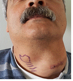
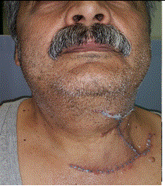
Figure 1. Clinical view of the patient. A) Preoperatively. B) Postoperatively
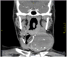
Figure 2. Preoperative CT. A) Axial view. B) Coronal view.
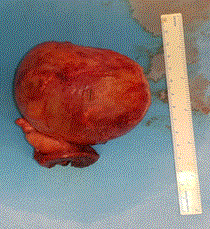
Figure 3. The specimen macroscopically.
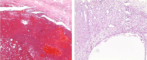
Figure 4. H&E shows a cavity with hemorrhagic content, surrounded by a thick fibrous capsule H&E and immunohistochemistry was negative for any lining.
A hematoma characterized as organized when surrounded by a fibrous capsule. The first stage of organized hematoma formation is the extravasation of red blood cells between vascular endothelial cells, followed by necrosis, hyalinization, neovascularization and fibrosis. Because of the slow blood flow, the newly formed vessels are dilated and rebleeded. On the other hand, the fibrous capsule around the hematoma prevents the absorption and facilitates its progressive expansion. Various causes can lead to the initial blood collection, such as bleeding diathesis, postoperative complication, radiation therapy and trauma [1,4,5].Our patient, despite our persistent question, did not report us a neck injury, while the hematological tests were normal
Many authors have reported several cases of organized hematoma in the head and the neck (Table 1). It is appeared in all age groups (2-85 years) with a slight predominance in males (57%). The vast majority is located in the paranasal sinuses, especially the maxillary sinus (90%), while other locations include the nasal cavity, the lower eyelid and the parapharyngeal space. In 1975 Helidonis and Myers [26] first reported an organized hematoma of the neck, in a man 55 years old, simulating carotid body tumor and displacing the internal and external carotid arteries. To our knowledge, we refer to the second case of organized hematoma in the carotid triangle as Helidonis and Myers have reported, but the first with such a large size that caused significant displacement of adjacent anatomical structures to the opposite side, especially the trachea.
Table 1. Summary of literature review
AUTHOR (year) |
PATIENTS No |
GENDER |
AGE |
LOCATION |
Kim et al. 2016 [6] |
23 |
M 15 F 8 |
18-75 y |
Antrum 15 p, Septum 4 p, Inferior turbinate 2 p, Ethmoid sinus 2 p |
Pang et al. 2016 [7] |
84 |
M 39 F 45 |
9-81 y |
Antrum 82 p, nasal cavity 2 p |
Lin et al. 2016 [8] |
1 |
F 1 |
81y |
Sphenoid sinus 1 p |
Oubahmane et al. 2016 [9] |
1 |
F 1 |
28y |
Lower eyelid 1 p |
Park and Kim 2015A [10] |
1 |
M 1 |
40 y |
Inferior turbinate 1 p |
Park and Kim 2015B [11] |
1 |
M1 |
55Y |
Antrum a1 p |
Imayoshi et al. 2015 [12] |
3 |
M 3 |
16-40y |
Antrum 3 p |
Choi et al. 2015 [13] |
7 |
M 10 F 7 |
17-74y |
Antrum 15 p, frontal sinus 1 p, sphenoid sinus 1 p |
Cho et al. 2015 [14] |
1 |
M 1 |
2y |
Antrum 1 p |
Yokoi et al. 2014 [15] |
5 |
M 3 F 2 |
19-68y |
Antrum 5 p |
Almasoud et al. 2014 [16] |
1 |
M1 |
26y |
Antrum 1 p |
Ohta et al. 2013 [17] |
5 |
M 3 F 2 |
14-56y |
Antrum 5 p |
Omura et al. 2010 [3] |
6 |
M 5 F 1 |
26-56y |
Antrum 5 p, nasal cavity 1 p |
Nakagawa et al. 2010 [18] |
1 |
F 1 |
85y |
Sphenoid sinus 1 p |
Hsu et al. 2009 [19] |
1 |
M 1 |
72y |
Parapharyngeal space 1p |
Suzuki et al. 2008 [20] |
3 |
M 2 F 1 |
50-62y |
Antrum 3 p |
Kim et al. 2008 [2] |
12 |
M 9 F 3 |
12-76y |
Antrum 12 p |
Song et al. 2007 [1] |
20 |
M14 F6 |
16-67 |
Antrum 20 p |
Nishiguchi et al. 2007 [21] |
2 |
M 2 |
22-76y |
Antrum 2 p |
Yoon et al 2006 [22] |
3 |
M 1 F 2 |
51-70y |
Antrum 3 p |
Lee et al. 2003 [23] |
8 |
M 5 F 3 |
18-67y |
Antrum 8 p |
Tabaee and Kacker 2002 [24] |
1 |
M 1 |
18y |
Antrum 1 p |
Unlu et al. 2001 [25] |
2 |
M 1 F 1 |
42-78y |
Antrum 2 p |
Helidonis and Myers 1975 [26] |
1 |
M1 |
55y |
Carotid triangle 1 p |
M: Males, F: Females, y: years, p: patients
The most tumors of the carotid triangle usually present as a palpable mass, painless and slow growing, whereas if the mass grown patient enough may complain of shortness of breath and dysphagia, as our patient who complained of discomfort in breathing [3]. Our findings on CT are consistent with those of other authors, such as heterogeneous or homogeneous lowly attenuated lesions with or without calcification. Here we would like to emphasize that the angiography is an additional examination choice when suspected vascular lesion, to define its blood supply and the site of origin [19,27].
The treatment of choice is surgical removal of the lesion. The surgical planning depends on its size and location as well as by its relation to the great vessels. If a small lesion is located in the maxillary sinus, usually the endoscopic sinus surgery is the treatment of choice. In other lesions, such as in our case, wide excision is used with open approach [19,16].
This research received no specific grant from any funding agency in the public, commercial, or not for profit sectors.
All authors declare they have no any conflict of interests
None acknowledgements
- Song HM, Jang YJ, Chung YS, Lee BJ (2007) Organizing hematoma of the maxillary sinus. Otolaryngol Head Neck Surg 136: 616-620. [Crossref]
- Kim EY, Kim HJ, Chung SK, Dhong HJ, Kim HY, et al. (2008) Sinonasal organized hematoma: CT and MR imaging findings. AJNR Am J Neuroradiol 29: 1204-1208. [Crossref]
- Kaman L, Singh R, Aggarwal R, Kumar R, Behera A, et al. (1999) Diagnostic and therapeutic approaches to carotid body tumours: report of three cases and review of the literature. Aust N Z J Surg 69: 852-855. [Crossref]
- Omura G, Watanabe K, Fujishiro Y, Ebihara Y, Nakao K, et al. (2010) Organized hematoma in the paranasal sinus and nasal cavity-imaging diagnosis and pathological findings. Auris Nasus Larynx 37:173-177. [Crossref]
- Urata S, Ohki M, Ts2021 Copyright OAT. All rights reservaematoma of the maxillary sinus: pathophysiological differences suggesting a new aetiological hypothesis. J Laryngol Otol 127: 519-524. [Crossref]
- Kim JS, Oh JS, Kwon SH (2016) The increasing incidence of paranasal organizing hematoma: a 20-year experience of 23 cases at a single center. Rhinology 54:176-182. [Crossref]
- Pang W, Hu L, Wang H, Sha Y, Ma N, et al. (2016) Organized Hematoma: An Analysis of 84 Cases with Emphasis on Difficult Prediction and Favorable Management. Otolaryngol Head Neck Surg 154: 626-633. [Crossref]
- Lin YH, Wang PC, Lin YS (2016) Sphenoid sinus organized hematoma with cranial neuropathies masquerading as a malignancy: A case report. Oncol Lett 11: 3571-3574. [Crossref]
- Oubahmane, T, Abou-elfadl M, Mahtar M, Kadiri F ( 2016) Unusual site of organized haematoma in a context of chronic myeloid leukaemia. Eur Ann Otorhinolaryngol Head Neck Dis 133: 221-222. [Crossref]
- Park SY, Kim KS (2015) Giant Organized Hematoma Originating From the Inferior Turbinate. Iran J Radiol 12: e12366. [Crossref]
- Park YK, Kim KS (2015) Organizing Hematoma of the Maxillary Sinus Mimicking Malignancy Diagnosed by Fluorodeoxyglucose Positron-Emission Tomography (FDG PET/CT): A Case Report. Iran J Radiol 30: e18924. [Crossref]
- Imayoshi S, Kanazawa T, Fukushima N, Nishino H (2015) Three cases of organized hematoma of the maxillary sinus: clinical features and immunohistological studies for vascular endothelial growth factor and vascular endothelial growth factor receptor 2 expressions. Case Rep Otolaryngol
- Choi SJ, Seo ST, Rha KS, Kim YM (2015) Sinonasal organized hematoma: Clinical features of seventeen cases and a systematic review. Laryngoscope 125: 2027-2033.
- Cho YA, Kwon IJ, Kim SM, Myoung H, Lee JH, et al. (2015) A rare pediatric variant of organized hematoma in the maxillary sinus. J Oral MaxfacSurg Med Pathol 27: 544-549.
- Yokoi H, Arakawa A, Matsumoto F, Yokoi N, Ikeda K, et al. (2014) Organized hematoma of the maxillary sinus: a clinicopathologic study of 5 cases. Ear Nose Throat J 93: E23-26. [Crossref]
- Almasoud M, Alhumaidan A, Ashoor M (2014) Maxillary sinus hematoma: Current pathogenesis and management. Egyptian J Ear, Nose, Throat and Allied Sciences 15: 37-40.
- Ohta N, Watanabe T, Ito T, Kubota T, Suzuki Y, et al. (2013) Clinical and pathological characteristics of organized hematoma. Int J Otolaryngol 2013: 539642. [Crossref]
- Nakagawa T, Kawai Y, Sakamoto T, Ito J (2010) Organised haematoma of the sphenoid sinus mimicking a pituitary tumour. J Laryngol Otol 124: 83-85. [Crossref]
- Hsu WS, Liu SF, Chu ST, Tseng HH (2009) An organizing hematoma in the parapharyngeal space. J Chin Med Assoc 72: 94-97. [Crossref]
- [Crossref] Suzuki H, Inaba T, Hiraki N, Hashida K, Wakasugi T, et al. (2008) Endoscopic sinus surgery for the treatment of organized hematoma of the maxillary sinus. Kurume Med J 55: 37-41.
- Nishiguchi T, Nakamura A, Mochizuki K, Tokuhara Y, Yamane H, et al. (2007) Expansile organized maxillary sinus hematoma: MR and CT findings and review of literature. AJNR Am J Neuroradiol 28: 1375-1377. [Crossref]
- Yoon TM, Kim JH, Cho YB (2006) Three cases of organized hematoma of the maxillary sinus. Eur Arch Otorhinolaryngol 263: 823-826. [Crossref]
- Lee BJ, Park HJ, Heo SC (2003) Organized hematoma of the maxillary sinus. Acta Otolaryngol 123: 869-872. [Crossref]
- Tabaee A, Kacker A (2002) Hematoma of the maxillary sinus presenting as a mass--a case report and review of literature. Int J Pediatr Otorhinolaryngol 65: 153-157. [Crossref]
- Unlu HH, Mutlu C, Ayhan S, Tarhan S (2001) Organized hematoma of the maxillary sinus mimicking tumor. Auris Nasus Larynx 28: 253-255. [Crossref]
- Helidonis E, Myers EN (1975) Organized hematoma of the neck simulating carotid body tumor. Int Surg 60: 519-520. [Crossref]
- Lee HK, Smoker WR, Lee BJ, Kim SJ, Cho KJ (2007) Organized hematoma of the maxillary sinus: CT findings. AJR Am J Roentgenol 188: W370-373. [Crossref]