Key words
epithelloid hemangiodendothelioma, interferon, autoimmune hepatitis, autoantibody
Case
A 23-year-old woman presented with complaints of jaundice, nausea with vomiting, and epigastric pain for ten days. She had a history of epithelioid hemangiodendothelioma (EHE) of the lungs, diagnosed six months prior to presentation. She had been treated with interferon alpha-2b (a type 1 interferon) for four months, but this was stopped two weeks prior to presentation due to lack of palliative benefit. She was taking oxycodone and polyethylene glycol.
On admission, she had a temperature of 36.1°C, a blood pressure of 115/75 mmHg, heart rate of 76 beats per minute, and an oxygen saturation of 96% on room air. She appeared in mild distress due to abominal pain. Her abdomen was soft but tender in in the right upper quadrant and epigastrium without rebound or guarding. Laboratory analysis was signifcant for an AST of 749 IU/L, ALT of 601 IU/L, alkaline phosphatase of 276 IU/L, total bilirubin of 11.1 mg/dL, and a direct bilirubin of 8.7 mg/dL. A right upper quandrant ultrasound revealed a few prominent lymph nodes in the territory of the porta hepatitis.
Her oncologist was concerned that there was hematogenous seeding of her epithelioid hemangiodendothelioma to the liver. Hepatitis antibodies, quantitative immunoglobulins, ceruloplasmin, ferritin, and acetaminophen level did not reveal an etiology of her liver function abnormalities. Anti-smooth muscle antibody (ASMA), liver-kidney-microsomal antibody (anti-LKM), antinuclear antibody (ANA), anti-mitochondrial antibody (AMA), and anti-soluble liver (anti-SLA) were nonreactive. Magnetic resonance cholangiopancreatography (MRCP) revealed nonspecific mild periportal edema (represented by arrows) and gallbladder wall thickening to 3.3 mm (with the upper limit of normal being 3mm) without obvious mass or biliary abnormality (Figure 1). After the initial negative workup, we proceeded to a liver biopsy. An intense lymphoplasmacytic portal infiltrate (encircled) was observed on the hematoxylin and eosin (H&E) stain (Figure 2). The trichome stain confirmed the diagnosis of autoimmune hepatitis as collagen stained a light blue (represented by arrows and a circle) suggesting a mild periportal fibrosis which is observed in all but the mildest cases of autoimmune hepatitis (Figure 3). Our patient’s hepatitis responded to a four month oral prednisone taper.
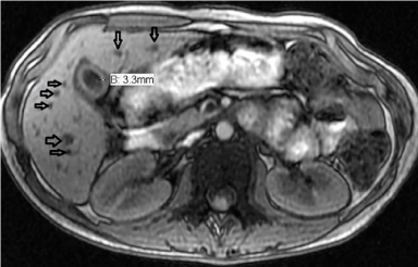
Figure 1. Mild periportal edema.
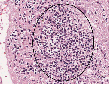
Figure 2. Lymphoplasmacytic portal infiltrate (encircled).
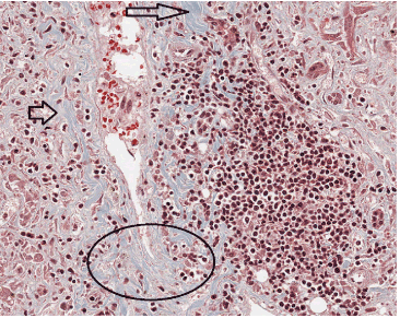
Figure 3. Mild periportal fibrosis.
Discussion
Our case of interferon induced autoimmune hepatitis was unusual in that the diagnosis was determined after cessation of interferon therapy and not associated with circulating autoantibodies, which are present in 80% of cases. Typically an asymptomatic rise in transaminases is noted in up to 25% patients treated with type 1 interferons and 80% of patients treated with high doses, although these abnormalities typically resolve with continued therapy or after dose reduction [1]. Type 1 interferons have a marked immunomodulatory effect and can lead to the development of autoimmune phenomena, exacerbate pre-existing autoimmunity, or unmask silent autoimmune processes [2]. Autoimmune hepatitis affects children and adults of all ages, but classically affects women between the ages of 15 and 40 and typically includes circulating autoantibodies and high serum globulin concentrations [3]. Diagnosis is made based on symptoms, positive autoantibodies (ASMA, anti-LKM, ANA, AMA, or anti-SLA), and liver biopsy confirmation. Glucocorticoids alone remain first line therapy as azathioprine may inherently affect liver function. Although a combination of low dose prednisone and azathioprine may be used as first line therapy in brittle diabetics, patients with osteoporosis, poorly controlled hypertension, and a history of psychosis as high dose steroids may not be tolerated by these patients.
Funding
None
References
- Quesada JR, Talpaz M, Rios A, Kurzrock R, Gutterman JU (1986) Clinical toxicity of interferons in cancer patients: a review. J Clin Oncol 4: 234-243. [Crossref]
- Papo T, Marcellin P, Bernuau J, Durand F, Poynard T, et al. (1992) Autoimmune chronic hepatitis exacerbated by alpha-interferon. Ann Intern Med 116: 51-53. [Crossref]
- Krawitt EL (2006) Autoimmune hepatitis. N Engl J Med 354: 54-66.